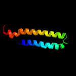
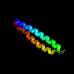
1 c2vs0B_
88.0
14
PDB header: cell invasionChain: B: PDB Molecule: virulence factor esxa;PDBTitle: structural analysis of homodimeric staphylococcal aureus2 virulence factor esxa
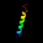
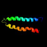
2 c3gvmA_
85.8
13
PDB header: viral proteinChain: A: PDB Molecule: putative uncharacterized protein sag1039;PDBTitle: structure of the homodimeric wxg-100 family protein from streptococcus2 agalactiae
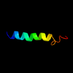
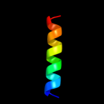
3 d1wa8b1
81.4
17
Fold: Ferritin-likeSuperfamily: EsxAB dimer-likeFamily: ESAT-6 like4 c3kdpG_
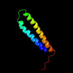
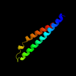
69.7
38
PDB header: hydrolaseChain: G: PDB Molecule: na+/k+ atpase gamma subunit transcript variant a;PDBTitle: crystal structure of the sodium-potassium pump
5 c3kdpH_
69.7
38
PDB header: hydrolaseChain: H: PDB Molecule: na+/k+ atpase gamma subunit transcript variant a;PDBTitle: crystal structure of the sodium-potassium pump
6 d1wa8a1
69.6
16
Fold: Ferritin-likeSuperfamily: EsxAB dimer-likeFamily: ESAT-6 like7 c1nfoA_
63.1
13
PDB header: lipid transportChain: A: PDB Molecule: apolipoprotein e2;PDBTitle: apolipoprotein e2 (apoe2, d154a mutation)
8 d1eq1a_
49.5
9
Fold: Apolipophorin-IIISuperfamily: Apolipophorin-IIIFamily: Apolipophorin-III9 c2jwaA_
35.6
21
PDB header: transferaseChain: A: PDB Molecule: receptor tyrosine-protein kinase erbb-2;PDBTitle: erbb2 transmembrane segment dimer spatial structure
10 c2kbbA_
32.0
15
PDB header: structural proteinChain: A: PDB Molecule: talin-1;PDBTitle: nmr structure of the talin rod domain, 1655-1822
11 d1gs9a_
30.2
6
Fold: Four-helical up-and-down bundleSuperfamily: ApolipoproteinFamily: Apolipoprotein12 c3hd7A_
28.6
12
PDB header: exocytosisChain: A: PDB Molecule: vesicle-associated membrane protein 2;PDBTitle: helical extension of the neuronal snare complex into the membrane,2 spacegroup c 1 2 1
13 c2l5bA_
26.5
33
PDB header: apoptosisChain: A: PDB Molecule: activator of apoptosis harakiri;PDBTitle: solution structure of the transmembrane domain of bcl-2 member2 harakiri in micelles
14 c3lf9A_
23.5
13
PDB header: immune systemChain: A: PDB Molecule: 4e10_d0_1is1a_001_c (t161);PDBTitle: crystal structure of hiv epitope-scaffold 4e10_d0_1is1a_001_c
15 d1v9da_
22.8
11
Fold: Formin homology 2 domain (FH2 domain)Superfamily: Formin homology 2 domain (FH2 domain)Family: Formin homology 2 domain (FH2 domain)16 c1iijA_
21.6
41
PDB header: signaling proteinChain: A: PDB Molecule: erbb-2 receptor protein-tyrosine kinase;PDBTitle: solution structure of the neu/erbb-2 membrane spanning2 segment
17 c2x43S_
21.2
10
PDB header: membrane proteinChain: S: PDB Molecule: sherp;PDBTitle: structural basis of molecular recognition by sherp at membrane2 surfaces
18 c3o3nB_
20.7
16
PDB header: lyaseChain: B: PDB Molecule: beta-subunit 2-hydroxyacyl-coa dehydratase;PDBTitle: (r)-2-hydroxyisocaproyl-coa dehydratase in complex with its substrate2 (r)-2-hydroxyisocaproyl-coa
19 c2dbiA_
20.3
6
PDB header: structural genomics, unknown functionChain: A: PDB Molecule: hypothetical protein ybiu;PDBTitle: crystal structure of a hypothetical protein jw0805 from2 escherichia coli
20 d1f16a_
20.0
26
Fold: Toxins' membrane translocation domainsSuperfamily: Bcl-2 inhibitors of programmed cell deathFamily: Bcl-2 inhibitors of programmed cell death21 c1av1B_
not modelled
19.9
16
PDB header: lipid transportChain: B: PDB Molecule: apolipoprotein a-i;PDBTitle: crystal structure of human apolipoprotein a-i
22 c3o4xF_
not modelled
19.5
9
PDB header: protein bindingChain: F: PDB Molecule: protein diaphanous homolog 1;PDBTitle: crystal structure of complex between amino and carboxy terminal2 fragments of mdia1
23 c3o4xE_
not modelled
19.5
9
PDB header: protein bindingChain: E: PDB Molecule: protein diaphanous homolog 1;PDBTitle: crystal structure of complex between amino and carboxy terminal2 fragments of mdia1
24 c2k14A_
not modelled
18.8
20
PDB header: unknown functionChain: A: PDB Molecule: yuaf protein;PDBTitle: solution structure of the soluble domain of the nfed2 protein yuaf from bacillus subtilis
25 c3lw5K_
not modelled
17.8
31
PDB header: photosynthesisChain: K: PDB Molecule: photosystem i reaction center subunit x psak;PDBTitle: improved model of plant photosystem i
26 c3r2pA_
not modelled
15.5
14
PDB header: lipid transportChain: A: PDB Molecule: apolipoprotein a-i;PDBTitle: 2.2 angstrom crystal structure of c terminal truncated human2 apolipoprotein a-i reveals the assembly of hdl by dimerization.
27 c2y44A_
not modelled
15.3
18
PDB header: membrane proteinChain: A: PDB Molecule: glutamic acid/alanine-rich protein;PDBTitle: crystal structure of garp from trypanosoma congolense
28 c2k9yB_
not modelled
15.2
28
PDB header: transferaseChain: B: PDB Molecule: ephrin type-a receptor 2;PDBTitle: epha2 dimeric structure in the lipidic bicelle at ph 5.0
29 c2z6eC_
not modelled
14.6
11
PDB header: protein fibril regulatorChain: C: PDB Molecule: disheveled-associated activator of morphogenesisPDBTitle: crystal structure of human daam1 fh2
30 d1nekd_
not modelled
14.4
7
Fold: Heme-binding four-helical bundleSuperfamily: Fumarate reductase respiratory complex transmembrane subunitsFamily: Succinate dehydrogenase/Fumarate reductase transmembrane subunits (SdhC/FrdC and SdhD/FrdD)31 c2k9yA_
not modelled
13.8
28
PDB header: transferaseChain: A: PDB Molecule: ephrin type-a receptor 2;PDBTitle: epha2 dimeric structure in the lipidic bicelle at ph 5.0
32 d1i6la_
not modelled
13.6
1
Fold: Adenine nucleotide alpha hydrolase-likeSuperfamily: Nucleotidylyl transferaseFamily: Class I aminoacyl-tRNA synthetases (RS), catalytic domain33 d2csga1
not modelled
13.3
8
Fold: Double-stranded beta-helixSuperfamily: Clavaminate synthase-likeFamily: YbiU-like34 c3e0sA_
not modelled
13.0
15
PDB header: structural genomics, unknown functionChain: A: PDB Molecule: uncharacterized protein;PDBTitle: crystal structure of an uncharacterized protein from2 chlorobium tepidum
35 c1p58F_
not modelled
12.4
18
PDB header: virusChain: F: PDB Molecule: envelope protein m;PDBTitle: complex organization of dengue virus membrane proteins as revealed by2 9.5 angstrom cryo-em reconstruction
36 c3peuB_
not modelled
11.5
13
PDB header: hydrolaseChain: B: PDB Molecule: nucleoporin gle1;PDBTitle: s. cerevisiae dbp5 l327v c-terminal domain bound to gle1 h337r and ip6
37 d1vcsa1
not modelled
10.7
15
Fold: STAT-likeSuperfamily: t-snare proteinsFamily: t-snare proteins38 c2j1dG_
not modelled
9.9
11
PDB header: protein bindingChain: G: PDB Molecule: disheveled-associated activator of morphogenesis 1;PDBTitle: crystallization of hdaam1 c-terminal fragment
39 c3sz3A_
not modelled
9.5
16
PDB header: ligaseChain: A: PDB Molecule: tryptophanyl-trna synthetase;PDBTitle: crystal structure of tryptophanyl-trna synthetase from vibrio cholerae2 with an endogenous tryptophan
40 d1lvfa_
not modelled
8.6
15
Fold: STAT-likeSuperfamily: t-snare proteinsFamily: t-snare proteins41 c2kncB_
not modelled
8.5
10
PDB header: cell adhesionChain: B: PDB Molecule: integrin beta-3;PDBTitle: platelet integrin alfaiib-beta3 transmembrane-cytoplasmic2 heterocomplex
42 c3prhB_
not modelled
8.3
3
PDB header: ligaseChain: B: PDB Molecule: tryptophanyl-trna synthetase;PDBTitle: tryptophanyl-trna synthetase val144pro mutant from b. subtilis
43 d1szia_
not modelled
8.2
17
Fold: Four-helical up-and-down bundleSuperfamily: Mannose-6-phosphate receptor binding protein 1 (Tip47), C-terminal domainFamily: Mannose-6-phosphate receptor binding protein 1 (Tip47), C-terminal domain44 d1ef1c_
not modelled
7.7
18
Fold: Non-globular all-alpha subunits of globular proteinsSuperfamily: Moesin tail domainFamily: Moesin tail domain45 d1ykhb1
not modelled
7.3
13
Fold: Mediator hinge subcomplex-likeSuperfamily: Mediator hinge subcomplex-likeFamily: CSE2-like46 c2pnvA_
not modelled
7.2
19
PDB header: membrane proteinChain: A: PDB Molecule: small conductance calcium-activated potassiumPDBTitle: crystal structure of the leucine zipper domain of small-2 conductance ca2+-activated k+ (skca) channel from rattus3 norvegicus
47 c2k1lA_
not modelled
7.2
17
PDB header: signaling proteinChain: A: PDB Molecule: ephrin type-a receptor 1;PDBTitle: nmr structures of dimeric transmembrane domain of the2 receptor tyrosine kinase epha1 in lipid bicelles at ph 6.3
48 c2k1lB_
not modelled
7.2
17
PDB header: signaling proteinChain: B: PDB Molecule: ephrin type-a receptor 1;PDBTitle: nmr structures of dimeric transmembrane domain of the2 receptor tyrosine kinase epha1 in lipid bicelles at ph 6.3
49 c2k1kA_
not modelled
7.2
17
PDB header: signaling proteinChain: A: PDB Molecule: ephrin type-a receptor 1;PDBTitle: nmr structures of dimeric transmembrane domain of the2 receptor tyrosine kinase epha1 in lipid bicelles at ph 4.3
50 c2k1kB_
not modelled
7.2
17
PDB header: signaling proteinChain: B: PDB Molecule: ephrin type-a receptor 1;PDBTitle: nmr structures of dimeric transmembrane domain of the2 receptor tyrosine kinase epha1 in lipid bicelles at ph 4.3
51 c3onjA_
not modelled
6.9
8
PDB header: protein transportChain: A: PDB Molecule: t-snare vti1;PDBTitle: crystal structure of yeast vti1p_habc domain
52 c2f42A_
not modelled
6.2
31
PDB header: chaperoneChain: A: PDB Molecule: stip1 homology and u-box containing protein 1;PDBTitle: dimerization and u-box domains of zebrafish c-terminal of hsp702 interacting protein
53 c1nohB_
not modelled
6.2
11
PDB header: viral proteinChain: B: PDB Molecule: head morphogenesis protein;PDBTitle: the structure of bacteriophage phi29 scaffolding protein2 gp7 after prohead assembly
54 d1ryka_
not modelled
5.6
6
Fold: SAM domain-likeSuperfamily: Hypothetical protein YjbJFamily: Hypothetical protein YjbJ55 d1q90l_
not modelled
5.5
40
Fold: Single transmembrane helixSuperfamily: PetL subunit of the cytochrome b6f complexFamily: PetL subunit of the cytochrome b6f complex56 c1q90L_
not modelled
5.5
40
PDB header: photosynthesisChain: L: PDB Molecule: cytochrome b6f complex subunit petl;PDBTitle: structure of the cytochrome b6f (plastohydroquinone : plastocyanin2 oxidoreductase) from chlamydomonas reinhardtii
57 c3zrwB_
not modelled
5.5
7
PDB header: signaling proteinChain: B: PDB Molecule: af1503 protein, osmolarity sensor protein envz;PDBTitle: the structure of the dimeric hamp-dhp fusion a291v mutant
58 c3n9iA_
not modelled
5.4
7
PDB header: ligaseChain: A: PDB Molecule: tryptophanyl-trna synthetase;PDBTitle: crystal structure of tryptophanyl-trna synthetase from yersinia pestis2 co92
59 d2ooca1
not modelled
5.3
11
Fold: Four-helical up-and-down bundleSuperfamily: Histidine-containing phosphotransfer domain, HPT domainFamily: SphA-like60 c1urqA_
not modelled
5.2
18
PDB header: transport proteinChain: A: PDB Molecule: m-tomosyn isoform;PDBTitle: crystal structure of neuronal q-snares in complex with2 r-snare motif of tomosyn
61 c2wukD_
not modelled
5.1
6
PDB header: cell cycleChain: D: PDB Molecule: septum site-determining protein diviva;PDBTitle: diviva n-terminal domain, f17a mutant
62 d1slqa_
not modelled
5.1
15
Fold: VP4 membrane interaction domainSuperfamily: VP4 membrane interaction domainFamily: VP4 membrane interaction domain63 c2kbvA_
not modelled
5.1
20
PDB header: membrane proteinChain: A: PDB Molecule: sodium/hydrogen exchanger 1;PDBTitle: structural and functional analysis of tm xi of the nhe12 isoform of the na+/h+ exchanger
64 c3a0hX_
not modelled
5.1
56
PDB header: electron transportChain: X: PDB Molecule: photosystem ii reaction center protein x;PDBTitle: crystal structure of i-substituted photosystem ii complex
65 c3a0bX_
not modelled
5.1
56
PDB header: electron transportChain: X: PDB Molecule: photosystem ii reaction center protein x;PDBTitle: crystal structure of br-substituted photosystem ii complex
66 c3a0bx_
not modelled
5.1
56
PDB header: electron transportChain: X: PDB Molecule: photosystem ii reaction center protein x;PDBTitle: crystal structure of br-substituted photosystem ii complex