1 c2y8pA_
100.0
37
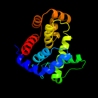
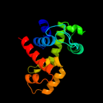
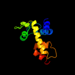
PDB header: lyaseChain: A: PDB Molecule: endo-type membrane-bound lytic murein transglycosylase a;PDBTitle: crystal structure of an outer membrane-anchored endolytic2 peptidoglycan lytic transglycosylase (mlte) from3 escherichia coli
2 c1slyA_
100.0
29
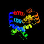
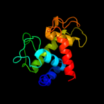
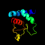
PDB header: glycosyltransferaseChain: A: PDB Molecule: 70-kda soluble lytic transglycosylase;PDBTitle: complex of the 70-kda soluble lytic transglycosylase with2 bulgecin a
3 d1qsaa2
100.0
30
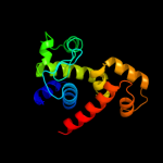
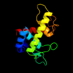
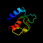
Fold: Lysozyme-likeSuperfamily: Lysozyme-likeFamily: Bacterial muramidase, catalytic domain4 c3mgwA_
100.0
23
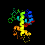
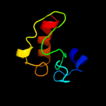
PDB header: hydrolaseChain: A: PDB Molecule: lysozyme g;PDBTitle: thermodynamics and structure of a salmon cold-active goose-type2 lysozyme
5 c3gxkB_
100.0
24
PDB header: hydrolaseChain: B: PDB Molecule: goose-type lysozyme 1;PDBTitle: the crystal structure of g-type lysozyme from atlantic cod2 (gadus morhua l.) in complex with nag oligomers sheds new3 light on substrate binding and the catalytic mechanism.4 native structure to 1.9
6 d1gbsa_
100.0
22
Fold: Lysozyme-likeSuperfamily: Lysozyme-likeFamily: G-type lysozyme7 c3bkhA_
99.0
18
PDB header: hydrolaseChain: A: PDB Molecule: lytic transglycosylase;PDBTitle: crystal structure of the bacteriophage phikz lytic2 transglycosylase, gp144
8 d1qusa_
99.0
23
Fold: Lysozyme-likeSuperfamily: Lysozyme-likeFamily: Bacterial muramidase, catalytic domain9 c1xsfA_
97.3
21
PDB header: cell cycle, hydrolaseChain: A: PDB Molecule: probable resuscitation-promoting factor rpfb;PDBTitle: solution structure of a resuscitation promoting factor2 domain from mycobacterium tuberculosis
10 d1xsfa1
97.2
20
Fold: Lysozyme-likeSuperfamily: Lysozyme-likeFamily: RPF-like11 c3ct5A_
96.9
23
PDB header: hydrolaseChain: A: PDB Molecule: morphogenesis protein 1;PDBTitle: crystal and cryoem structural studies of a cell wall degrading enzyme2 in the bacteriophage phi29 tail
12 c3eo5A_
96.6
20
PDB header: cell adhesionChain: A: PDB Molecule: resuscitation-promoting factor rpfb;PDBTitle: crystal structure of the resuscitation promoting factor rpfb
13 c2fbdB_
96.5
25
PDB header: hydrolaseChain: B: PDB Molecule: lysozyme 1;PDBTitle: the crystallographic structure of the digestive lysozyme 1 from musca2 domestica at 1.90 ang.
14 d1gd6a_
96.4
32
Fold: Lysozyme-likeSuperfamily: Lysozyme-likeFamily: C-type lysozyme15 d1hhla_
96.2
30
Fold: Lysozyme-likeSuperfamily: Lysozyme-likeFamily: C-type lysozyme16 d1iiza_
96.2
29
Fold: Lysozyme-likeSuperfamily: Lysozyme-likeFamily: C-type lysozyme17 d2vb1a1
96.2
30
Fold: Lysozyme-likeSuperfamily: Lysozyme-likeFamily: C-type lysozyme18 d1ghla_
96.1
30
Fold: Lysozyme-likeSuperfamily: Lysozyme-likeFamily: C-type lysozyme19 d1jsea_
95.8
28
Fold: Lysozyme-likeSuperfamily: Lysozyme-likeFamily: C-type lysozyme20 d1lmqa_
95.8
30
Fold: Lysozyme-likeSuperfamily: Lysozyme-likeFamily: C-type lysozyme21 c3csqC_
not modelled
95.7
21
PDB header: hydrolaseChain: C: PDB Molecule: morphogenesis protein 1;PDBTitle: crystal and cryoem structural studies of a cell wall2 degrading enzyme in the bacteriophage phi29 tail
22 c2goiC_
not modelled
95.6
25
PDB header: cell adhesion, sugar binding proteinChain: C: PDB Molecule: sperm lysozyme-like protein 1;PDBTitle: crystal structure of mouse sperm c-type lysozyme-like2 protein 1
23 d1qqya_
not modelled
95.5
28
Fold: Lysozyme-likeSuperfamily: Lysozyme-likeFamily: C-type lysozyme24 c2z2fA_
not modelled
95.4
25
PDB header: hydrolaseChain: A: PDB Molecule: lysozyme c-2;PDBTitle: x-ray crystal structure of bovine stomach lysozyme
25 d1juga_
not modelled
95.4
23
Fold: Lysozyme-likeSuperfamily: Lysozyme-likeFamily: C-type lysozyme26 d1lsga1
not modelled
95.3
31
Fold: Lysozyme-likeSuperfamily: Lysozyme-likeFamily: C-type lysozyme27 c3fi7A_
not modelled
95.0
19
PDB header: hydrolaseChain: A: PDB Molecule: lmo1076 protein;PDBTitle: crystal structure of the autolysin auto (lmo1076) from listeria2 monocytogenes, catalytic domain
28 d1ivma_
not modelled
94.8
23
Fold: Lysozyme-likeSuperfamily: Lysozyme-likeFamily: C-type lysozyme29 c2zycA_
not modelled
94.6
16
PDB header: hydrolaseChain: A: PDB Molecule: peptidoglycan hydrolase flgj;PDBTitle: crystal structure of peptidoglycan hydrolase from2 sphingomonas sp. a1
30 d2nwdx1
not modelled
94.5
23
Fold: Lysozyme-likeSuperfamily: Lysozyme-likeFamily: C-type lysozyme31 d2eqla_
not modelled
93.5
31
Fold: Lysozyme-likeSuperfamily: Lysozyme-likeFamily: C-type lysozyme32 d1f6sa_
not modelled
93.5
21
Fold: Lysozyme-likeSuperfamily: Lysozyme-likeFamily: C-type lysozyme33 d1b9oa_
not modelled
93.1
31
Fold: Lysozyme-likeSuperfamily: Lysozyme-likeFamily: C-type lysozyme34 d1hfxa_
not modelled
92.3
28
Fold: Lysozyme-likeSuperfamily: Lysozyme-likeFamily: C-type lysozyme35 d1yroa1
not modelled
92.3
28
Fold: Lysozyme-likeSuperfamily: Lysozyme-likeFamily: C-type lysozyme36 d1fkqa_
not modelled
92.2
21
Fold: Lysozyme-likeSuperfamily: Lysozyme-likeFamily: C-type lysozyme37 d1alca_
not modelled
91.5
31
Fold: Lysozyme-likeSuperfamily: Lysozyme-likeFamily: C-type lysozyme38 d1nvma1
not modelled
37.3
30
Fold: RuvA C-terminal domain-likeSuperfamily: post-HMGL domain-likeFamily: DmpG/LeuA communication domain-like39 d3efxd1
not modelled
22.9
15
Fold: OB-foldSuperfamily: Bacterial enterotoxinsFamily: Bacterial AB5 toxins, B-subunits40 c3lxuX_
not modelled
18.7
19
PDB header: hydrolaseChain: X: PDB Molecule: tripeptidyl-peptidase 2;PDBTitle: crystal structure of tripeptidyl peptidase 2 (tpp ii)
41 d157la_
not modelled
11.5
12
Fold: Lysozyme-likeSuperfamily: Lysozyme-likeFamily: Phage lysozyme42 d1t8fa_
not modelled
11.3
9
Fold: Lysozyme-likeSuperfamily: Lysozyme-likeFamily: Phage lysozyme43 c1mgtA_
not modelled
11.1
28
PDB header: transferaseChain: A: PDB Molecule: protein (o6-methylguanine-dna methyltransferase);PDBTitle: crystal structure of o6-methylguanine-dna methyltransferase from2 hyperthermophilic archaeon pyrococcus kodakaraensis strain kod1
44 d1jwya1
not modelled
9.2
23
Fold: SH3-like barrelSuperfamily: Myosin S1 fragment, N-terminal domainFamily: Myosin S1 fragment, N-terminal domain45 d1jtma_
not modelled
9.0
10
Fold: Lysozyme-likeSuperfamily: Lysozyme-likeFamily: Phage lysozyme46 d176la_
not modelled
8.7
10
Fold: Lysozyme-likeSuperfamily: Lysozyme-likeFamily: Phage lysozyme47 d189la_
not modelled
8.6
10
Fold: Lysozyme-likeSuperfamily: Lysozyme-likeFamily: Phage lysozyme48 c2dbtA_
not modelled
8.5
23
PDB header: hydrolaseChain: A: PDB Molecule: chitinase c;PDBTitle: crystal structure of chitinase c from streptomyces griseus2 hut6037
49 d2cvza1
not modelled
8.4
22
Fold: 6-phosphogluconate dehydrogenase C-terminal domain-likeSuperfamily: 6-phosphogluconate dehydrogenase C-terminal domain-likeFamily: Hydroxyisobutyrate and 6-phosphogluconate dehydrogenase domain50 d2fp1a1
not modelled
8.1
14
Fold: Chorismate mutase IISuperfamily: Chorismate mutase IIFamily: Secreted chorismate mutase-like51 c2cooA_
not modelled
7.3
18
PDB header: transferaseChain: A: PDB Molecule: lipoamide acyltransferase component of branched-PDBTitle: solution structure of the e3_binding domain of2 dihydrolipoamide branched chaintransacylase
52 d1mgta1
not modelled
6.7
28
Fold: DNA/RNA-binding 3-helical bundleSuperfamily: Methylated DNA-protein cysteine methyltransferase, C-terminal domainFamily: Methylated DNA-protein cysteine methyltransferase, C-terminal domain53 d191la_
not modelled
6.4
10
Fold: Lysozyme-likeSuperfamily: Lysozyme-likeFamily: Phage lysozyme54 c1fi4A_
not modelled
5.8
14
PDB header: lyaseChain: A: PDB Molecule: mevalonate 5-diphosphate decarboxylase;PDBTitle: the x-ray crystal structure of mevalonate 5-diphosphate decarboxylase2 at 2.3 angstrom resolution.
55 c1umwB_
not modelled
5.8
40
PDB header: kinaseChain: B: PDB Molecule: serine/threonine-protein kinase plk;PDBTitle: structure of a human plk1 polo-box domain/phosphopeptide2 complex
56 c3hbhA_
not modelled
5.7
16
PDB header: hydrolaseChain: A: PDB Molecule: class iv chitinase chia4-pa2;PDBTitle: class iv chitinase structure from picea abies at 2.25a
57 c2eq9C_
not modelled
5.5
18
PDB header: oxidoreductaseChain: C: PDB Molecule: pyruvate dehydrogenase complex, dihydrolipoamidePDBTitle: crystal structure of lipoamide dehydrogenase from thermus thermophilus2 hb8 with psbdb
58 d2cyua1
not modelled
5.4
24
Fold: Peripheral subunit-binding domain of 2-oxo acid dehydrogenase complexSuperfamily: Peripheral subunit-binding domain of 2-oxo acid dehydrogenase complexFamily: Peripheral subunit-binding domain of 2-oxo acid dehydrogenase complex59 c2cjlA_
not modelled
5.2
21
PDB header: hydrolaseChain: A: PDB Molecule: secreted chitinase;PDBTitle: crystal structure and enzymatic properties of a bacterial2 family 19 chitinase reveal differences with plant enzymes
60 d1w4ha1
not modelled
5.1
24
Fold: Peripheral subunit-binding domain of 2-oxo acid dehydrogenase complexSuperfamily: Peripheral subunit-binding domain of 2-oxo acid dehydrogenase complexFamily: Peripheral subunit-binding domain of 2-oxo acid dehydrogenase complex