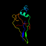
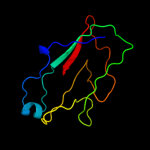
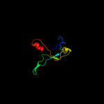
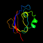
1 c2w07B_
94.8
19
PDB header: cell adhesionChain: B: PDB Molecule: minor pilin subunit papf;PDBTitle: structural determinants of polymerization reactivity of the2 p pilus adaptor subunit papf
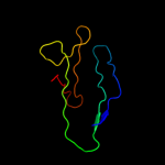
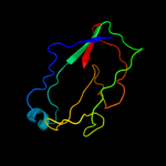
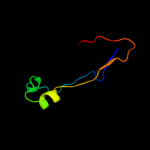
2 c3bfwA_
94.7
15
PDB header: structural protein/structural proteinChain: A: PDB Molecule: protein fimg;PDBTitle: crystal structure of truncated fimg (fimgt) in complex with the donor2 strand peptide of fimf (dsf)
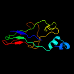
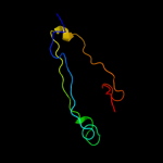
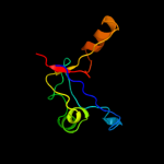
3 c3jwnK_
94.1
17
PDB header: protein binding/cell adhesionChain: K: PDB Molecule: protein fimf;PDBTitle: complex of fimc, fimf, fimg and fimh
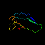
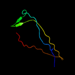
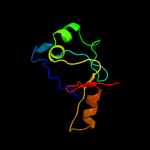
4 c1klfP_
93.9
13
PDB header: chaperone/adhesin complexChain: P: PDB Molecule: fimh protein;PDBTitle: fimh adhesin-fimc chaperone complex with d-mannose
5 c3jwnE_
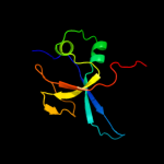
93.8
16
PDB header: protein binding/cell adhesionChain: E: PDB Molecule: protein fimf;PDBTitle: complex of fimc, fimf, fimg and fimh
6 d1ze3h1
93.3
12
Fold: Common fold of diphtheria toxin/transcription factors/cytochrome fSuperfamily: Bacterial adhesinsFamily: Pilus subunits7 c3jwnL_
93.3
17
PDB header: protein binding/cell adhesionChain: L: PDB Molecule: protein fimf;PDBTitle: complex of fimc, fimf, fimg and fimh
8 c3jwnF_
92.9
17
PDB header: protein binding/cell adhesionChain: F: PDB Molecule: protein fimf;PDBTitle: complex of fimc, fimf, fimg and fimh
9 d1n12a_
87.9
14
Fold: Common fold of diphtheria toxin/transcription factors/cytochrome fSuperfamily: Bacterial adhesinsFamily: Pilus subunits10 c2jtyA_
77.9
14
PDB header: structural proteinChain: A: PDB Molecule: type-1 fimbrial protein, a chain;PDBTitle: self-complemented variant of fima, the main subunit of type 1 pilus
11 d2uy6b1
70.7
13
Fold: Common fold of diphtheria toxin/transcription factors/cytochrome fSuperfamily: Bacterial adhesinsFamily: Pilus subunits12 d2j2zb1
65.5
15
Fold: Common fold of diphtheria toxin/transcription factors/cytochrome fSuperfamily: Bacterial adhesinsFamily: Pilus subunits13 d1pdkb_
60.7
9
Fold: Common fold of diphtheria toxin/transcription factors/cytochrome fSuperfamily: Bacterial adhesinsFamily: Pilus subunits14 c2wmpB_
47.1
25
PDB header: chaperoneChain: B: PDB Molecule: papg protein;PDBTitle: structure of the e. coli chaperone papd in complex with the pilin2 domain of the papgii adhesin
15 c2vsvB_
41.4
14
PDB header: protein-bindingChain: B: PDB Molecule: rhophilin-2;PDBTitle: crystal structure of the pdz domain of human rhophilin-2
16 c3qbtH_
34.1
11
PDB header: protein transport/hydrolaseChain: H: PDB Molecule: inositol polyphosphate 5-phosphatase ocrl-1;PDBTitle: crystal structure of ocrl1 540-678 in complex with rab8a:gppnhp
17 d1r6ja_
29.6
28
Fold: PDZ domain-likeSuperfamily: PDZ domain-likeFamily: PDZ domain18 c1r6jA_
29.6
28
PDB header: membrane proteinChain: A: PDB Molecule: syntenin 1;PDBTitle: ultrahigh resolution crystal structure of syntenin pdz2
19 c1nteA_
29.6
28
PDB header: signaling proteinChain: A: PDB Molecule: syntenin 1;PDBTitle: crystal structure analysis of the second pdz domain of2 syntenin
20 c3khfA_
21.2
16
PDB header: transferaseChain: A: PDB Molecule: microtubule-associated serine/threonine-proteinPDBTitle: the crystal structure of the pdz domain of human microtubule2 associated serine/threonine kinase 3 (mast3)
21 c2kjmA_
not modelled
18.4
50
PDB header: rna binding proteinChain: A: PDB Molecule: histone rna hairpin-binding protein;PDBTitle: solution structure of slbp rna binding domain fragment
22 c3qglD_
not modelled
17.8
16
PDB header: protein bindingChain: D: PDB Molecule: sorting nexin-27;PDBTitle: crystal structure of pdz domain of sorting nexin 27 (snx27) in complex2 with the eseskv peptide corresponding to the c-terminal tail of girk3
23 d1eaqa_
not modelled
16.5
55
Fold: Common fold of diphtheria toxin/transcription factors/cytochrome fSuperfamily: p53-like transcription factorsFamily: RUNT domain24 d1ljma_
not modelled
16.0
55
Fold: Common fold of diphtheria toxin/transcription factors/cytochrome fSuperfamily: p53-like transcription factorsFamily: RUNT domain25 c1y7nA_
not modelled
15.9
22
PDB header: protein transportChain: A: PDB Molecule: amyloid beta a4 precursor protein-binding familyPDBTitle: solution structure of the second pdz domain of the human2 neuronal adaptor x11alpha
26 d1ry4a_
not modelled
11.8
20
Fold: PDZ domain-likeSuperfamily: PDZ domain-likeFamily: PDZ domain27 c3l4fD_
not modelled
10.9
18
PDB header: signaling protein/protein bindingChain: D: PDB Molecule: sh3 and multiple ankyrin repeat domains proteinPDBTitle: crystal structure of betapix coiled-coil domain and shank2 pdz complex
28 d1auua_
not modelled
10.6
25
Fold: GroES-likeSuperfamily: SacY-like RNA-binding domainFamily: BglG-like antiterminator proteins29 c1u39A_
not modelled
10.2
16
PDB header: protein transportChain: A: PDB Molecule: amyloid beta a4 precursor protein-binding,PDBTitle: auto-inhibition mechanism of x11s/mints family scaffold2 proteins revealed by the closed conformation of the tandem3 pdz domains
30 d1hcza1
not modelled
10.1
20
Fold: Common fold of diphtheria toxin/transcription factors/cytochrome fSuperfamily: Cytochrome f, large domainFamily: Cytochrome f, large domain31 d2oz4a1
not modelled
9.9
19
Fold: Immunoglobulin-like beta-sandwichSuperfamily: ImmunoglobulinFamily: C2 set domains32 c2jtvA_
not modelled
9.9
22
PDB header: structural genomicsChain: A: PDB Molecule: protein of unknown function;PDBTitle: solution structure of protein rpa3401, northeast structural genomics2 consortium target rpt7, ontario center for structural proteomics3 target rp3384
33 d1uf1a_
not modelled
9.8
12
Fold: PDZ domain-likeSuperfamily: PDZ domain-likeFamily: PDZ domain34 c7mdhA_
not modelled
9.3
10
PDB header: chloroplastic malate dehydrogenaseChain: A: PDB Molecule: protein (malate dehydrogenase);PDBTitle: structural basis for light acitvation of a chloroplast enzyme. the2 structure of sorghum nadp-malate dehydrogenase in its oxidized form
35 c2eaqA_
not modelled
9.3
13
PDB header: metal binding proteinChain: A: PDB Molecule: lim domain only protein 7;PDBTitle: crystal structure of pdz domain of kiaa0858 (lim), ms07932 from homo sapiens
36 d1b8pa2
not modelled
9.0
10
Fold: LDH C-terminal domain-likeSuperfamily: LDH C-terminal domain-likeFamily: Lactate & malate dehydrogenases, C-terminal domain37 c2yzyA_
not modelled
8.9
8
PDB header: structural genomics, unknown functionChain: A: PDB Molecule: putative uncharacterized protein ttha1012;PDBTitle: crystal structure of uncharacterized conserved protein from thermus2 thermophilus hb8
38 d1kwaa_
not modelled
8.5
21
Fold: PDZ domain-likeSuperfamily: PDZ domain-likeFamily: PDZ domain39 d1yq2a3
not modelled
8.3
29
Fold: Galactose-binding domain-likeSuperfamily: Galactose-binding domain-likeFamily: beta-Galactosidase/glucuronidase, N-terminal domain40 d1y7na1
not modelled
8.1
21
Fold: PDZ domain-likeSuperfamily: PDZ domain-likeFamily: PDZ domain41 c2k8pA_
not modelled
8.0
18
PDB header: signaling proteinChain: A: PDB Molecule: sclerostin;PDBTitle: characterisation of the structural features and2 interactions of sclerostin: molecular insight into a key3 regulator of wnt-mediated bone formation
42 d7mdha2
not modelled
8.0
10
Fold: LDH C-terminal domain-likeSuperfamily: LDH C-terminal domain-likeFamily: Lactate & malate dehydrogenases, C-terminal domain43 d1wioa2
not modelled
7.4
43
Fold: Immunoglobulin-like beta-sandwichSuperfamily: ImmunoglobulinFamily: V set domains (antibody variable domain-like)44 c2z17A_
not modelled
7.3
17
PDB header: protein bindingChain: A: PDB Molecule: pleckstrin homology sec7 and coiled-coil domains-PDBTitle: crystal sturcture of pdz domain from human pleckstrin2 homology, sec7
45 d1civa2
not modelled
7.3
9
Fold: LDH C-terminal domain-likeSuperfamily: LDH C-terminal domain-likeFamily: Lactate & malate dehydrogenases, C-terminal domain46 d1jz8a3
not modelled
7.3
11
Fold: Galactose-binding domain-likeSuperfamily: Galactose-binding domain-likeFamily: beta-Galactosidase/glucuronidase, N-terminal domain47 c2d8iA_
not modelled
7.2
16
PDB header: immune system, signaling proteinChain: A: PDB Molecule: t-cell lymphoma invasion and metastasis 1PDBTitle: solution structure of the pdz domain of t-cell lymphoma2 invasion and metastasis 1 varian
48 c2w95B_
not modelled
7.1
12
PDB header: cell adhesionChain: B: PDB Molecule: discoidin-1 subunit a;PDBTitle: structure of the discoidin i from dictyostelium discoideum2 in complex with galnac at 1.75 angstrom resolution
49 c2fc6A_
not modelled
6.9
27
PDB header: transcriptionChain: A: PDB Molecule: target of egr1, member 1;PDBTitle: solution structure of the zf-ccch domain of target of egr1,2 member 1 (nuclear)
50 c2jmrA_
not modelled
6.8
13
PDB header: cell adhesionChain: A: PDB Molecule: fimf;PDBTitle: nmr structure of the e. coli type 1 pilus subunit fimf
51 d2fc6a1
not modelled
6.4
44
Fold: CCCH zinc fingerSuperfamily: CCCH zinc fingerFamily: CCCH zinc finger52 c2kv8A_
not modelled
6.3
19
PDB header: signaling proteinChain: A: PDB Molecule: regulator of g-protein signaling 12;PDBTitle: solution structure ofrgs12 pdz domain
53 d1gv0a2
not modelled
6.2
8
Fold: LDH C-terminal domain-likeSuperfamily: LDH C-terminal domain-likeFamily: Lactate & malate dehydrogenases, C-terminal domain54 c1wziA_
not modelled
6.1
14
PDB header: oxidoreductaseChain: A: PDB Molecule: malate dehydrogenase;PDBTitle: structural basis for alteration of cofactor specificity of2 malate dehydrogenase from thermus flavus
55 d1be9a_
not modelled
6.0
13
Fold: PDZ domain-likeSuperfamily: PDZ domain-likeFamily: PDZ domain56 d1y7ta2
not modelled
5.9
14
Fold: LDH C-terminal domain-likeSuperfamily: LDH C-terminal domain-likeFamily: Lactate & malate dehydrogenases, C-terminal domain57 d2zjrp1
not modelled
5.7
15
Fold: Ribosomal protein L22Superfamily: Ribosomal protein L22Family: Ribosomal protein L2258 c2xzzA_
not modelled
5.6
10
PDB header: transferaseChain: A: PDB Molecule: protein-glutamine gamma-glutamyltransferase k;PDBTitle: crystal structure of the human transglutaminase 1 beta-barrel domain
59 c2nncB_
not modelled
5.4
10
PDB header: ligand binding proteinChain: B: PDB Molecule: sulfur covalently-binding protein;PDBTitle: structure of the sulfur carrier protein soxy from chlorobium limicola2 f thiosulfatophilum
60 c1b8vA_
not modelled
5.3
12
PDB header: oxidoreductaseChain: A: PDB Molecule: protein (malate dehydrogenase);PDBTitle: malate dehydrogenase from aquaspirillum arcticum
61 d1hyha2
not modelled
5.1
15
Fold: LDH C-terminal domain-likeSuperfamily: LDH C-terminal domain-likeFamily: Lactate & malate dehydrogenases, C-terminal domain62 c1xt5A_
not modelled
5.1
4
PDB header: immune systemChain: A: PDB Molecule: variable region-containing chitin-bindingPDBTitle: crystal structure of vcbp3, domain 1, from branchiostoma2 floridae