| 1 | c3giaA_ |
|
 |
99.8 |
13 |
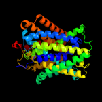
PDB header:transport protein
Chain: A: PDB Molecule:uncharacterized protein mj0609;
PDBTitle: crystal structure of apct transporter
|
| 2 | c3lrcC_ |
|
 |
99.5 |
13 |
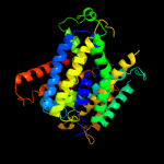
PDB header:transport protein
Chain: C: PDB Molecule:arginine/agmatine antiporter;
PDBTitle: structure of e. coli adic (p1)
|
| 3 | c2jlnA_ |
|
 |
99.1 |
11 |
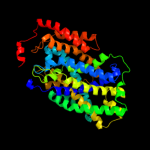
PDB header:membrane protein
Chain: A: PDB Molecule:mhp1;
PDBTitle: structure of mhp1, a nucleobase-cation-symport-1 family2 transporter
|
| 4 | c2h3oA_ |
|
 |
36.4 |
25 |
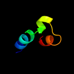
PDB header:membrane protein
Chain: A: PDB Molecule:merf;
PDBTitle: structure of merft, a membrane protein with two trans-2 membrane helices
|
| 5 | c1wazA_ |
|
 |
31.5 |
25 |
PDB header:transport protein
Chain: A: PDB Molecule:merf;
PDBTitle: nmr structure determination of the bacterial mercury2 transporter, merf, in micelles
|
| 6 | d2h8pc1 |
|
 |
22.8 |
16 |
Fold:Voltage-gated potassium channels
Superfamily:Voltage-gated potassium channels
Family:Voltage-gated potassium channels |
| 7 | c2l2tA_ |
|
 |
17.9 |
34 |
PDB header:membrane protein
Chain: A: PDB Molecule:receptor tyrosine-protein kinase erbb-4;
PDBTitle: solution nmr structure of the erbb4 dimeric membrane domain
|
| 8 | c2zt9H_ |
|
 |
14.1 |
30 |
PDB header:photosynthesis
Chain: H: PDB Molecule:cytochrome b6-f complex subunit 8;
PDBTitle: crystal structure of the cytochrome b6f complex from nostoc sp. pcc2 7120
|
| 9 | d1gsma1 |
|
 |
11.8 |
32 |
Fold:Immunoglobulin-like beta-sandwich
Superfamily:Immunoglobulin
Family:I set domains |
| 10 | d1pe4a_ |
|
 |
11.8 |
60 |
Fold:Knottins (small inhibitors, toxins, lectins)
Superfamily:Scorpion toxin-like
Family:Long-chain scorpion toxins |
| 11 | d2e74h1 |
|
 |
11.5 |
30 |
Fold:Single transmembrane helix
Superfamily:PetN subunit of the cytochrome b6f complex
Family:PetN subunit of the cytochrome b6f complex |
| 12 | c1vf5U_ |
|
 |
11.5 |
30 |
PDB header:photosynthesis
Chain: U: PDB Molecule:protein pet n;
PDBTitle: crystal structure of cytochrome b6f complex from m.laminosus
|
| 13 | c1vf5H_ |
|
 |
11.5 |
30 |
PDB header:photosynthesis
Chain: H: PDB Molecule:protein pet n;
PDBTitle: crystal structure of cytochrome b6f complex from m.laminosus
|
| 14 | c2d2cH_ |
|
 |
11.5 |
30 |
PDB header:photosynthesis
Chain: H: PDB Molecule:cytochrome b6-f complex subunit viii;
PDBTitle: crystal structure of cytochrome b6f complex with dbmib from2 m. laminosus
|
| 15 | c2d2cU_ |
|
 |
11.5 |
30 |
PDB header:photosynthesis
Chain: U: PDB Molecule:cytochrome b6-f complex subunit viii;
PDBTitle: crystal structure of cytochrome b6f complex with dbmib from2 m. laminosus
|
| 16 | c2e75H_ |
|
 |
10.6 |
30 |
PDB header:photosynthesis
Chain: H: PDB Molecule:cytochrome b6-f complex subunit 8;
PDBTitle: crystal structure of the cytochrome b6f complex with 2-nonyl-4-2 hydroxyquinoline n-oxide (nqno) from m.laminosus
|
| 17 | c2e76H_ |
|
 |
10.6 |
30 |
PDB header:photosynthesis
Chain: H: PDB Molecule:cytochrome b6-f complex subunit 8;
PDBTitle: crystal structure of the cytochrome b6f complex with tridecyl-2 stigmatellin (tds) from m.laminosus
|
| 18 | c2e74H_ |
|
 |
10.6 |
30 |
PDB header:photosynthesis
Chain: H: PDB Molecule:cytochrome b6-f complex subunit 8;
PDBTitle: crystal structure of the cytochrome b6f complex from m.laminosus
|
| 19 | c2kn8A_ |
|
 |
9.1 |
30 |
PDB header:protein binding, dna binding protein
Chain: A: PDB Molecule:dna cleavage and packaging protein large subunit, ul89;
PDBTitle: nmr structure of the c-terminal domain of pul89
|
| 20 | c3iefA_ |
|
 |
7.3 |
31 |
PDB header:transferase, rna binding protein
Chain: A: PDB Molecule:trna (guanine-n(1)-)-methyltransferase;
PDBTitle: crystal structure of trna guanine-n1-methyltransferase from2 bartonella henselae using mpcs.
|
| 21 | d1p9pa_ |
|
not modelled |
7.0 |
25 |
Fold:alpha/beta knot
Superfamily:alpha/beta knot
Family:tRNA(m1G37)-methyltransferase TrmD |
| 22 | c2l35A_ |
|
not modelled |
6.9 |
24 |
PDB header:protein binding
Chain: A: PDB Molecule:dap12-nkg2c_tm;
PDBTitle: structure of the dap12-nkg2c transmembrane heterotrimer
|
| 23 | d1qaza_ |
|
not modelled |
6.7 |
6 |
Fold:alpha/alpha toroid
Superfamily:Chondroitin AC/alginate lyase
Family:Alginate lyase A1-III |
| 24 | c1oy5B_ |
|
not modelled |
6.7 |
25 |
PDB header:transferase
Chain: B: PDB Molecule:trna (guanine-n(1)-)-methyltransferase;
PDBTitle: crystal structure of trna (m1g37) methyltransferase from aquifex2 aeolicus
|
| 25 | d1oy5a_ |
|
not modelled |
6.7 |
25 |
Fold:alpha/beta knot
Superfamily:alpha/beta knot
Family:tRNA(m1G37)-methyltransferase TrmD |
| 26 | d1bf2a1 |
|
not modelled |
6.4 |
28 |
Fold:Immunoglobulin-like beta-sandwich
Superfamily:E set domains
Family:E-set domains of sugar-utilizing enzymes |
| 27 | c3quvB_ |
|
not modelled |
6.4 |
25 |
PDB header:transferase
Chain: B: PDB Molecule:trna (guanine-n(1)-)-methyltransferase;
PDBTitle: crystal structure of a trna-guanine-n1-methyltransferase from2 mycobacterium abscessus
|
| 28 | d1uala_ |
|
not modelled |
6.4 |
38 |
Fold:alpha/beta knot
Superfamily:alpha/beta knot
Family:tRNA(m1G37)-methyltransferase TrmD |
| 29 | c3ky7A_ |
|
not modelled |
6.3 |
44 |
PDB header:transferase
Chain: A: PDB Molecule:trna (guanine-n(1)-)-methyltransferase;
PDBTitle: 2.35 angstrom resolution crystal structure of a putative trna2 (guanine-7-)-methyltransferase (trmd) from staphylococcus aureus3 subsp. aureus mrsa252
|
| 30 | d2axtz1 |
|
not modelled |
6.2 |
28 |
Fold:Transmembrane helix hairpin
Superfamily:PsbZ-like
Family:PsbZ-like |
| 31 | d1oz2a3 |
|
not modelled |
6.1 |
12 |
Fold:SH3-like barrel
Superfamily:Tudor/PWWP/MBT
Family:MBT repeat |
| 32 | c2xq2A_ |
|
not modelled |
5.8 |
14 |
PDB header:transport protein
Chain: A: PDB Molecule:sodium/glucose cotransporter;
PDBTitle: structure of the k294a mutant of vsglt
|
| 33 | c3knuD_ |
|
not modelled |
5.5 |
19 |
PDB header:transferase
Chain: D: PDB Molecule:trna (guanine-n(1)-)-methyltransferase;
PDBTitle: crystal structure of trna (guanine-n1)-methyltransferase from2 anaplasma phagocytophilum
|
| 34 | c1nyjC_ |
|
not modelled |
5.5 |
40 |
PDB header:viral protein
Chain: C: PDB Molecule:matrix protein m2;
PDBTitle: the closed state structure of m2 protein h+ channel by2 solid state nmr spectroscopy
|
| 35 | c1nyjB_ |
|
not modelled |
5.5 |
40 |
PDB header:viral protein
Chain: B: PDB Molecule:matrix protein m2;
PDBTitle: the closed state structure of m2 protein h+ channel by2 solid state nmr spectroscopy
|
| 36 | c2kqtC_ |
|
not modelled |
5.5 |
40 |
PDB header:transport protein
Chain: C: PDB Molecule:m2 protein;
PDBTitle: solid-state nmr structure of the m2 transmembrane peptide of the2 influenza a virus in dmpc lipid bilayers bound to deuterated3 amantadine
|
| 37 | c2kqtA_ |
|
not modelled |
5.5 |
40 |
PDB header:transport protein
Chain: A: PDB Molecule:m2 protein;
PDBTitle: solid-state nmr structure of the m2 transmembrane peptide of the2 influenza a virus in dmpc lipid bilayers bound to deuterated3 amantadine
|
| 38 | c2kqtB_ |
|
not modelled |
5.5 |
40 |
PDB header:transport protein
Chain: B: PDB Molecule:m2 protein;
PDBTitle: solid-state nmr structure of the m2 transmembrane peptide of the2 influenza a virus in dmpc lipid bilayers bound to deuterated3 amantadine
|
| 39 | c1nyjD_ |
|
not modelled |
5.5 |
40 |
PDB header:viral protein
Chain: D: PDB Molecule:matrix protein m2;
PDBTitle: the closed state structure of m2 protein h+ channel by2 solid state nmr spectroscopy
|
| 40 | c2kqtD_ |
|
not modelled |
5.5 |
40 |
PDB header:transport protein
Chain: D: PDB Molecule:m2 protein;
PDBTitle: solid-state nmr structure of the m2 transmembrane peptide of the2 influenza a virus in dmpc lipid bilayers bound to deuterated3 amantadine
|
| 41 | c1nyjA_ |
|
not modelled |
5.5 |
40 |
PDB header:viral protein
Chain: A: PDB Molecule:matrix protein m2;
PDBTitle: the closed state structure of m2 protein h+ channel by2 solid state nmr spectroscopy
|
| 42 | c1mp6A_ |
|
not modelled |
5.5 |
40 |
PDB header:membrane protein
Chain: A: PDB Molecule:matrix protein m2;
PDBTitle: structure of the transmembrane region of the m2 protein h+2 channel by solid state nmr spectroscopy
|
| 43 | c3c66B_ |
|
not modelled |
5.4 |
18 |
PDB header:transferase
Chain: B: PDB Molecule:poly(a) polymerase;
PDBTitle: yeast poly(a) polymerase in complex with fip1 residues 80-105
|
| 44 | d1moua_ |
|
not modelled |
5.1 |
13 |
Fold:GFP-like
Superfamily:GFP-like
Family:Fluorescent proteins |