Phyre Home Retrieve Phyre Job Id
Emaill.a.kelley@imperial.ac.uk
DescriptionP0AEV4
DateThu Jan 5 11:24:20 GMT 2012
Unique Job IDe9215e6cc75ae905

Summary 

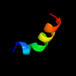
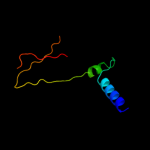
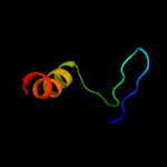
Top model
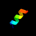
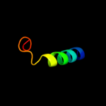
Image coloured by rainbow N → C terminus
Model (left) based on template c2wpvG_
Top template information
PDB header:protein binding
Chain: G: PDB Molecule:upf0363 protein yor164c;
PDBTitle: crystal structure of s. cerevisiae get4-get5 complex
Confidence and coverage
Confidence: 42.2% Coverage: 11%
17 residues ( 11% of your sequence) have been modelled with 42.2% confidence by the single highest scoring template.
You may wish to submit your sequence to Phyrealarm. This will automatically scan your sequence every week for new potential templates as they appear in the Phyre2 library.
Please note: You must be registered and logged in to use Phyrealarm.
3D viewing
Interactive 3D view in Jmol

Sequence analysis 

Secondary structure and disorder prediction 

   1........10.........20.........30.........40.........50.........60
Sequence  MTIWEISEKADYIAQRHRRLQDQWHIYCNSLVQGITLSKARLHHAMSCAPDKELCFVLFE
Secondary structure 


SS confidence 


Disorder  ???????


Disorder confidence 


   .........70.........80.........90.........100.........110.........120
Sequence  HFRIYVTLADGFNSHTIEYYVETKDGEDKQRIAQAQLSIDGMIDGKVNIRDREQVLEHYL
Secondary structure 


SS confidence 


Disorder 


?????????


?


?


?


?


Disorder confidence 


   .........130.........140.........150...
Sequence  EKIAGVYDSLYTAIENNVPVNLSQLVKGQSPAA
Secondary structure 


SS confidence 


Disorder 


????????
Disorder confidence 


Confidence Key
High(9)                    Low (0)
?Disordered
Alpha helix
Beta strand

Domain analysis 

Hover over an aligned region to see model and summary info

Please note, only up to the top 20 hits are modelled to reduce computer load

RankAligned region

PDB 2wpv chain G

3D model

Region: 111 - 127
Aligned: 17
Modelled: 17
Confidence: 42.2%
Identity: 29%
PDB header:protein binding
Chain: G: PDB Molecule:upf0363 protein yor164c;
PDBTitle: crystal structure of s. cerevisiae get4-get5 complex

Phyre2

PDB 3lpz chain A

3D model

Region: 111 - 127
Aligned: 17
Modelled: 17
Confidence: 21.7%
Identity: 29%
PDB header:protein transport
Chain: A: PDB Molecule:get4 (yor164c homolog);
PDBTitle: crystal structure of c. therm. get4

Phyre2

PDB 2qgo chain A

3D model

Region: 98 - 117
Aligned: 20
Modelled: 20
Confidence: 20.3%
Identity: 35%
PDB header:biosynthetic protein
Chain: A: PDB Molecule:putative fe-s biosynthesis protein;
PDBTitle: crystal structure of a putative fe-s biosynthesis protein from2 lactobacillus acidophilus

Phyre2

PDB 3kcp chain A

3D model

Region: 104 - 120
Aligned: 17
Modelled: 17
Confidence: 17.2%
Identity: 29%
PDB header:structural protein
Chain: A: PDB Molecule:cellulosomal-scaffolding protein a;
PDBTitle: crystal structure of interacting clostridium thermocellum2 multimodular components

Phyre2

PDB 3iz5 chain F

3D model

Region: 17 - 81
Aligned: 56
Modelled: 65
Confidence: 16.8%
Identity: 16%
PDB header:ribosome
Chain: F: PDB Molecule:60s ribosomal protein l9 (l6p);
PDBTitle: localization of the large subunit ribosomal proteins into a 5.5 a2 cryo-em map of triticum aestivum translating 80s ribosome

Phyre2

PDB 2idb chain A domain 2

3D model

Region: 64 - 121
Aligned: 57
Modelled: 58
Confidence: 15.7%
Identity: 18%
Fold: UbiD C-terminal domain-like
Superfamily: UbiD C-terminal domain-like
Family: UbiD C-terminal domain-like

Phyre2

PDB 1or7 chain C

3D model

Region: 16 - 28
Aligned: 13
Modelled: 13
Confidence: 14.6%
Identity: 23%
Fold: N-terminal, cytoplasmic domain of anti-sigmaE factor RseA
Superfamily: N-terminal, cytoplasmic domain of anti-sigmaE factor RseA
Family: N-terminal, cytoplasmic domain of anti-sigmaE factor RseA

Phyre2

PDB 1or7 chain C

3D model

Region: 16 - 28
Aligned: 13
Modelled: 13
Confidence: 14.6%
Identity: 23%
PDB header:transcription
Chain: C: PDB Molecule:sigma-e factor negative regulatory protein;
PDBTitle: crystal structure of escherichia coli sigmae with the cytoplasmic2 domain of its anti-sigma rsea

Phyre2

PDB 3bes chain L domain 1

3D model

Region: 119 - 143
Aligned: 25
Modelled: 25
Confidence: 13.6%
Identity: 16%
Fold: 4-helical cytokines
Superfamily: 4-helical cytokines
Family: Interferons/interleukin-10 (IL-10)

Phyre2

PDB 3udi chain A

3D model

Region: 72 - 123
Aligned: 18
Modelled: 18
Confidence: 13.1%
Identity: 39%
PDB header:penicillin-binding protein/antibiotic
Chain: A: PDB Molecule:penicillin-binding protein 1a;
PDBTitle: crystal structure of acinetobacter baumannii pbp1a in complex with2 penicillin g

Phyre2

PDB 2e9h chain A

3D model

Region: 93 - 122
Aligned: 30
Modelled: 30
Confidence: 12.1%
Identity: 20%
PDB header:translation
Chain: A: PDB Molecule:eukaryotic translation initiation factor 5;
PDBTitle: solution structure of the eif-5_eif-2b domain from human2 eukaryotic translation initiation factor 5

Phyre2

PDB 2e32 chain A

3D model

Region: 41 - 73
Aligned: 32
Modelled: 33
Confidence: 11.0%
Identity: 22%
PDB header:ligase
Chain: A: PDB Molecule:f-box only protein 2;
PDBTitle: structural basis for selection of glycosylated substrate by2 scffbs1 ubiquitin ligase

Phyre2

PDB 2dci chain A

3D model

Region: 99 - 107
Aligned: 9
Modelled: 7
Confidence: 10.6%
Identity: 56%
PDB header:viral protein
Chain: A: PDB Molecule:hemagglutinin;
PDBTitle: nmr structure of influenza ha fusion peptide mutant w14a in2 dpc in ph5

Phyre2

PDB 2olv chain A

3D model

Region: 112 - 123
Aligned: 12
Modelled: 12
Confidence: 9.9%
Identity: 33%
PDB header:transferase
Chain: A: PDB Molecule:penicillin-binding protein 2;
PDBTitle: structural insight into the transglycosylation step of bacterial cell2 wall biosynthesis : donor ligand complex

Phyre2

PDB 3dwk chain C

3D model

Region: 111 - 123
Aligned: 13
Modelled: 13
Confidence: 8.9%
Identity: 31%
PDB header:transferase
Chain: C: PDB Molecule:penicillin-binding protein 2;
PDBTitle: identification of dynamic structural motifs involved in2 peptidoglycan glycosyltransfer

Phyre2

PDB 1xoo chain A

3D model

Region: 100 - 107
Aligned: 8
Modelled: 6
Confidence: 8.7%
Identity: 63%
PDB header:viral protein
Chain: A: PDB Molecule:hemagglutinin;
PDBTitle: nmr structure of g1s mutant of influenza hemagglutinin2 fusion peptide in dpc micelles at ph 5

Phyre2

PDB 1xop chain A

3D model

Region: 100 - 107
Aligned: 8
Modelled: 6
Confidence: 8.7%
Identity: 63%
PDB header:viral protein
Chain: A: PDB Molecule:hemagglutinin;
PDBTitle: nmr structure of g1v mutant of influenza hemagglutinin2 fusion peptide in dpc micelles at ph 5

Phyre2

PDB 3hlk chain B

3D model

Region: 60 - 104
Aligned: 45
Modelled: 45
Confidence: 8.7%
Identity: 22%
PDB header:hydrolase
Chain: B: PDB Molecule:acyl-coenzyme a thioesterase 2, mitochondrial;
PDBTitle: crystal structure of human mitochondrial acyl-coa2 thioesterase (acot2)

Phyre2

PDB 1eku chain A

3D model

Region: 120 - 143
Aligned: 24
Modelled: 24
Confidence: 8.1%
Identity: 17%
PDB header:immune system
Chain: A: PDB Molecule:interferon gamma;
PDBTitle: crystal structure of a biologically active single chain2 mutant of human ifn-gamma

Phyre2

PDB 2oq0 chain A domain 1

3D model

Region: 44 - 60
Aligned: 17
Modelled: 17
Confidence: 8.1%
Identity: 24%
Fold: OB-fold
Superfamily: HIN-2000 domain-like
Family: HIN-200/IF120x domain

Phyre2
1

c2wpvG_
2

c3lpzA_
3

c2qgoA_
4

c3kcpA_
5

c3iz5F_
6

d2idba2
7

d1or7c_
8

c1or7C_
9

d3besl1
10

c3udiA_
11

c2e9hA_
12

c2e32A_
13

c2dciA_
14

c2olvA_
15

c3dwkC_
16

c1xooA_
17

c1xopA_
18

c3hlkB_
19

c1ekuA_
20

d2oq0a1
21


22


23


24


25


26


27


28


29


30


31


32


33


34


35


36


37


38


39


40


41


42


43


44


45


Detailed template information 

#
Template Alignment Coverage3D Model Confidence
% i.d. Template Information
1c2wpvG_


42.2 29 PDB header:protein binding
Chain: G: PDB Molecule:upf0363 protein yor164c;
PDBTitle: crystal structure of s. cerevisiae get4-get5 complex
2c3lpzA_


21.7 29 PDB header:protein transport
Chain: A: PDB Molecule:get4 (yor164c homolog);
PDBTitle: crystal structure of c. therm. get4
3c2qgoA_


20.3 35 PDB header:biosynthetic protein
Chain: A: PDB Molecule:putative fe-s biosynthesis protein;
PDBTitle: crystal structure of a putative fe-s biosynthesis protein from2 lactobacillus acidophilus
4c3kcpA_


17.2 29 PDB header:structural protein
Chain: A: PDB Molecule:cellulosomal-scaffolding protein a;
PDBTitle: crystal structure of interacting clostridium thermocellum2 multimodular components
5c3iz5F_


16.8 16 PDB header:ribosome
Chain: F: PDB Molecule:60s ribosomal protein l9 (l6p);
PDBTitle: localization of the large subunit ribosomal proteins into a 5.5 a2 cryo-em map of triticum aestivum translating 80s ribosome
6d2idba2


15.7 18 Fold:UbiD C-terminal domain-like
Superfamily:UbiD C-terminal domain-like
Family:UbiD C-terminal domain-like
7d1or7c_


14.6 23 Fold:N-terminal, cytoplasmic domain of anti-sigmaE factor RseA
Superfamily:N-terminal, cytoplasmic domain of anti-sigmaE factor RseA
Family:N-terminal, cytoplasmic domain of anti-sigmaE factor RseA
8c1or7C_


14.6 23 PDB header:transcription
Chain: C: PDB Molecule:sigma-e factor negative regulatory protein;
PDBTitle: crystal structure of escherichia coli sigmae with the cytoplasmic2 domain of its anti-sigma rsea
9d3besl1


13.6 16 Fold:4-helical cytokines
Superfamily:4-helical cytokines
Family:Interferons/interleukin-10 (IL-10)
10c3udiA_


13.1 39 PDB header:penicillin-binding protein/antibiotic
Chain: A: PDB Molecule:penicillin-binding protein 1a;
PDBTitle: crystal structure of acinetobacter baumannii pbp1a in complex with2 penicillin g
11c2e9hA_


12.1 20 PDB header:translation
Chain: A: PDB Molecule:eukaryotic translation initiation factor 5;
PDBTitle: solution structure of the eif-5_eif-2b domain from human2 eukaryotic translation initiation factor 5
12c2e32A_


11.0 22 PDB header:ligase
Chain: A: PDB Molecule:f-box only protein 2;
PDBTitle: structural basis for selection of glycosylated substrate by2 scffbs1 ubiquitin ligase
13c2dciA_


10.6 56 PDB header:viral protein
Chain: A: PDB Molecule:hemagglutinin;
PDBTitle: nmr structure of influenza ha fusion peptide mutant w14a in2 dpc in ph5
14c2olvA_


9.9 33 PDB header:transferase
Chain: A: PDB Molecule:penicillin-binding protein 2;
PDBTitle: structural insight into the transglycosylation step of bacterial cell2 wall biosynthesis : donor ligand complex
15c3dwkC_


8.9 31 PDB header:transferase
Chain: C: PDB Molecule:penicillin-binding protein 2;
PDBTitle: identification of dynamic structural motifs involved in2 peptidoglycan glycosyltransfer
16c1xooA_


8.7 63 PDB header:viral protein
Chain: A: PDB Molecule:hemagglutinin;
PDBTitle: nmr structure of g1s mutant of influenza hemagglutinin2 fusion peptide in dpc micelles at ph 5
17c1xopA_


8.7 63 PDB header:viral protein
Chain: A: PDB Molecule:hemagglutinin;
PDBTitle: nmr structure of g1v mutant of influenza hemagglutinin2 fusion peptide in dpc micelles at ph 5
18c3hlkB_


8.7 22 PDB header:hydrolase
Chain: B: PDB Molecule:acyl-coenzyme a thioesterase 2, mitochondrial;
PDBTitle: crystal structure of human mitochondrial acyl-coa2 thioesterase (acot2)
19c1ekuA_


8.1 17 PDB header:immune system
Chain: A: PDB Molecule:interferon gamma;
PDBTitle: crystal structure of a biologically active single chain2 mutant of human ifn-gamma
20d2oq0a1


8.1 24 Fold:OB-fold
Superfamily:HIN-2000 domain-like
Family:HIN-200/IF120x domain
21c2l4gA_


not modelled 8.0 63 PDB header:viral protein
Chain: A: PDB Molecule:haemagglutinin;
PDBTitle: influenza haemagglutinin fusion peptide mutant g13a
22d1iyjb3


not modelled 8.0 30 Fold:OB-fold
Superfamily:Nucleic acid-binding proteins
Family:Single strand DNA-binding domain, SSB
23c2jrdA_


not modelled 7.9 63 PDB header:viral protein
Chain: A: PDB Molecule:hemagglutinin;
PDBTitle: influenza hemagglutinin fusion domain mutant f9a
24d1wi9a_


not modelled 7.5 24 Fold:DNA/RNA-binding 3-helical bundle
Superfamily:"Winged helix" DNA-binding domain
Family:PCI domain (PINT motif)
25c2x49A_


not modelled 7.4 18 PDB header:protein transport
Chain: A: PDB Molecule:invasion protein inva;
PDBTitle: crystal structure of the c-terminal domain of inva
26c2kk1A_


not modelled 7.1 26 PDB header:transferase
Chain: A: PDB Molecule:tyrosine-protein kinase abl2;
PDBTitle: solution structure of c-terminal domain of tyrosine-protein2 kinase abl2 from homo sapiens, northeast structural3 genomics consortium (nesg) target hr5537a
27d2yrba1


not modelled 6.9 23 Fold:C2 domain-like
Superfamily:C2 domain (Calcium/lipid-binding domain, CaLB)
Family:PLC-like (P variant)
28d2doda1


not modelled 6.8 32 Fold:Another 3-helical bundle
Superfamily:FF domain
Family:FF domain
29d1e9ya1


not modelled 6.8 11 Fold:beta-clip
Superfamily:Urease, beta-subunit
Family:Urease, beta-subunit
30c1iboA_


not modelled 6.6 63 PDB header:viral protein
Chain: A: PDB Molecule:hemagglutinin ha2 chain peptide;
PDBTitle: nmr structure of hemagglutinin fusion peptide in dpc2 micelles at ph 7.4
31c1ibnA_


not modelled 6.6 63 PDB header:viral protein
Chain: A: PDB Molecule:hemagglutinin ha2 chain peptide;
PDBTitle: nmr structure of hemagglutinin fusion peptide in dpc2 micelles at ph 5
32d1j5ya2


not modelled 6.6 50 Fold:HPr-like
Superfamily:Putative transcriptional regulator TM1602, C-terminal domain
Family:Putative transcriptional regulator TM1602, C-terminal domain
33c3a5iB_


not modelled 6.2 41 PDB header:protein transport
Chain: B: PDB Molecule:flagellar biosynthesis protein flha;
PDBTitle: structure of the cytoplasmic domain of flha
34c2oznB_


not modelled 6.1 26 PDB header:toxin
Chain: B: PDB Molecule:hyalurononglucosaminidase;
PDBTitle: the cohesin-dockerin complex of nagj and nagh from clostridium2 perfringens
35c2bgcA_


not modelled 6.1 19 PDB header:transcription
Chain: A: PDB Molecule:prfa;
PDBTitle: prfa-g145s, a constitutive active mutant of the2 transcriptional regulator in l.monocytogenes
36c3kz5E_


not modelled 5.9 33 PDB header:dna binding protein
Chain: E: PDB Molecule:protein sopb;
PDBTitle: structure of cdomain
37c3mixA_


not modelled 5.8 29 PDB header:protein transport
Chain: A: PDB Molecule:flagellar biosynthesis protein flha;
PDBTitle: crystal structure of the cytosolic domain of b. subtilis flha
38c3mydA_


not modelled 5.7 29 PDB header:protein transport
Chain: A: PDB Molecule:flagellar biosynthesis protein flha;
PDBTitle: structure of the cytoplasmic domain of flha from helicobacter pylori
39d2oqoa1


not modelled 5.4 38 Fold:Lysozyme-like
Superfamily:Lysozyme-like
Family:PBP transglycosylase domain-like
40d2f9yb1


not modelled 5.3 30 Fold:ClpP/crotonase
Superfamily:ClpP/crotonase
Family:Biotin dependent carboxylase carboxyltransferase domain
41c2f9yB_


not modelled 5.3 30 PDB header:ligase
Chain: B: PDB Molecule:acetyl-coenzyme a carboxylase carboxyl transferase subunit
PDBTitle: the crystal structure of the carboxyltransferase subunit of acc from2 escherichia coli
42c3p0dD_


not modelled 5.3 37 PDB header:hydrolase
Chain: D: PDB Molecule:glycoside hydrolase family 9;
PDBTitle: crystal structure of a multimodular ternary protein complex from2 clostridium thermocellum
43c2zibA_


not modelled 5.3 29 PDB header:antifreeze protein
Chain: A: PDB Molecule:type ii antifreeze protein;
PDBTitle: crystal structure analysis of calcium-independent type ii2 antifreeze protein
44d2olua1


not modelled 5.3 31 Fold:Lysozyme-like
Superfamily:Lysozyme-like
Family:PBP transglycosylase domain-like
45d2cqna1


not modelled 5.2 4 Fold:Another 3-helical bundle
Superfamily:FF domain
Family:FF domain

Binding site prediction 

Due to computational demand, binding site predictions are not run for batch jobs

If you want to predict binding sites, please manually submit your model of choice to 3DLigandSite


Phyre is for academic use only

Please cite: Protein structure prediction on the web: a case study using the Phyre server
Kelley LA and Sternberg MJE. Nature Protocols 4, 363 - 371 (2009) [pdf] [Import into BibTeX]
 
If you use the binding site predictions from 3DLigandSite, please also cite:
3DLigandSite: predicting ligand-binding sites using similar structures.
Wass MN, Kelley LA and Sternberg MJ Nucleic Acids Research 38, W469-73 (2010) [PubMed]
 
© Structural Bioinformatics Group
Imperial College London
Lawrence Kelley, Benjamin Jefferys
Disclaimer
Terms and Conditions
Component software
Template detection: HHpred 1.51
Secondary structure prediction: Psi-pred 2.5
Disorder prediction: Disopred 2.4
Transmembrane prediction: Memsat_SVM
Multi-template modelling and ab initio: Poing 1.0