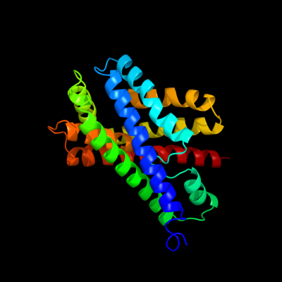
1 c3klzE_
100.0
27
PDB header: membrane proteinChain: E: PDB Molecule: putative formate transporter 1;PDBTitle: pentameric formate channel with formate bound
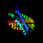
2 c3kcvG_
100.0
28
PDB header: transport proteinChain: G: PDB Molecule: probable formate transporter 1;PDBTitle: structure of formate channel
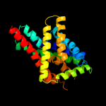
3 c3llqB_
95.5
9
PDB header: membrane proteinChain: B: PDB Molecule: aquaporin z 2;PDBTitle: aquaporin structure from plant pathogen agrobacterium tumerfaciens
4 c1ldaA_
93.7
10
PDB header: transport proteinChain: A: PDB Molecule: glycerol uptake facilitator protein;PDBTitle: crystal structure of the e. coli glycerol facilitator (glpf) without2 substrate glycerol
5 c3d9sB_
93.1
9
PDB header: membrane proteinChain: B: PDB Molecule: aquaporin-5;PDBTitle: human aquaporin 5 (aqp5) - high resolution x-ray structure
6 d1j4na_
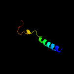
91.2
11
Fold: Aquaporin-likeSuperfamily: Aquaporin-likeFamily: Aquaporin-like7 d1fx8a_
88.9
10
Fold: Aquaporin-likeSuperfamily: Aquaporin-likeFamily: Aquaporin-like8 d1rc2a_
87.5
12
Fold: Aquaporin-likeSuperfamily: Aquaporin-likeFamily: Aquaporin-like9 c1ymgA_
78.5
12
PDB header: membrane proteinChain: A: PDB Molecule: lens fiber major intrinsic protein;PDBTitle: the channel architecture of aquaporin o at 2.2 angstrom resolution
10 d1ymga1
78.5
12
Fold: Aquaporin-likeSuperfamily: Aquaporin-likeFamily: Aquaporin-like11 c2kncA_
73.0
21
PDB header: cell adhesionChain: A: PDB Molecule: integrin alpha-iib;PDBTitle: platelet integrin alfaiib-beta3 transmembrane-cytoplasmic2 heterocomplex
12 c3c02A_
62.1
11
PDB header: membrane proteinChain: A: PDB Molecule: aquaglyceroporin;PDBTitle: x-ray structure of the aquaglyceroporin from plasmodium falciparum
13 c2b5fD_
36.3
10
PDB header: transport protein,membrane proteinChain: D: PDB Molecule: aquaporin;PDBTitle: crystal structure of the spinach aquaporin sopip2;1 in an2 open conformation to 3.9 resolution
14 c2w2eA_
35.9
9
PDB header: membrane proteinChain: A: PDB Molecule: aquaporin;PDBTitle: 1.15 angstrom crystal structure of p.pastoris aquaporin,2 aqy1, in a closed conformation at ph 3.5
15 c3gd8A_
34.3
14
PDB header: membrane proteinChain: A: PDB Molecule: aquaporin-4;PDBTitle: crystal structure of human aquaporin 4 at 1.8 and its mechanism of2 conductance
16 c3iyzA_
29.1
15
PDB header: transport proteinChain: A: PDB Molecule: aquaporin-4;PDBTitle: structure of aquaporin-4 s180d mutant at 10.0 a resolution from2 electron micrograph
17 d1pv7a_
16.5
15
Fold: MFS general substrate transporterSuperfamily: MFS general substrate transporterFamily: LacY-like proton/sugar symporter18 d1c17m_
15.9
22
Fold: F1F0 ATP synthase subunit ASuperfamily: F1F0 ATP synthase subunit AFamily: F1F0 ATP synthase subunit A19 d2nr9a1
11.4
14
Fold: Rhomboid-likeSuperfamily: Rhomboid-likeFamily: Rhomboid-like20 c2rddB_
9.8
14
PDB header: membrane protein/transport proteinChain: B: PDB Molecule: upf0092 membrane protein yajc;PDBTitle: x-ray crystal structure of acrb in complex with a novel2 transmembrane helix.
21 c2jy0A_
not modelled
9.7
39
PDB header: membrane protein, viral proteinChain: A: PDB Molecule: protease ns2-3;PDBTitle: solution nmr structure of hcv ns2 protein, membrane segment2 (1-27)
22 c2f2bA_
not modelled
7.5
13
PDB header: membrane proteinChain: A: PDB Molecule: aquaporin aqpm;PDBTitle: crystal structure of integral membrane protein aquaporin aqpm at 1.68a2 resolution
23 c3qnqD_
not modelled
7.4
9
PDB header: membrane protein, transport proteinChain: D: PDB Molecule: pts system, cellobiose-specific iic component;PDBTitle: crystal structure of the transporter chbc, the iic component from the2 n,n'-diacetylchitobiose-specific phosphotransferase system
24 d2foka2
not modelled
6.2
30
Fold: DNA/RNA-binding 3-helical bundleSuperfamily: "Winged helix" DNA-binding domainFamily: Restriction endonuclease FokI, N-terminal (recognition) domain25 d2c1wa1
not modelled
6.1
50
Fold: EndoU-likeSuperfamily: EndoU-likeFamily: Eukaryotic EndoU ribonuclease26 c2jp3A_
not modelled
6.1
14
PDB header: transcriptionChain: A: PDB Molecule: fxyd domain-containing ion transport regulator 4;PDBTitle: solution structure of the human fxyd4 (chif) protein in sds2 micelles
27 c2vv5D_
not modelled
5.9
12
PDB header: membrane proteinChain: D: PDB Molecule: small-conductance mechanosensitive channel;PDBTitle: the open structure of mscs
28 c2voyG_
not modelled
5.9
50
PDB header: hydrolaseChain: G: PDB Molecule: sarcoplasmic/endoplasmic reticulum calciumPDBTitle: cryoem model of copa, the copper transporting atpase from2 archaeoglobus fulgidus