Phyre Home Retrieve Phyre Job Id
Emaill.a.kelley@imperial.ac.uk
DescriptionP0A8N0
DateThu Jan 5 11:08:13 GMT 2012
Unique Job IDd55e0eeb54e337ff

Summary 

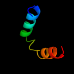
Top model
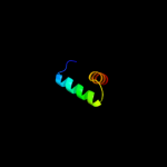
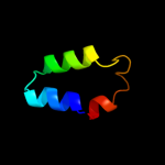
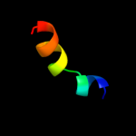
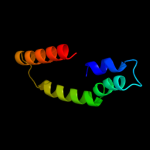
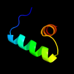
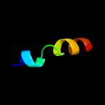
Image coloured by rainbow N → C terminus
Model (left) based on template c3kk4B_
Top template information
PDB header:structural genomics, unknown function
Chain: B: PDB Molecule:uncharacterized protein bp1543;
PDBTitle: uncharacterized protein bp1543 from bordetella pertussis tohama i
Confidence and coverage
Confidence: 87.0% Coverage: 23%
34 residues ( 23% of your sequence) have been modelled with 87.0% confidence by the single highest scoring template.
You may wish to submit your sequence to Phyrealarm. This will automatically scan your sequence every week for new potential templates as they appear in the Phyre2 library.
Please note: You must be registered and logged in to use Phyrealarm.
3D viewing
Interactive 3D view in Jmol

Sequence analysis 

Secondary structure and disorder prediction 

   1........10.........20.........30.........40.........50.........60
Sequence  MKYQQLENLESGWKWKYLVKKHREGELITRYIEASAAQEAVDVLLSLENEPVLVNGWIDK
Secondary structure 


SS confidence 


Disorder  ??


????


??


Disorder confidence 


   .........70.........80.........90.........100.........110.........120
Sequence  HMNPELVNRMKQTIRARRKRHFNAEHQHTRKKSIDLEFIVWQRLAGLAQRRGKTLSETIV
Secondary structure 


SS confidence 


Disorder 


???


??????


??


Disorder confidence 


   .........130.........140.........150
Sequence  QLIEDAENKEKYANKMSSLKQDLQALLGKE
Secondary structure 


SS confidence 


Disorder 


??


????
Disorder confidence 


Confidence Key
High(9)                    Low (0)
?Disordered
Alpha helix
Beta strand

Domain analysis 

Hover over an aligned region to see model and summary info

Please note, only up to the top 20 hits are modelled to reduce computer load

RankAligned region

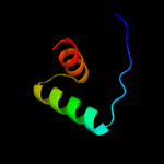
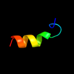
PDB 3kk4 chain B

3D model

Region: 93 - 126
Aligned: 34
Modelled: 34
Confidence: 87.0%
Identity: 29%
PDB header:structural genomics, unknown function
Chain: B: PDB Molecule:uncharacterized protein bp1543;
PDBTitle: uncharacterized protein bp1543 from bordetella pertussis tohama i

Phyre2

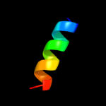
PDB 2ay0 chain A domain 1

3D model

Region: 89 - 125
Aligned: 37
Modelled: 37
Confidence: 32.6%
Identity: 24%
Fold: Ribbon-helix-helix
Superfamily: Ribbon-helix-helix
Family: PutA pre-N-terminal region-like

Phyre2

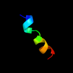
PDB 2aua chain A domain 1

3D model

Region: 64 - 78
Aligned: 15
Modelled: 15
Confidence: 22.7%
Identity: 40%
Fold: ADP-ribosylation
Superfamily: ADP-ribosylation
Family: BC2332-like

Phyre2

PDB 3nsn chain A

3D model

Region: 95 - 110
Aligned: 16
Modelled: 16
Confidence: 21.0%
Identity: 44%
PDB header:hydrolase
Chain: A: PDB Molecule:n-acetylglucosaminidase;
PDBTitle: crystal structure of insect beta-n-acetyl-d-hexosaminidase ofhex12 complexed with tmg-chitotriomycin

Phyre2

PDB 1ug3 chain A domain 2

3D model

Region: 38 - 76
Aligned: 38
Modelled: 39
Confidence: 20.2%
Identity: 18%
Fold: alpha-alpha superhelix
Superfamily: ARM repeat
Family: MIF4G domain-like

Phyre2

PDB 1qba chain A domain 3

3D model

Region: 95 - 111
Aligned: 17
Modelled: 17
Confidence: 14.4%
Identity: 24%
Fold: TIM beta/alpha-barrel
Superfamily: (Trans)glycosidases
Family: beta-N-acetylhexosaminidase catalytic domain

Phyre2

PDB 1qba chain A

3D model

Region: 95 - 110
Aligned: 16
Modelled: 16
Confidence: 13.8%
Identity: 25%
PDB header:glycosyl hydrolase
Chain: A: PDB Molecule:chitobiase;
PDBTitle: bacterial chitobiase, glycosyl hydrolase family 20

Phyre2

PDB 2pnv chain A

3D model

Region: 122 - 148
Aligned: 27
Modelled: 27
Confidence: 12.3%
Identity: 22%
PDB header:membrane protein
Chain: A: PDB Molecule:small conductance calcium-activated potassium
PDBTitle: crystal structure of the leucine zipper domain of small-2 conductance ca2+-activated k+ (skca) channel from rattus3 norvegicus

Phyre2

PDB 3rcn chain A

3D model

Region: 95 - 110
Aligned: 16
Modelled: 16
Confidence: 12.2%
Identity: 19%
PDB header:hydrolase
Chain: A: PDB Molecule:beta-n-acetylhexosaminidase;
PDBTitle: crystal structure of beta-n-acetylhexosaminidase from arthrobacter2 aurescens

Phyre2

PDB 2qzc chain B

3D model

Region: 38 - 101
Aligned: 62
Modelled: 64
Confidence: 11.5%
Identity: 16%
PDB header:lyase
Chain: B: PDB Molecule:transcriptional activator tena-1;
PDBTitle: crystal structure of a putative tena-like thiaminase (tena-1, sso2206)2 from sulfolobus solfataricus p2 at 1.50 a resolution

Phyre2

PDB 2rbf chain B

3D model

Region: 93 - 124
Aligned: 32
Modelled: 32
Confidence: 10.7%
Identity: 25%
PDB header:oxidoreductase/dna
Chain: B: PDB Molecule:bifunctional protein puta;
PDBTitle: structure of the ribbon-helix-helix domain of escherichia coli puta2 (puta52) complexed with operator dna (o2)

Phyre2

PDB 1m04 chain A

3D model

Region: 95 - 111
Aligned: 17
Modelled: 17
Confidence: 10.6%
Identity: 29%
PDB header:hydrolase
Chain: A: PDB Molecule:beta-n-acetylhexosaminidase;
PDBTitle: mutant streptomyces plicatus beta-hexosaminidase (d313n) in complex2 with product (glcnac)

Phyre2

PDB 2ahm chain A domain 1

3D model

Region: 8 - 24
Aligned: 17
Modelled: 17
Confidence: 10.5%
Identity: 35%
Fold: immunoglobulin/albumin-binding domain-like
Superfamily: Coronavirus NSP7-like
Family: Coronavirus NSP7-like

Phyre2

PDB 1ysy chain A domain 1

3D model

Region: 8 - 24
Aligned: 17
Modelled: 17
Confidence: 10.2%
Identity: 35%
Fold: immunoglobulin/albumin-binding domain-like
Superfamily: Coronavirus NSP7-like
Family: Coronavirus NSP7-like

Phyre2

PDB 1jak chain A domain 1

3D model

Region: 95 - 110
Aligned: 16
Modelled: 16
Confidence: 9.4%
Identity: 31%
Fold: TIM beta/alpha-barrel
Superfamily: (Trans)glycosidases
Family: beta-N-acetylhexosaminidase catalytic domain

Phyre2

PDB 2o59 chain B

3D model

Region: 23 - 78
Aligned: 56
Modelled: 56
Confidence: 8.4%
Identity: 23%
PDB header:isomerase/dna
Chain: B: PDB Molecule:dna topoisomerase 3;
PDBTitle: structure of e. coli topoisomerase iii in complex with an 8-2 base single stranded oligonucleotide. frozen in glycerol3 ph 8.0

Phyre2

PDB 2p62 chain A domain 1

3D model

Region: 112 - 143
Aligned: 32
Modelled: 32
Confidence: 8.3%
Identity: 31%
Fold: PH0156-like
Superfamily: PH0156-like
Family: PH0156-like

Phyre2

PDB 2p58 chain A

3D model

Region: 98 - 106
Aligned: 9
Modelled: 9
Confidence: 7.9%
Identity: 44%
PDB header:transport protein/chaperone
Chain: A: PDB Molecule:putative type iii secretion protein ysce;
PDBTitle: structure of the yersinia pestis type iii secretion system2 needle protein yscf in complex with its chaperones3 ysce/yscg

Phyre2

PDB 1i7d chain A

3D model

Region: 23 - 78
Aligned: 56
Modelled: 56
Confidence: 7.9%
Identity: 23%
Fold: Prokaryotic type I DNA topoisomerase
Superfamily: Prokaryotic type I DNA topoisomerase
Family: Prokaryotic type I DNA topoisomerase

Phyre2

PDB 1v4a chain A domain 1

3D model

Region: 69 - 101
Aligned: 33
Modelled: 31
Confidence: 7.5%
Identity: 21%
Fold: Four-helical up-and-down bundle
Superfamily: Nucleotidyltransferase substrate binding subunit/domain
Family: Glutamine synthase adenylyltransferase GlnE, domain 2

Phyre2
1

c3kk4B_
2

d2ay0a1
3

d2auaa1
4

c3nsnA_
5

d1ug3a2
6

d1qbaa3
7

c1qbaA_
8

c2pnvA_
9

c3rcnA_
10

c2qzcB_
11

c2rbfB_
12

c1m04A_
13

d2ahma1
14

d1ysya1
15

d1jaka1
16

c2o59B_
17

d2p62a1
18

c2p58A_
19

d1i7da_
20

d1v4aa1
21


22


23


24


25


26


27


28


29


30


31


Detailed template information 

#
Template Alignment Coverage3D Model Confidence
% i.d. Template Information
1c3kk4B_


87.0 29 PDB header:structural genomics, unknown function
Chain: B: PDB Molecule:uncharacterized protein bp1543;
PDBTitle: uncharacterized protein bp1543 from bordetella pertussis tohama i
2d2ay0a1


32.6 24 Fold:Ribbon-helix-helix
Superfamily:Ribbon-helix-helix
Family:PutA pre-N-terminal region-like
3d2auaa1


22.7 40 Fold:ADP-ribosylation
Superfamily:ADP-ribosylation
Family:BC2332-like
4c3nsnA_


21.0 44 PDB header:hydrolase
Chain: A: PDB Molecule:n-acetylglucosaminidase;
PDBTitle: crystal structure of insect beta-n-acetyl-d-hexosaminidase ofhex12 complexed with tmg-chitotriomycin
5d1ug3a2


20.2 18 Fold:alpha-alpha superhelix
Superfamily:ARM repeat
Family:MIF4G domain-like
6d1qbaa3


14.4 24 Fold:TIM beta/alpha-barrel
Superfamily:(Trans)glycosidases
Family:beta-N-acetylhexosaminidase catalytic domain
7c1qbaA_


13.8 25 PDB header:glycosyl hydrolase
Chain: A: PDB Molecule:chitobiase;
PDBTitle: bacterial chitobiase, glycosyl hydrolase family 20
8c2pnvA_


12.3 22 PDB header:membrane protein
Chain: A: PDB Molecule:small conductance calcium-activated potassium
PDBTitle: crystal structure of the leucine zipper domain of small-2 conductance ca2+-activated k+ (skca) channel from rattus3 norvegicus
9c3rcnA_


12.2 19 PDB header:hydrolase
Chain: A: PDB Molecule:beta-n-acetylhexosaminidase;
PDBTitle: crystal structure of beta-n-acetylhexosaminidase from arthrobacter2 aurescens
10c2qzcB_


11.5 16 PDB header:lyase
Chain: B: PDB Molecule:transcriptional activator tena-1;
PDBTitle: crystal structure of a putative tena-like thiaminase (tena-1, sso2206)2 from sulfolobus solfataricus p2 at 1.50 a resolution
11c2rbfB_


10.7 25 PDB header:oxidoreductase/dna
Chain: B: PDB Molecule:bifunctional protein puta;
PDBTitle: structure of the ribbon-helix-helix domain of escherichia coli puta2 (puta52) complexed with operator dna (o2)
12c1m04A_


10.6 29 PDB header:hydrolase
Chain: A: PDB Molecule:beta-n-acetylhexosaminidase;
PDBTitle: mutant streptomyces plicatus beta-hexosaminidase (d313n) in complex2 with product (glcnac)
13d2ahma1


10.5 35 Fold:immunoglobulin/albumin-binding domain-like
Superfamily:Coronavirus NSP7-like
Family:Coronavirus NSP7-like
14d1ysya1


10.2 35 Fold:immunoglobulin/albumin-binding domain-like
Superfamily:Coronavirus NSP7-like
Family:Coronavirus NSP7-like
15d1jaka1


9.4 31 Fold:TIM beta/alpha-barrel
Superfamily:(Trans)glycosidases
Family:beta-N-acetylhexosaminidase catalytic domain
16c2o59B_


8.4 23 PDB header:isomerase/dna
Chain: B: PDB Molecule:dna topoisomerase 3;
PDBTitle: structure of e. coli topoisomerase iii in complex with an 8-2 base single stranded oligonucleotide. frozen in glycerol3 ph 8.0
17d2p62a1


8.3 31 Fold:PH0156-like
Superfamily:PH0156-like
Family:PH0156-like
18c2p58A_


7.9 44 PDB header:transport protein/chaperone
Chain: A: PDB Molecule:putative type iii secretion protein ysce;
PDBTitle: structure of the yersinia pestis type iii secretion system2 needle protein yscf in complex with its chaperones3 ysce/yscg
19d1i7da_


7.9 23 Fold:Prokaryotic type I DNA topoisomerase
Superfamily:Prokaryotic type I DNA topoisomerase
Family:Prokaryotic type I DNA topoisomerase
20d1v4aa1


7.5 21 Fold:Four-helical up-and-down bundle
Superfamily:Nucleotidyltransferase substrate binding subunit/domain
Family:Glutamine synthase adenylyltransferase GlnE, domain 2
21d2cpga_


not modelled 7.2 28 Fold:Ribbon-helix-helix
Superfamily:Ribbon-helix-helix
Family:CopG-like
22c3fshC_


not modelled 7.0 39 PDB header:ligase
Chain: C: PDB Molecule:autocrine motility factor receptor, isoform 2;
PDBTitle: crystal structure of the ubiquitin conjugating enzyme2 ube2g2 bound to the g2br domain of ubiquitin ligase gp78
23c3lmyA_


not modelled 6.5 25 PDB header:hydrolase
Chain: A: PDB Molecule:beta-hexosaminidase subunit beta;
PDBTitle: the crystal structure of beta-hexosaminidase b in complex with2 pyrimethamine
24c3h8kB_


not modelled 6.4 39 PDB header:ligase
Chain: B: PDB Molecule:autocrine motility factor receptor, isoform 2;
PDBTitle: crystal structure of ube2g2 complxed with the g2br domain of2 gp78 at 1.8-a resolution
25d1nowa1


not modelled 6.2 24 Fold:TIM beta/alpha-barrel
Superfamily:(Trans)glycosidases
Family:beta-N-acetylhexosaminidase catalytic domain
26c1zw3B_


not modelled 5.7 58 PDB header:protein binding
Chain: B: PDB Molecule:talin 1;
PDBTitle: vinculin head (0-258) in complex with the talin rod2 residues 1630-1652
27c1nouA_


not modelled 5.5 24 PDB header:hydrolase
Chain: A: PDB Molecule:beta-hexosaminidase beta chain;
PDBTitle: native human lysosomal beta-hexosaminidase isoform b
28c2ykqC_


not modelled 5.4 21 PDB header:rna-binding protein
Chain: C: PDB Molecule:line-1 orf1p;
PDBTitle: structure of the human line-1 orf1p trimer
29d1ukfa_


not modelled 5.4 11 Fold:Cysteine proteinases
Superfamily:Cysteine proteinases
Family:Avirulence protein Avrpph3
30d1g73a_


not modelled 5.3 37 Fold:Spectrin repeat-like
Superfamily:Smac/diablo
Family:Smac/diablo
31d1fewa_


not modelled 5.0 37 Fold:Spectrin repeat-like
Superfamily:Smac/diablo
Family:Smac/diablo

Binding site prediction 

Due to computational demand, binding site predictions are not run for batch jobs

If you want to predict binding sites, please manually submit your model of choice to 3DLigandSite


Phyre is for academic use only

Please cite: Protein structure prediction on the web: a case study using the Phyre server
Kelley LA and Sternberg MJE. Nature Protocols 4, 363 - 371 (2009) [pdf] [Import into BibTeX]
 
If you use the binding site predictions from 3DLigandSite, please also cite:
3DLigandSite: predicting ligand-binding sites using similar structures.
Wass MN, Kelley LA and Sternberg MJ Nucleic Acids Research 38, W469-73 (2010) [PubMed]
 
© Structural Bioinformatics Group
Imperial College London
Lawrence Kelley, Benjamin Jefferys
Disclaimer
Terms and Conditions
Component software
Template detection: HHpred 1.51
Secondary structure prediction: Psi-pred 2.5
Disorder prediction: Disopred 2.4
Transmembrane prediction: Memsat_SVM
Multi-template modelling and ab initio: Poing 1.0