Phyre Home Retrieve Phyre Job Id
Emaill.a.kelley@imperial.ac.uk
DescriptionP52130
DateThu Jan 5 12:05:35 GMT 2012
Unique Job IDd4a6387acab167c6

Summary 

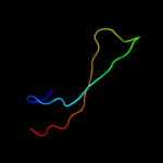
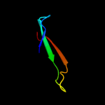
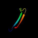
Top model
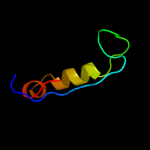
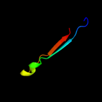
Image coloured by rainbow N → C terminus
Model (left) based on template d1v8ha1
Top template information
Fold:Immunoglobulin-like beta-sandwich
Superfamily:E set domains
Family:SoxZ-like
Confidence and coverage
Confidence: 29.3% Coverage: 24%
30 residues ( 24% of your sequence) have been modelled with 29.3% confidence by the single highest scoring template.
You may wish to submit your sequence to Phyrealarm. This will automatically scan your sequence every week for new potential templates as they appear in the Phyre2 library.
Please note: You must be registered and logged in to use Phyrealarm.
3D viewing
Interactive 3D view in Jmol

Sequence analysis 

Secondary structure and disorder prediction 

   1........10.........20.........30.........40.........50.........60
Sequence  MFEITGINVSGALKAVVMATGFENPLSSVNEIETKLSALLGSETTGEILFDLLCANGPEW
Secondary structure 


SS confidence 


Disorder  ?????


?


???


Disorder confidence 


   .........70.........80.........90.........100.........110.........120
Sequence  NRFVTLEMKYGRIMLDTAKIIDEQDVPTHILSKLTFTLRNHPEYLEASVLSPDDVRQVLS
Secondary structure 


SS confidence 


Disorder 


?


??


??

??


??
Disorder confidence 


   ...
Sequence  MDF
Secondary structure 


SS confidence 


Disorder  ???
Disorder confidence 


Confidence Key
High(9)                    Low (0)
?Disordered
Alpha helix
Beta strand

Domain analysis 

Hover over an aligned region to see model and summary info

Please note, only up to the top 20 hits are modelled to reduce computer load

RankAligned region

PDB 1v8h chain A domain 1

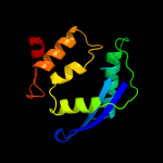
3D model

Region: 43 - 72
Aligned: 30
Modelled: 30
Confidence: 29.3%
Identity: 13%
Fold: Immunoglobulin-like beta-sandwich
Superfamily: E set domains
Family: SoxZ-like

Phyre2

PDB 1mgt chain A

3D model

Region: 1 - 119
Aligned: 113
Modelled: 119
Confidence: 16.1%
Identity: 12%
PDB header:transferase
Chain: A: PDB Molecule:protein (o6-methylguanine-dna methyltransferase);
PDBTitle: crystal structure of o6-methylguanine-dna methyltransferase from2 hyperthermophilic archaeon pyrococcus kodakaraensis strain kod1

Phyre2

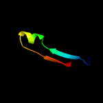
PDB 3lf4 chain B

3D model

Region: 54 - 80
Aligned: 25
Modelled: 27
Confidence: 9.9%
Identity: 20%
PDB header:fluorescent protein
Chain: B: PDB Molecule:fluorescent timer precursor blue102;
PDBTitle: crystal structure of fluorescent timer precursor blue102

Phyre2

PDB 2gw4 chain D

3D model

Region: 54 - 80
Aligned: 25
Modelled: 27
Confidence: 9.6%
Identity: 28%
PDB header:luminescent protein
Chain: D: PDB Molecule:kaede;
PDBTitle: crystal structure of stony coral fluorescent protein kaede, red form

Phyre2

PDB 2zmw chain C

3D model

Region: 54 - 80
Aligned: 25
Modelled: 27
Confidence: 8.8%
Identity: 32%
PDB header:luminescent protein
Chain: C: PDB Molecule:fluorescent protein;
PDBTitle: crystal structure of monomeric kusabira-orange (mko),2 orange-emitting gfp-like protein, at ph 6.0

Phyre2

PDB 1mou chain A

3D model

Region: 54 - 80
Aligned: 25
Modelled: 27
Confidence: 8.8%
Identity: 16%
Fold: GFP-like
Superfamily: GFP-like
Family: Fluorescent proteins

Phyre2

PDB 1w2t chain E

3D model

Region: 52 - 82
Aligned: 31
Modelled: 31
Confidence: 8.3%
Identity: 16%
PDB header:hydrolase
Chain: E: PDB Molecule:beta fructosidase;
PDBTitle: beta-fructosidase from thermotoga maritima in complex with2 raffinose

Phyre2

PDB 3evp chain A

3D model

Region: 54 - 80
Aligned: 25
Modelled: 27
Confidence: 8.0%
Identity: 20%
PDB header:signaling protein
Chain: A: PDB Molecule:circular-permutated green fluorescent protein;
PDBTitle: crystal structure of circular-permutated egfp

Phyre2

PDB 3cfh chain B

3D model

Region: 50 - 80
Aligned: 29
Modelled: 31
Confidence: 7.8%
Identity: 21%
PDB header:fluorescent protein
Chain: B: PDB Molecule:gfp-like photoswitchable fluorescent protein;
PDBTitle: photoswitchable red fluorescent protein psrfp, off-state

Phyre2

PDB 2rh7 chain A domain 1

3D model

Region: 54 - 80
Aligned: 25
Modelled: 27
Confidence: 7.8%
Identity: 16%
Fold: GFP-like
Superfamily: GFP-like
Family: Fluorescent proteins

Phyre2

PDB 1lc0 chain A domain 2

3D model

Region: 57 - 75
Aligned: 19
Modelled: 19
Confidence: 7.8%
Identity: 26%
Fold: FwdE/GAPDH domain-like
Superfamily: Glyceraldehyde-3-phosphate dehydrogenase-like, C-terminal domain
Family: Biliverdin reductase

Phyre2

PDB 2b5e chain A domain 3

3D model

Region: 10 - 50
Aligned: 36
Modelled: 41
Confidence: 7.7%
Identity: 19%
Fold: Thioredoxin fold
Superfamily: Thioredoxin-like
Family: PDI-like

Phyre2

PDB 2z6z chain A

3D model

Region: 54 - 80
Aligned: 25
Modelled: 27
Confidence: 7.7%
Identity: 28%
PDB header:fluorescent protein
Chain: A: PDB Molecule:fluorescent protein dronpa;
PDBTitle: crystal structure of a photoswitchable gfp-like protein2 dronpa in the bright-state

Phyre2

PDB 1xqm chain A

3D model

Region: 54 - 80
Aligned: 25
Modelled: 27
Confidence: 7.7%
Identity: 20%
Fold: GFP-like
Superfamily: GFP-like
Family: Fluorescent proteins

Phyre2

PDB 1ggx chain A

3D model

Region: 54 - 80
Aligned: 25
Modelled: 27
Confidence: 7.5%
Identity: 20%
Fold: GFP-like
Superfamily: GFP-like
Family: Fluorescent proteins

Phyre2

PDB 2c9j chain G

3D model

Region: 54 - 80
Aligned: 25
Modelled: 27
Confidence: 7.5%
Identity: 24%
PDB header:luminescent protein
Chain: G: PDB Molecule:green fluorescent protein fp512;
PDBTitle: structure of the fluorescent protein cmfp512 at 1.35a from2 cerianthus membranaceus

Phyre2

PDB 2otb chain B

3D model

Region: 54 - 80
Aligned: 25
Modelled: 27
Confidence: 7.5%
Identity: 24%
PDB header:fluorescent protein
Chain: B: PDB Molecule:gfp-like fluorescent chromoprotein cfp484;
PDBTitle: crystal structure of a monomeric cyan fluorescent protein2 in the fluorescent state

Phyre2

PDB 3ai5 chain A

3D model

Region: 54 - 80
Aligned: 25
Modelled: 27
Confidence: 7.3%
Identity: 20%
PDB header:fluorescent protein, transcription
Chain: A: PDB Molecule:yeast enhanced green fluorescent protein, ubiquitin;
PDBTitle: crystal structure of yeast enhanced green fluorescent protein-2 ubiquitin fusion protein

Phyre2

PDB 3gb3 chain B

3D model

Region: 54 - 80
Aligned: 25
Modelled: 27
Confidence: 7.3%
Identity: 16%
PDB header:fluorescent protein
Chain: B: PDB Molecule:killerred;
PDBTitle: x-ray structure of genetically encoded photosensitizer2 killerred in native form

Phyre2

PDB 1yzw chain B

3D model

Region: 54 - 80
Aligned: 25
Modelled: 27
Confidence: 7.3%
Identity: 24%
PDB header:luminescent protein
Chain: B: PDB Molecule:gfp-like non-fluorescent chromoprotein;
PDBTitle: the 2.1a crystal structure of the far-red fluorescent2 protein hcred: inherent conformational flexibility of the3 chromophore

Phyre2
1

d1v8ha1
2

c1mgtA_
3

c3lf4B_
4

c2gw4D_
5

c2zmwC_
6

d1moua_
7

c1w2tE_
8

c3evpA_
9

c3cfhB_
10

d2rh7a1
11

d1lc0a2
12

d2b5ea3
13

c2z6zA_
14

d1xqma_
15

d1ggxa_
16

c2c9jG_
17

c2otbB_
18

c3ai5A_
19

c3gb3B_
20

c1yzwB_
21


22


23


24


25


26


27


28


29


30


31


32


33


34


35


Detailed template information 

#
Template Alignment Coverage3D Model Confidence
% i.d. Template Information
1d1v8ha1


29.3 13 Fold:Immunoglobulin-like beta-sandwich
Superfamily:E set domains
Family:SoxZ-like
2c1mgtA_


16.1 12 PDB header:transferase
Chain: A: PDB Molecule:protein (o6-methylguanine-dna methyltransferase);
PDBTitle: crystal structure of o6-methylguanine-dna methyltransferase from2 hyperthermophilic archaeon pyrococcus kodakaraensis strain kod1
3c3lf4B_


9.9 20 PDB header:fluorescent protein
Chain: B: PDB Molecule:fluorescent timer precursor blue102;
PDBTitle: crystal structure of fluorescent timer precursor blue102
4c2gw4D_


9.6 28 PDB header:luminescent protein
Chain: D: PDB Molecule:kaede;
PDBTitle: crystal structure of stony coral fluorescent protein kaede, red form
5c2zmwC_


8.8 32 PDB header:luminescent protein
Chain: C: PDB Molecule:fluorescent protein;
PDBTitle: crystal structure of monomeric kusabira-orange (mko),2 orange-emitting gfp-like protein, at ph 6.0
6d1moua_


8.8 16 Fold:GFP-like
Superfamily:GFP-like
Family:Fluorescent proteins
7c1w2tE_


8.3 16 PDB header:hydrolase
Chain: E: PDB Molecule:beta fructosidase;
PDBTitle: beta-fructosidase from thermotoga maritima in complex with2 raffinose
8c3evpA_


8.0 20 PDB header:signaling protein
Chain: A: PDB Molecule:circular-permutated green fluorescent protein;
PDBTitle: crystal structure of circular-permutated egfp
9c3cfhB_


7.8 21 PDB header:fluorescent protein
Chain: B: PDB Molecule:gfp-like photoswitchable fluorescent protein;
PDBTitle: photoswitchable red fluorescent protein psrfp, off-state
10d2rh7a1


7.8 16 Fold:GFP-like
Superfamily:GFP-like
Family:Fluorescent proteins
11d1lc0a2


7.8 26 Fold:FwdE/GAPDH domain-like
Superfamily:Glyceraldehyde-3-phosphate dehydrogenase-like, C-terminal domain
Family:Biliverdin reductase
12d2b5ea3


7.7 19 Fold:Thioredoxin fold
Superfamily:Thioredoxin-like
Family:PDI-like
13c2z6zA_


7.7 28 PDB header:fluorescent protein
Chain: A: PDB Molecule:fluorescent protein dronpa;
PDBTitle: crystal structure of a photoswitchable gfp-like protein2 dronpa in the bright-state
14d1xqma_


7.7 20 Fold:GFP-like
Superfamily:GFP-like
Family:Fluorescent proteins
15d1ggxa_


7.5 20 Fold:GFP-like
Superfamily:GFP-like
Family:Fluorescent proteins
16c2c9jG_


7.5 24 PDB header:luminescent protein
Chain: G: PDB Molecule:green fluorescent protein fp512;
PDBTitle: structure of the fluorescent protein cmfp512 at 1.35a from2 cerianthus membranaceus
17c2otbB_


7.5 24 PDB header:fluorescent protein
Chain: B: PDB Molecule:gfp-like fluorescent chromoprotein cfp484;
PDBTitle: crystal structure of a monomeric cyan fluorescent protein2 in the fluorescent state
18c3ai5A_


7.3 20 PDB header:fluorescent protein, transcription
Chain: A: PDB Molecule:yeast enhanced green fluorescent protein, ubiquitin;
PDBTitle: crystal structure of yeast enhanced green fluorescent protein-2 ubiquitin fusion protein
19c3gb3B_


7.3 16 PDB header:fluorescent protein
Chain: B: PDB Molecule:killerred;
PDBTitle: x-ray structure of genetically encoded photosensitizer2 killerred in native form
20c1yzwB_


7.3 24 PDB header:luminescent protein
Chain: B: PDB Molecule:gfp-like non-fluorescent chromoprotein;
PDBTitle: the 2.1a crystal structure of the far-red fluorescent2 protein hcred: inherent conformational flexibility of the3 chromophore
21d1vkna2


not modelled 7.1 30 Fold:FwdE/GAPDH domain-like
Superfamily:Glyceraldehyde-3-phosphate dehydrogenase-like, C-terminal domain
Family:GAPDH-like
22c3rwaE_


not modelled 7.1 20 PDB header:fluorescent protein
Chain: E: PDB Molecule:fluorescent protein fp480;
PDBTitle: crystal structure of circular-permutated mkate
23d1ne2a_


not modelled 6.9 18 Fold:S-adenosyl-L-methionine-dependent methyltransferases
Superfamily:S-adenosyl-L-methionine-dependent methyltransferases
Family:Ta1320-like
24c3akoG_


not modelled 6.9 20 PDB header:fluorescent protein
Chain: G: PDB Molecule:venus;
PDBTitle: crystal structure of the reassembled venus
25d2q49a2


not modelled 6.9 40 Fold:FwdE/GAPDH domain-like
Superfamily:Glyceraldehyde-3-phosphate dehydrogenase-like, C-terminal domain
Family:GAPDH-like
26c2oxgE_


not modelled 6.5 14 PDB header:transport protein
Chain: E: PDB Molecule:soxz protein;
PDBTitle: the soxyz complex of paracoccus pantotrophus
27c2g3dB_


not modelled 6.4 20 PDB header:luminescent protein
Chain: B: PDB Molecule:green fluorescent protein;
PDBTitle: structure of s65g y66a gfp variant after spontaneous2 peptide hydrolysis
28c3cglE_


not modelled 5.9 24 PDB header:fluorescent protein
Chain: E: PDB Molecule:gfp-like fluorescent chromoprotein dsfp483;
PDBTitle: crystal structure and raman studies of dsfp483, a cyan fluorescent2 protein from discosoma striata
29c3t9aA_


not modelled 5.9 25 PDB header:transferase
Chain: A: PDB Molecule:inositol pyrophosphate kinase;
PDBTitle: crystal structure of the catalytic domain of human diphosphoinositol2 pentakisphosphate kinase 2 (ppip5k2) in complex with amppnp at ph 7.0
30c2ib5H_


not modelled 5.8 20 PDB header:luminescent protein
Chain: H: PDB Molecule:chromo protein;
PDBTitle: structural characterization of a blue chromoprotein and its yellow2 mutant from the sea anemone cnidopus japonicus
31d1nh8a2


not modelled 5.7 25 Fold:Ferredoxin-like
Superfamily:GlnB-like
Family:ATP phosphoribosyltransferase (ATP-PRTase, HisG), regulatory C-terminal domain
32d1ckma2


not modelled 5.4 40 Fold:ATP-grasp
Superfamily:DNA ligase/mRNA capping enzyme, catalytic domain
Family:mRNA capping enzyme
33d2v0ea1


not modelled 5.4 30 Fold:GYF/BRK domain-like
Superfamily:BRK domain-like
Family:BRK domain-like
34d1mywa_


not modelled 5.3 20 Fold:GFP-like
Superfamily:GFP-like
Family:Fluorescent proteins
35d1uisa_


not modelled 5.3 16 Fold:GFP-like
Superfamily:GFP-like
Family:Fluorescent proteins

Binding site prediction 

Due to computational demand, binding site predictions are not run for batch jobs

If you want to predict binding sites, please manually submit your model of choice to 3DLigandSite


Phyre is for academic use only

Please cite: Protein structure prediction on the web: a case study using the Phyre server
Kelley LA and Sternberg MJE. Nature Protocols 4, 363 - 371 (2009) [pdf] [Import into BibTeX]
 
If you use the binding site predictions from 3DLigandSite, please also cite:
3DLigandSite: predicting ligand-binding sites using similar structures.
Wass MN, Kelley LA and Sternberg MJ Nucleic Acids Research 38, W469-73 (2010) [PubMed]
 
© Structural Bioinformatics Group
Imperial College London
Lawrence Kelley, Benjamin Jefferys
Disclaimer
Terms and Conditions
Component software
Template detection: HHpred 1.51
Secondary structure prediction: Psi-pred 2.5
Disorder prediction: Disopred 2.4
Transmembrane prediction: Memsat_SVM
Multi-template modelling and ab initio: Poing 1.0