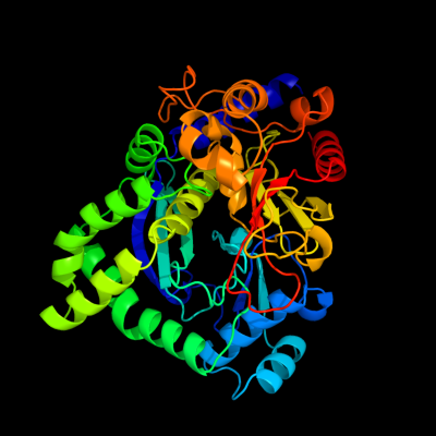
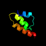
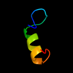
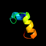
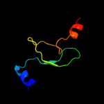
1 c3clqC_
100.0
65
PDB header: structural genomics, unknown functionChain: C: PDB Molecule: uncharacterized protein;PDBTitle: crystal structure of a conserved protein of unknown function from2 enterococcus faecalis v583
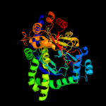
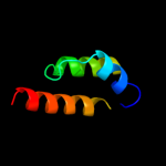
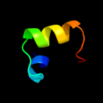
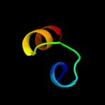
2 d1pdza2
74.3
35
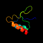
Fold: Enolase N-terminal domain-likeSuperfamily: Enolase N-terminal domain-likeFamily: Enolase N-terminal domain-like3 d2akza2
68.4
24
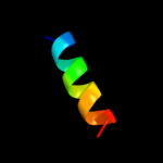
Fold: Enolase N-terminal domain-likeSuperfamily: Enolase N-terminal domain-likeFamily: Enolase N-terminal domain-like4 d2al1a2
65.9
22
Fold: Enolase N-terminal domain-likeSuperfamily: Enolase N-terminal domain-likeFamily: Enolase N-terminal domain-like5 d1wo8a1
60.8
19
Fold: Methylglyoxal synthase-likeSuperfamily: Methylglyoxal synthase-likeFamily: Methylglyoxal synthase, MgsA6 c1vjtA_
51.7
9
PDB header: hydrolaseChain: A: PDB Molecule: alpha-glucosidase;PDBTitle: crystal structure of alpha-glucosidase (tm0752) from thermotoga2 maritima at 2.50 a resolution
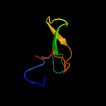
7 c2cqjA_
50.2
17
PDB header: rna binding proteinChain: A: PDB Molecule: u3 small nucleolar ribonucleoprotein proteinPDBTitle: solution structure of the s4 domain of u3 small nucleolar2 ribonucleoprotein protein imp3 homolog
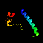
8 d1alua_
41.0
15
Fold: 4-helical cytokinesSuperfamily: 4-helical cytokinesFamily: Long-chain cytokines9 d2fyma2
40.7
15
Fold: Enolase N-terminal domain-likeSuperfamily: Enolase N-terminal domain-likeFamily: Enolase N-terminal domain-like10 c2re2A_
31.9
32
PDB header: oxidoreductaseChain: A: PDB Molecule: uncharacterized protein ta1041;PDBTitle: crystal structure of a putative iron-molybdenum cofactor (femo-co)2 dinitrogenase (ta1041m) from thermoplasma acidophilum dsm 1728 at3 1.30 a resolution
11 d1h3fa2
21.9
21
Fold: Alpha-L RNA-binding motifSuperfamily: Alpha-L RNA-binding motifFamily: Tyrosyl-tRNA synthetase (TyrRS), C-terminal domain12 d1c06a_
21.5
36
Fold: Alpha-L RNA-binding motifSuperfamily: Alpha-L RNA-binding motifFamily: Ribosomal protein S413 c2y9xG_
20.4
30
PDB header: oxidoreductaseChain: G: PDB Molecule: lectin-like fold protein;PDBTitle: crystal structure of ppo3, a tyrosinase from agaricus bisporus, in2 deoxy-form that contains additional unknown lectin-like subunit,3 with inhibitor tropolone
14 c2yvqA_
20.2
18
PDB header: ligaseChain: A: PDB Molecule: carbamoyl-phosphate synthase;PDBTitle: crystal structure of mgs domain of carbamoyl-phosphate2 synthetase from homo sapiens
15 d2pw4a1
19.7
18
Fold: Jann2411-likeSuperfamily: Jann2411-likeFamily: Jann2411-like16 d1b93a_
19.4
23
Fold: Methylglyoxal synthase-likeSuperfamily: Methylglyoxal synthase-likeFamily: Methylglyoxal synthase, MgsA17 c3hp7A_
19.2
17
PDB header: structural genomics, unknown functionChain: A: PDB Molecule: hemolysin, putative;PDBTitle: putative hemolysin from streptococcus thermophilus.
18 c2htmB_
17.0
19
PDB header: biosynthetic proteinChain: B: PDB Molecule: thiazole biosynthesis protein thig;PDBTitle: crystal structure of ttha0676 from thermus thermophilus hb8
19 c2l58A_
16.6
40
PDB header: apoptosisChain: A: PDB Molecule: activator of apoptosis harakiri;PDBTitle: solution structure of the cytosolic fragment 22-53 of bcl-2 member2 harakiri
20 d2gy9d1
16.4
24
Fold: Alpha-L RNA-binding motifSuperfamily: Alpha-L RNA-binding motifFamily: Ribosomal protein S421 d1twda_
not modelled
12.7
17
Fold: TIM beta/alpha-barrelSuperfamily: CutC-likeFamily: CutC-like22 d1nuba2
not modelled
11.7
50
Fold: Knottins (small inhibitors, toxins, lectins)Superfamily: EGF/LamininFamily: Follistatin (FS) module N-terminal domain, FS-N23 c2xzmD_
not modelled
11.6
18
PDB header: ribosomeChain: D: PDB Molecule: ribosomal protein s4 containing protein;PDBTitle: crystal structure of the eukaryotic 40s ribosomal2 subunit in complex with initiation factor 1. this file3 contains the 40s subunit and initiation factor for4 molecule 1
24 c2ldkA_
not modelled
11.5
14
PDB header: structural genomics, unknown functionChain: A: PDB Molecule: uncharacterized protein;PDBTitle: solution nmr structure of protein aaur_3427 from arthrobacter2 aurescens, northeast structural genomics consortium target aar96
25 d1jh3a_
not modelled
10.5
25
Fold: Alpha-L RNA-binding motifSuperfamily: Alpha-L RNA-binding motifFamily: Tyrosyl-tRNA synthetase (TyrRS), C-terminal domain26 d3cuma1
not modelled
10.2
19
Fold: 6-phosphogluconate dehydrogenase C-terminal domain-likeSuperfamily: 6-phosphogluconate dehydrogenase C-terminal domain-likeFamily: Hydroxyisobutyrate and 6-phosphogluconate dehydrogenase domain27 c3msuA_
not modelled
10.0
20
PDB header: transferaseChain: A: PDB Molecule: citrate synthase;PDBTitle: crystal structure of citrate synthase from francisella tularensis
28 c3d0jA_
not modelled
9.9
12
PDB header: structural genomics, unknown functionChain: A: PDB Molecule: uncharacterized protein ca_c3497;PDBTitle: crystal structure of conserved protein of unknown function ca_c34972 from clostridium acetobutylicum atcc 824
29 c2d5nB_
not modelled
9.8
17
PDB header: hydrolase, oxidoreductaseChain: B: PDB Molecule: riboflavin biosynthesis protein ribd;PDBTitle: crystal structure of a bifunctional deaminase and reductase2 involved in riboflavin biosynthesis
30 d2hsja1
not modelled
9.8
18
Fold: Flavodoxin-likeSuperfamily: SGNH hydrolaseFamily: Acetylhydrolase31 c2bibA_
not modelled
9.8
17
PDB header: hydrolaseChain: A: PDB Molecule: teichoic acid phosphorylcholine esterase/ choline bindingPDBTitle: crystal structure of the complete modular teichioic acid2 phosphorylcholine esterase pce (cbpe) from streptococcus3 pneumoniae
32 c3iefA_
not modelled
9.8
33
PDB header: transferase, rna binding proteinChain: A: PDB Molecule: trna (guanine-n(1)-)-methyltransferase;PDBTitle: crystal structure of trna guanine-n1-methyltransferase from2 bartonella henselae using mpcs.
33 d2o34a1
not modelled
9.7
57
Fold: T-foldSuperfamily: ApbE-likeFamily: DVU1097-like34 c2bs5A_
not modelled
9.4
25
PDB header: sugar binding proteinChain: A: PDB Molecule: fucose-binding lectin protein;PDBTitle: lectin from ralstonia solanacearum complexed with 2-2 fucosyllactose
35 d1p9ka_
not modelled
8.6
24
Fold: Alpha-L RNA-binding motifSuperfamily: Alpha-L RNA-binding motifFamily: YbcJ-like36 c1u8xX_
not modelled
8.4
21
PDB header: hydrolaseChain: X: PDB Molecule: maltose-6'-phosphate glucosidase;PDBTitle: crystal structure of glva from bacillus subtilis, a metal-requiring,2 nad-dependent 6-phospho-alpha-glucosidase
37 c3fefB_
not modelled
8.3
24
PDB header: hydrolaseChain: B: PDB Molecule: putative glucosidase lpld;PDBTitle: crystal structure of putative glucosidase lpld from2 bacillus subtilis
38 d1vmda_
not modelled
8.3
19
Fold: Methylglyoxal synthase-likeSuperfamily: Methylglyoxal synthase-likeFamily: Methylglyoxal synthase, MgsA39 d2isba1
not modelled
7.8
17
Fold: The "swivelling" beta/beta/alpha domainSuperfamily: FumA C-terminal domain-likeFamily: FumA C-terminal domain-like40 d1vjta1
not modelled
7.7
7
Fold: NAD(P)-binding Rossmann-fold domainsSuperfamily: NAD(P)-binding Rossmann-fold domainsFamily: LDH N-terminal domain-like41 c3n5mD_
not modelled
7.4
20
PDB header: transferaseChain: D: PDB Molecule: adenosylmethionine-8-amino-7-oxononanoate aminotransferase;PDBTitle: crystals structure of a bacillus anthracis aminotransferase
42 d2uubd1
not modelled
7.3
35
Fold: Alpha-L RNA-binding motifSuperfamily: Alpha-L RNA-binding motifFamily: Ribosomal protein S443 c2i7uA_
not modelled
7.2
43
PDB header: de novo protein/ligand binding proteinChain: A: PDB Molecule: four-alpha-helix bundle;PDBTitle: structural and dynamical analysis of a four-alpha-helix2 bundle with designed anesthetic binding pockets
44 d1vk4a_
not modelled
7.0
15
Fold: Ribokinase-likeSuperfamily: Ribokinase-likeFamily: Ribokinase-like45 c1s1hD_
not modelled
6.8
32
PDB header: ribosomeChain: D: PDB Molecule: 40s ribosomal protein s9-a;PDBTitle: structure of the ribosomal 80s-eef2-sordarin complex from2 yeast obtained by docking atomic models for rna and protein3 components into a 11.7 a cryo-em map. this file, 1s1h,4 contains 40s subunit. the 60s ribosomal subunit is in file5 1s1i.
46 d2jeka1
not modelled
6.5
32
Fold: Rv1873-likeSuperfamily: Rv1873-likeFamily: Rv1873-like47 c2iunD_
not modelled
6.4
25
PDB header: viral proteinChain: D: PDB Molecule: avian adenovirus celo long fibre;PDBTitle: structure of the c-terminal head domain of the avian2 adenovirus celo long fibre (p21 crystal form)
48 c3bbnD_
not modelled
6.3
25
PDB header: ribosomeChain: D: PDB Molecule: ribosomal protein s4;PDBTitle: homology model for the spinach chloroplast 30s subunit2 fitted to 9.4a cryo-em map of the 70s chlororibosome.
49 c1oy5B_
not modelled
6.3
32
PDB header: transferaseChain: B: PDB Molecule: trna (guanine-n(1)-)-methyltransferase;PDBTitle: crystal structure of trna (m1g37) methyltransferase from aquifex2 aeolicus
50 d1oy5a_
not modelled
6.3
32
Fold: alpha/beta knotSuperfamily: alpha/beta knotFamily: tRNA(m1G37)-methyltransferase TrmD51 c1s6yA_
not modelled
6.3
31
PDB header: hydrolaseChain: A: PDB Molecule: 6-phospho-beta-glucosidase;PDBTitle: 2.3a crystal structure of phospho-beta-glucosidase
52 d1s4da_
not modelled
6.1
19
Fold: Tetrapyrrole methylaseSuperfamily: Tetrapyrrole methylaseFamily: Tetrapyrrole methylase53 c2k4mA_
not modelled
6.1
29
PDB header: structural genomics, unknown functionChain: A: PDB Molecule: upf0146 protein mth_1000;PDBTitle: solution nmr structure of m. thermoautotrophicum protein2 mth_1000, northeast structural genomics consortium target3 tr8
54 c3btpA_
not modelled
6.0
43
PDB header: dna binding protein, chaperoneChain: A: PDB Molecule: single-strand dna-binding protein;PDBTitle: crystal structure of agrobacterium tumefaciens vire2 in complex with2 its chaperone vire1: a novel fold and implications for dna binding
55 c3brtC_
not modelled
5.9
33
PDB header: transferase/transcriptionChain: C: PDB Molecule: inhibitor of nuclear factor kappa-b kinasePDBTitle: nemo/ikk association domain structure
56 c2akmA_
not modelled
5.9
23
PDB header: lyaseChain: A: PDB Molecule: gamma enolase;PDBTitle: fluoride inhibition of enolase: crystal structure of the2 inhibitory complex
57 d1t6la2
not modelled
5.8
56
Fold: DNA clampSuperfamily: DNA clampFamily: DNA polymerase processivity factor58 c3p04A_
not modelled
5.8
19
PDB header: structural genomics, unknown functionChain: A: PDB Molecule: uncharacterized bcr;PDBTitle: crystal structure of the bcr protein from corynebacterium glutamicum.2 northeast structural genomics consortium target cgr8
59 c3bboG_
not modelled
5.7
46
PDB header: ribosomeChain: G: PDB Molecule: ribosomal protein l4;PDBTitle: homology model for the spinach chloroplast 50s subunit2 fitted to 9.4a cryo-em map of the 70s chlororibosome
60 c2q7aA_
not modelled
5.5
43
PDB header: heme binding proteinChain: A: PDB Molecule: cell surface heme-binding protein;PDBTitle: crystal structure of the cell surface heme transfer protein shp
61 c2w40C_
not modelled
5.4
18
PDB header: transferaseChain: C: PDB Molecule: glycerol kinase, putative;PDBTitle: crystal structure of plasmodium falciparum glycerol kinase2 with bound glycerol
62 d1a7ha_
not modelled
5.3
23
Fold: gamma-Crystallin-likeSuperfamily: gamma-Crystallin-likeFamily: Crystallins/Ca-binding development proteins63 d1s0aa_
not modelled
5.3
32
Fold: PLP-dependent transferase-likeSuperfamily: PLP-dependent transferasesFamily: GABA-aminotransferase-like64 c3fd0B_
not modelled
5.1
20
PDB header: lyaseChain: B: PDB Molecule: putative cystathionine beta-lyase involved in aluminumPDBTitle: crystal structure of putative cystathionine beta-lyase involved in2 aluminum resistance (np_470671.1) from listeria innocua at 2.12 a3 resolution
65 d2cr9a1
not modelled
5.1
21
Fold: WGR domain-likeSuperfamily: WGR domain-likeFamily: WGR domain66 c2lf2A_
not modelled
5.1
26
PDB header: structural genomics, unknown functionChain: A: PDB Molecule: uncharacterized protein;PDBTitle: solution nmr structure of the ahsa1-like protein chu_1110 from2 cytophaga hutchinsonii, northeast structural genomics consortium3 target chr152