Phyre Home Retrieve Phyre Job Id
Emaill.a.kelley@imperial.ac.uk
DescriptionP76536
DateThu Jan 5 12:24:05 GMT 2012
Unique Job IDbf1d669316f34b13

Summary 

Top model
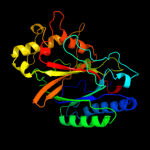
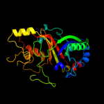
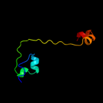
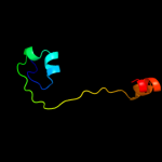
Image coloured by rainbow N → C terminus
Model (left) based on template d2iiza1
Top template information
Fold:Ferredoxin-like
Superfamily:Dimeric alpha+beta barrel
Family:Dyp-type peroxidase-like
Confidence and coverage
Confidence:100.0% Coverage: 99%
297 residues ( 99% of your sequence) have been modelled with 100.0% confidence by the single highest scoring template.
3D viewing
Interactive 3D view in Jmol

Sequence analysis 

Secondary structure and disorder prediction 

   1........10.........20.........30.........40.........50.........60
Sequence  MSQVQSGILPEHCRAAIWIEANVKGEVDALRAASKTFADKLATFEAKFPDAHLGAVVAFG
Secondary structure 


SS confidence 


Disorder  ???????


Disorder confidence 


   .........70.........80.........90.........100.........110.........120
Sequence  NNTWRALSGGVGAEELKDFPGYGKGLAPTTQFDVLIHILSLRHDVNFSVAQAAMEAFGDC
Secondary structure 


SS confidence 


Disorder 


??
?


?
????


Disorder confidence 


   .........130.........140.........150.........160.........170.........180
Sequence  IEVKEEIHGFRWVEERDLSGFVDGTENPAGEETRREVAVIKDGVDAGGSYVFVQRWEHNL
Secondary structure 


SS confidence 


Disorder 


?
?


?
???


Disorder confidence 


   .........190.........200.........210.........220.........230.........240
Sequence  KQLNRMSVHDQEMVIGRTKEANEEIDGDERPETSHLTRVDLKEDGKGLKIVRQSLPYGTA
Secondary structure 


SS confidence 


Disorder 


???????


????????


??
Disorder confidence 


   .........250.........260.........270.........280.........290.........
Sequence  SGTHGLYFCAYCARLHNIEQQLLSMFGDTDGKRDAMLRFTKPVTGGYYFAPSLDKLMAL
Secondary structure 


SS confidence 


Disorder 


???


???
Disorder confidence 


Confidence Key
High(9)                    Low (0)
?Disordered
Alpha helix
Beta strand

Domain analysis 

Hover over an aligned region to see model and summary info

Please note, only up to the top 20 hits are modelled to reduce computer load

RankAligned region

PDB 2iiz chain A domain 1

3D model

Region: 2 - 299
Aligned: 297
Modelled: 294
Confidence: 100.0%
Identity: 37%
Fold: Ferredoxin-like
Superfamily: Dimeric alpha+beta barrel
Family: Dyp-type peroxidase-like

Phyre2

PDB 3qns chain A

3D model

Region: 4 - 299
Aligned: 296
Modelled: 296
Confidence: 100.0%
Identity: 32%
PDB header:oxidoreductase
Chain: A: PDB Molecule:dyp peroxidase;
PDBTitle: dypb from rhodococcus jostii rha1, crystal form 2

Phyre2

PDB 2gvk chain A domain 1

3D model

Region: 5 - 299
Aligned: 292
Modelled: 292
Confidence: 100.0%
Identity: 33%
Fold: Ferredoxin-like
Superfamily: Dimeric alpha+beta barrel
Family: Dyp-type peroxidase-like

Phyre2

PDB 2wx6 chain B

3D model

Region: 3 - 292
Aligned: 278
Modelled: 285
Confidence: 100.0%
Identity: 22%
PDB header:oxidoreductase
Chain: B: PDB Molecule:peroxidase ycdb;
PDBTitle: x-ray crystallographic structure of e. coli apo-efeb

Phyre2

PDB 2d3q chain A domain 1

3D model

Region: 1 - 299
Aligned: 297
Modelled: 299
Confidence: 100.0%
Identity: 24%
Fold: Ferredoxin-like
Superfamily: Dimeric alpha+beta barrel
Family: Dyp-type peroxidase-like

Phyre2

PDB 3nn4 chain C

3D model

Region: 89 - 265
Aligned: 143
Modelled: 160
Confidence: 81.8%
Identity: 10%
PDB header:oxidoreductase
Chain: C: PDB Molecule:chlorite dismutase;
PDBTitle: structure of chlorite dismutase from candidatus nitrospira defluvii2 r173k mutant

Phyre2

PDB 2vxh chain F

3D model

Region: 90 - 265
Aligned: 144
Modelled: 154
Confidence: 81.1%
Identity: 13%
PDB header:oxidoreductase
Chain: F: PDB Molecule:chlorite dismutase;
PDBTitle: the crystal structure of chlorite dismutase: a detox enzyme2 producing molecular oxygen

Phyre2

PDB 3dtz chain B

3D model

Region: 89 - 129
Aligned: 41
Modelled: 41
Confidence: 62.2%
Identity: 12%
PDB header:structural genomics, unknown function
Chain: B: PDB Molecule:putative chlorite dismutase ta0507;
PDBTitle: crystal structure of putative chlorite dismutase ta0507

Phyre2

PDB 1t0t chain V

3D model

Region: 92 - 129
Aligned: 38
Modelled: 38
Confidence: 47.8%
Identity: 18%
Fold: Ferredoxin-like
Superfamily: Dimeric alpha+beta barrel
Family: Chlorite dismutase-like

Phyre2

PDB 2hpg chain B

3D model

Region: 139 - 194
Aligned: 56
Modelled: 56
Confidence: 40.3%
Identity: 13%
PDB header:ligand binding protein
Chain: B: PDB Molecule:abc transporter, periplasmic substrate-binding
PDBTitle: the crystal structure of a thermophilic trap periplasmic2 binding protein

Phyre2

PDB 2pfy chain A

3D model

Region: 140 - 194
Aligned: 55
Modelled: 55
Confidence: 29.7%
Identity: 9%
PDB header:transport protein
Chain: A: PDB Molecule:putative exported protein;
PDBTitle: crystal structure of dctp7, a bordetella pertussis2 extracytoplasmic solute receptor binding pyroglutamic acid

Phyre2

PDB 3b50 chain A

3D model

Region: 140 - 194
Aligned: 55
Modelled: 55
Confidence: 28.6%
Identity: 16%
PDB header:transport protein
Chain: A: PDB Molecule:sialic acid-binding periplasmic protein siap;
PDBTitle: structure of h. influenzae sialic acid binding protein2 bound to neu5ac.

Phyre2

PDB 3gyy chain C

3D model

Region: 140 - 194
Aligned: 55
Modelled: 55
Confidence: 24.6%
Identity: 22%
PDB header:transport protein
Chain: C: PDB Molecule:periplasmic substrate binding protein;
PDBTitle: the ectoine binding protein of the teaabc trap transporter teaa in the2 apo-state

Phyre2

PDB 3fxb chain B

3D model

Region: 140 - 194
Aligned: 55
Modelled: 55
Confidence: 23.4%
Identity: 22%
PDB header:transport protein
Chain: B: PDB Molecule:trap dicarboxylate transporter, dctp subunit;
PDBTitle: crystal structure of the ectoine-binding protein ueha

Phyre2

PDB 2zzx chain D

3D model

Region: 140 - 194
Aligned: 55
Modelled: 55
Confidence: 20.7%
Identity: 16%
PDB header:transport protein
Chain: D: PDB Molecule:abc transporter, solute-binding protein;
PDBTitle: crystal structure of a periplasmic substrate binding protein in2 complex with lactate

Phyre2

PDB 1vdh chain A

3D model

Region: 92 - 129
Aligned: 38
Modelled: 38
Confidence: 20.1%
Identity: 16%
Fold: Ferredoxin-like
Superfamily: Dimeric alpha+beta barrel
Family: Chlorite dismutase-like

Phyre2

PDB 2hzk chain B

3D model

Region: 140 - 194
Aligned: 55
Modelled: 55
Confidence: 19.5%
Identity: 11%
PDB header:ligand binding, transport protein
Chain: B: PDB Molecule:trap-t family sorbitol/mannitol transporter, periplasmic
PDBTitle: crystal structures of a sodium-alpha-keto acid binding subunit from a2 trap transporter in its open form

Phyre2

PDB 2pfz chain A

3D model

Region: 140 - 194
Aligned: 55
Modelled: 55
Confidence: 17.5%
Identity: 9%
PDB header:transport protein
Chain: A: PDB Molecule:putative exported protein;
PDBTitle: crystal structure of dctp6, a bordetella pertussis2 extracytoplasmic solute receptor binding pyroglutamic acid

Phyre2

PDB 2pie chain A domain 1

3D model

Region: 166 - 211
Aligned: 44
Modelled: 46
Confidence: 9.5%
Identity: 18%
Fold: SMAD/FHA domain
Superfamily: SMAD/FHA domain
Family: FHA domain

Phyre2

PDB 3b0v chain D

3D model

Region: 94 - 123
Aligned: 30
Modelled: 30
Confidence: 9.5%
Identity: 10%
PDB header:oxidoreductase/rna
Chain: D: PDB Molecule:trna-dihydrouridine synthase;
PDBTitle: trna-dihydrouridine synthase from thermus thermophilus in complex with2 trna

Phyre2
1

d2iiza1
2

c3qnsA_
3

d2gvka1
4

c2wx6B_
5

d2d3qa1
6

c3nn4C_
7

c2vxhF_
8

c3dtzB_
9

d1t0tv_
10

c2hpgB_
11

c2pfyA_
12

c3b50A_
13

c3gyyC_
14

c3fxbB_
15

c2zzxD_
16

d1vdha_
17

c2hzkB_
18

c2pfzA_
19

d2piea1
20

c3b0vD_
21


22


23


24


25


26


27


28


29


30


31


32


33


34


35


36


37


Detailed template information 

#
Template Alignment Coverage3D Model Confidence
% i.d. Template Information
1d2iiza1


100.0 37 Fold:Ferredoxin-like
Superfamily:Dimeric alpha+beta barrel
Family:Dyp-type peroxidase-like
2c3qnsA_


100.0 32 PDB header:oxidoreductase
Chain: A: PDB Molecule:dyp peroxidase;
PDBTitle: dypb from rhodococcus jostii rha1, crystal form 2
3d2gvka1


100.0 33 Fold:Ferredoxin-like
Superfamily:Dimeric alpha+beta barrel
Family:Dyp-type peroxidase-like
4c2wx6B_


100.0 22 PDB header:oxidoreductase
Chain: B: PDB Molecule:peroxidase ycdb;
PDBTitle: x-ray crystallographic structure of e. coli apo-efeb
5d2d3qa1


100.0 24 Fold:Ferredoxin-like
Superfamily:Dimeric alpha+beta barrel
Family:Dyp-type peroxidase-like
6c3nn4C_


81.8 10 PDB header:oxidoreductase
Chain: C: PDB Molecule:chlorite dismutase;
PDBTitle: structure of chlorite dismutase from candidatus nitrospira defluvii2 r173k mutant
7c2vxhF_


81.1 13 PDB header:oxidoreductase
Chain: F: PDB Molecule:chlorite dismutase;
PDBTitle: the crystal structure of chlorite dismutase: a detox enzyme2 producing molecular oxygen
8c3dtzB_


62.2 12 PDB header:structural genomics, unknown function
Chain: B: PDB Molecule:putative chlorite dismutase ta0507;
PDBTitle: crystal structure of putative chlorite dismutase ta0507
9d1t0tv_


47.8 18 Fold:Ferredoxin-like
Superfamily:Dimeric alpha+beta barrel
Family:Chlorite dismutase-like
10c2hpgB_


40.3 13 PDB header:ligand binding protein
Chain: B: PDB Molecule:abc transporter, periplasmic substrate-binding
PDBTitle: the crystal structure of a thermophilic trap periplasmic2 binding protein
11c2pfyA_


29.7 9 PDB header:transport protein
Chain: A: PDB Molecule:putative exported protein;
PDBTitle: crystal structure of dctp7, a bordetella pertussis2 extracytoplasmic solute receptor binding pyroglutamic acid
12c3b50A_


28.6 16 PDB header:transport protein
Chain: A: PDB Molecule:sialic acid-binding periplasmic protein siap;
PDBTitle: structure of h. influenzae sialic acid binding protein2 bound to neu5ac.
13c3gyyC_


24.6 22 PDB header:transport protein
Chain: C: PDB Molecule:periplasmic substrate binding protein;
PDBTitle: the ectoine binding protein of the teaabc trap transporter teaa in the2 apo-state
14c3fxbB_


23.4 22 PDB header:transport protein
Chain: B: PDB Molecule:trap dicarboxylate transporter, dctp subunit;
PDBTitle: crystal structure of the ectoine-binding protein ueha
15c2zzxD_


20.7 16 PDB header:transport protein
Chain: D: PDB Molecule:abc transporter, solute-binding protein;
PDBTitle: crystal structure of a periplasmic substrate binding protein in2 complex with lactate
16d1vdha_


20.1 16 Fold:Ferredoxin-like
Superfamily:Dimeric alpha+beta barrel
Family:Chlorite dismutase-like
17c2hzkB_


19.5 11 PDB header:ligand binding, transport protein
Chain: B: PDB Molecule:trap-t family sorbitol/mannitol transporter, periplasmic
PDBTitle: crystal structures of a sodium-alpha-keto acid binding subunit from a2 trap transporter in its open form
18c2pfzA_


17.5 9 PDB header:transport protein
Chain: A: PDB Molecule:putative exported protein;
PDBTitle: crystal structure of dctp6, a bordetella pertussis2 extracytoplasmic solute receptor binding pyroglutamic acid
19d2piea1


9.5 18 Fold:SMAD/FHA domain
Superfamily:SMAD/FHA domain
Family:FHA domain
20c3b0vD_


9.5 10 PDB header:oxidoreductase/rna
Chain: D: PDB Molecule:trna-dihydrouridine synthase;
PDBTitle: trna-dihydrouridine synthase from thermus thermophilus in complex with2 trna
21d1uoua3


not modelled 9.0 12 Fold:alpha/beta-Hammerhead
Superfamily:Pyrimidine nucleoside phosphorylase C-terminal domain
Family:Pyrimidine nucleoside phosphorylase C-terminal domain
22d1sfea2


not modelled 8.4 18 Fold:Ribonuclease H-like motif
Superfamily:Methylated DNA-protein cysteine methyltransferase domain
Family:Methylated DNA-protein cysteine methyltransferase domain
23d2cg4a2


not modelled 8.1 11 Fold:Ferredoxin-like
Superfamily:Dimeric alpha+beta barrel
Family:Lrp/AsnC-like transcriptional regulator C-terminal domain
24d1vjja1


not modelled 7.9 11 Fold:Immunoglobulin-like beta-sandwich
Superfamily:E set domains
Family:Transglutaminase N-terminal domain
25d2q3za1


not modelled 7.6 17 Fold:Immunoglobulin-like beta-sandwich
Superfamily:E set domains
Family:Transglutaminase N-terminal domain
26d1g0da1


not modelled 7.4 28 Fold:Immunoglobulin-like beta-sandwich
Superfamily:E set domains
Family:Transglutaminase N-terminal domain
27c2e1aD_


not modelled 7.1 8 PDB header:transcription
Chain: D: PDB Molecule:75aa long hypothetical regulatory protein asnc;
PDBTitle: crystal structure of ffrp-dm1
28d1s6na_


not modelled 6.5 29 Fold:Immunoglobulin-like beta-sandwich
Superfamily:E set domains
Family:Class II viral fusion proteins C-terminal domain
29c2vpnB_


not modelled 6.3 26 PDB header:transport
Chain: B: PDB Molecule:periplasmic substrate binding protein;
PDBTitle: high-resolution structure of the periplasmic ectoine-2 binding protein from teaabc trap-transporter of halomonas3 elongata
30c2qdrA_


not modelled 6.3 29 PDB header:unknown function
Chain: A: PDB Molecule:uncharacterized protein;
PDBTitle: crystal structure of a putative dioxygenase (npun_f5605) from nostoc2 punctiforme pcc 73102 at 2.60 a resolution
31d2i7pa2


not modelled 6.2 22 Fold:Ribonuclease H-like motif
Superfamily:Actin-like ATPase domain
Family:Fumble-like
32d4tsva_


not modelled 6.1 7 Fold:TNF-like
Superfamily:TNF-like
Family:TNF-like
33c1ohfB_


not modelled 5.7 39 PDB header:virus
Chain: B: PDB Molecule:nudaurelia capensis omega virus capsid protein;
PDBTitle: the refined structure of nudaurelia capensis omega virus
34c2h0pA_


not modelled 5.7 18 PDB header:viral protein
Chain: A: PDB Molecule:envelope glycoprotein;
PDBTitle: nmr structure of the dengue-4 virus envelope protein domain2 iii
35d1ok8a1


not modelled 5.7 26 Fold:Immunoglobulin-like beta-sandwich
Superfamily:E set domains
Family:Class II viral fusion proteins C-terminal domain
36d1ex0a1


not modelled 5.3 17 Fold:Immunoglobulin-like beta-sandwich
Superfamily:E set domains
Family:Transglutaminase N-terminal domain
37c1fuiB_


not modelled 5.1 15 PDB header:isomerase
Chain: B: PDB Molecule:l-fucose isomerase;
PDBTitle: l-fucose isomerase from escherichia coli

Binding site prediction 

Due to computational demand, binding site predictions are not run for batch jobs

If you want to predict binding sites, please manually submit your model of choice to 3DLigandSite


Phyre is for academic use only

Please cite: Protein structure prediction on the web: a case study using the Phyre server
Kelley LA and Sternberg MJE. Nature Protocols 4, 363 - 371 (2009) [pdf] [Import into BibTeX]
 
If you use the binding site predictions from 3DLigandSite, please also cite:
3DLigandSite: predicting ligand-binding sites using similar structures.
Wass MN, Kelley LA and Sternberg MJ Nucleic Acids Research 38, W469-73 (2010) [PubMed]
 
© Structural Bioinformatics Group
Imperial College London
Lawrence Kelley, Benjamin Jefferys
Disclaimer
Terms and Conditions
Component software
Template detection: HHpred 1.51
Secondary structure prediction: Psi-pred 2.5
Disorder prediction: Disopred 2.4
Transmembrane prediction: Memsat_SVM
Multi-template modelling and ab initio: Poing 1.0