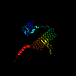
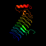
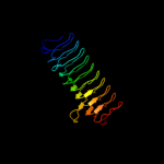
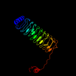
| 1 | c3eh0C_
|
|
 |
100.0 |
100 |
PDB header:transferase
Chain: C: PDB Molecule:udp-3-o-[3-hydroxymyristoyl] glucosamine n-
PDBTitle: crystal structure of lpxd from escherichia coli
|
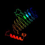
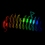
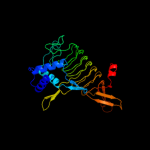
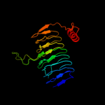
| 2 | c3pmoA_
|
|
 |
100.0 |
51 |
PDB header:transferase
Chain: A: PDB Molecule:udp-3-o-[3-hydroxymyristoyl] glucosamine n-acyltransferase;
PDBTitle: the structure of lpxd from pseudomonas aeruginosa at 1.3 a resolution
|
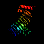
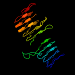
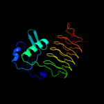
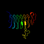
| 3 | c2iu9C_
|
|
 |
100.0 |
33 |
PDB header:transferase
Chain: C: PDB Molecule:udp-3-o-[3-hydroxymyristoyl] glucosamine
PDBTitle: chlamydia trachomatis lpxd with 100mm udpglcnac (complex ii)
|
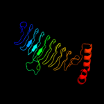
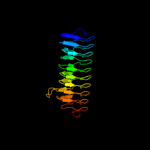
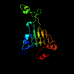
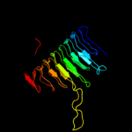
| 4 | d1j2za_
|
|
 |
100.0 |
24 |
Fold:Single-stranded left-handed beta-helix
Superfamily:Trimeric LpxA-like enzymes
Family:UDP N-acetylglucosamine acyltransferase |
| 5 | c3i3aC_
|
|
 |
100.0 |
22 |
PDB header:transferase
Chain: C: PDB Molecule:acyl-[acyl-carrier-protein]--udp-n-
PDBTitle: structural basis for the sugar nucleotide and acyl chain2 selectivity of leptospira interrogans lpxa
|
| 6 | d2jf2a1
|
|
 |
100.0 |
25 |
Fold:Single-stranded left-handed beta-helix
Superfamily:Trimeric LpxA-like enzymes
Family:UDP N-acetylglucosamine acyltransferase |
| 7 | c3r0sA_
|
|
 |
100.0 |
28 |
PDB header:transferase
Chain: A: PDB Molecule:acyl-[acyl-carrier-protein]--udp-n-acetylglucosamine o-
PDBTitle: udp-n-acetylglucosamine acyltransferase from campylobacter jejuni
|
| 8 | c3fsbB_
|
|
 |
100.0 |
25 |
PDB header:transferase
Chain: B: PDB Molecule:qdtc;
PDBTitle: crystal structure of qdtc, the dtdp-3-amino-3,6-dideoxy-d-2 glucose n-acetyl transferase from thermoanaerobacterium3 thermosaccharolyticum in complex with coa and dtdp-3-amino-4 quinovose
|
| 9 | d2f9ca1
|
|
 |
100.0 |
13 |
Fold:Single-stranded left-handed beta-helix
Superfamily:Trimeric LpxA-like enzymes
Family:YdcK-like |
| 10 | c3c8vA_
|
|
 |
100.0 |
15 |
PDB header:transferase
Chain: A: PDB Molecule:putative acetyltransferase;
PDBTitle: crystal structure of putative acetyltransferase (yp_390128.1) from2 desulfovibrio desulfuricans g20 at 2.28 a resolution
|
| 11 | d2oi6a1
|
|
 |
100.0 |
21 |
Fold:Single-stranded left-handed beta-helix
Superfamily:Trimeric LpxA-like enzymes
Family:GlmU C-terminal domain-like |
| 12 | d1g97a1
|
|
 |
100.0 |
20 |
Fold:Single-stranded left-handed beta-helix
Superfamily:Trimeric LpxA-like enzymes
Family:GlmU C-terminal domain-like |
| 13 | c3eg4A_
|
|
 |
100.0 |
16 |
PDB header:transferase
Chain: A: PDB Molecule:2,3,4,5-tetrahydropyridine-2,6-dicarboxylate n-
PDBTitle: crystal structure of 2,3,4,5-tetrahydropyridine-2-2 carboxylate n-succinyltransferase from brucella melitensis3 biovar abortus 2308
|
| 14 | d3bswa1
|
|
 |
100.0 |
22 |
Fold:Single-stranded left-handed beta-helix
Superfamily:Trimeric LpxA-like enzymes
Family:PglD-like |
| 15 | d1mr7a_
|
|
 |
100.0 |
20 |
Fold:Single-stranded left-handed beta-helix
Superfamily:Trimeric LpxA-like enzymes
Family:Galactoside acetyltransferase-like |
| 16 | c1hm8A_
|
|
 |
100.0 |
20 |
PDB header:transferase
Chain: A: PDB Molecule:udp-n-acetylglucosamine-1-phosphate uridyltransferase;
PDBTitle: crystal structure of s.pneumoniae n-acetylglucosamine-1-phosphate2 uridyltransferase, glmu, bound to acetyl coenzyme a
|
| 17 | c3jqyB_
|
|
 |
100.0 |
18 |
PDB header:transferase
Chain: B: PDB Molecule:polysialic acid o-acetyltransferase;
PDBTitle: crystal strucutre of the polysia specific acetyltransferase neuo
|
| 18 | c3mqhD_
|
|
 |
100.0 |
19 |
PDB header:transferase
Chain: D: PDB Molecule:lipopolysaccharides biosynthesis acetyltransferase;
PDBTitle: crystal structure of the 3-n-acetyl transferase wlbb from bordetella2 petrii in complex with coa and udp-3-amino-2-acetamido-2,3-dideoxy3 glucuronic acid
|
| 19 | c2wlgA_
|
|
 |
99.9 |
21 |
PDB header:transferase
Chain: A: PDB Molecule:polysialic acid o-acetyltransferase;
PDBTitle: crystallographic analysis of the polysialic acid o-2 acetyltransferase oatwy
|
| 20 | c2v0hA_
|
|
 |
99.9 |
16 |
PDB header:transferase
Chain: A: PDB Molecule:bifunctional protein glmu;
PDBTitle: characterization of substrate binding and catalysis of the2 potential antibacterial target n-acetylglucosamine-1-3 phosphate uridyltransferase (glmu)
|
| 21 | c3cj8B_ |
|
not modelled |
99.9 |
22 |
PDB header:transferase
Chain: B: PDB Molecule:2,3,4,5-tetrahydropyridine-2-carboxylate n-
PDBTitle: crystal structure of 2,3,4,5-tetrahydropyridine-2-carboxylate n-2 succinyltransferase from enterococcus faecalis v583
|
| 22 | d1qrea_ |
|
not modelled |
99.9 |
17 |
Fold:Single-stranded left-handed beta-helix
Superfamily:Trimeric LpxA-like enzymes
Family:gamma-carbonic anhydrase-like |
| 23 | c1qreA_ |
|
not modelled |
99.9 |
17 |
PDB header:lyase
Chain: A: PDB Molecule:carbonic anhydrase;
PDBTitle: a closer look at the active site of gamma-carbonic anhydrases: high2 resolution crystallographic studies of the carbonic anhydrase from3 methanosarcina thermophila
|
| 24 | c3r3rA_ |
|
not modelled |
99.9 |
19 |
PDB header:transferase
Chain: A: PDB Molecule:ferripyochelin binding protein;
PDBTitle: structure of the yrda ferripyochelin binding protein from salmonella2 enterica
|
| 25 | c2oi6A_ |
|
not modelled |
99.9 |
20 |
PDB header:transferase
Chain: A: PDB Molecule:bifunctional protein glmu;
PDBTitle: e. coli glmu- complex with udp-glcnac, coa and glcn-1-po4
|
| 26 | d1v3wa_ |
|
not modelled |
99.9 |
20 |
Fold:Single-stranded left-handed beta-helix
Superfamily:Trimeric LpxA-like enzymes
Family:gamma-carbonic anhydrase-like |
| 27 | c3ectA_ |
|
not modelled |
99.9 |
18 |
PDB header:transferase
Chain: A: PDB Molecule:hexapeptide-repeat containing-acetyltransferase;
PDBTitle: crystal structure of the hexapeptide-repeat containing-2 acetyltransferase vca0836 from vibrio cholerae
|
| 28 | d1xata_ |
|
not modelled |
99.9 |
13 |
Fold:Single-stranded left-handed beta-helix
Superfamily:Trimeric LpxA-like enzymes
Family:Galactoside acetyltransferase-like |
| 29 | c3srtB_ |
|
not modelled |
99.9 |
26 |
PDB header:transferase
Chain: B: PDB Molecule:maltose o-acetyltransferase;
PDBTitle: the crystal structure of a maltose o-acetyltransferase from2 clostridium difficile 630
|
| 30 | d1krra_ |
|
not modelled |
99.9 |
24 |
Fold:Single-stranded left-handed beta-helix
Superfamily:Trimeric LpxA-like enzymes
Family:Galactoside acetyltransferase-like |
| 31 | c2ic7A_ |
|
not modelled |
99.9 |
19 |
PDB header:transferase
Chain: A: PDB Molecule:maltose transacetylase;
PDBTitle: crystal structure of maltose transacetylase from2 geobacillus kaustophilus
|
| 32 | c3fttA_ |
|
not modelled |
99.9 |
32 |
PDB header:transferase
Chain: A: PDB Molecule:putative acetyltransferase sacol2570;
PDBTitle: crystal structure of the galactoside o-acetyltransferase2 from staphylococcus aureus
|
| 33 | c3eevC_ |
|
not modelled |
99.9 |
16 |
PDB header:transferase
Chain: C: PDB Molecule:chloramphenicol acetyltransferase;
PDBTitle: crystal structure of chloramphenicol acetyltransferase vca0300 from2 vibrio cholerae o1 biovar eltor
|
| 34 | d1xhda_ |
|
not modelled |
99.9 |
25 |
Fold:Single-stranded left-handed beta-helix
Superfamily:Trimeric LpxA-like enzymes
Family:gamma-carbonic anhydrase-like |
| 35 | d3tdta_ |
|
not modelled |
99.9 |
20 |
Fold:Single-stranded left-handed beta-helix
Superfamily:Trimeric LpxA-like enzymes
Family:Tetrahydrodipicolinate-N-succinlytransferase, THDP-succinlytransferase, DapD |
| 36 | d1ocxa_ |
|
not modelled |
99.9 |
26 |
Fold:Single-stranded left-handed beta-helix
Superfamily:Trimeric LpxA-like enzymes
Family:Galactoside acetyltransferase-like |
| 37 | c3r1wA_ |
|
not modelled |
99.9 |
25 |
PDB header:lyase
Chain: A: PDB Molecule:carbonic anhydrase;
PDBTitle: crystal structure of a carbonic anhydrase from a crude oil degrading2 psychrophilic library
|
| 38 | c3ixcA_ |
|
not modelled |
99.9 |
17 |
PDB header:transferase
Chain: A: PDB Molecule:hexapeptide transferase family protein;
PDBTitle: crystal structure of hexapeptide transferase family protein from2 anaplasma phagocytophilum
|
| 39 | d1t3da_ |
|
not modelled |
99.9 |
26 |
Fold:Single-stranded left-handed beta-helix
Superfamily:Trimeric LpxA-like enzymes
Family:Serine acetyltransferase |
| 40 | c1t3dB_ |
|
not modelled |
99.9 |
28 |
PDB header:transferase
Chain: B: PDB Molecule:serine acetyltransferase;
PDBTitle: crystal structure of serine acetyltransferase from e.coli at 2.2a
|
| 41 | d1ssqa_ |
|
not modelled |
99.8 |
24 |
Fold:Single-stranded left-handed beta-helix
Superfamily:Trimeric LpxA-like enzymes
Family:Serine acetyltransferase |
| 42 | c2ggqA_ |
|
not modelled |
99.8 |
21 |
PDB header:transferase
Chain: A: PDB Molecule:401aa long hypothetical glucose-1-phosphate
PDBTitle: complex of hypothetical glucose-1-phosphate thymidylyltransferase from2 sulfolobus tokodaii
|
| 43 | c3mc4A_ |
|
not modelled |
99.8 |
20 |
PDB header:transferase
Chain: A: PDB Molecule:ww/rsp5/wwp domain:bacterial transferase
PDBTitle: crystal structure of ww/rsp5/wwp domain: bacterial2 transferase hexapeptide repeat: serine o-acetyltransferase3 from brucella melitensis
|
| 44 | c3q1xA_ |
|
not modelled |
99.8 |
19 |
PDB header:transferase
Chain: A: PDB Molecule:serine acetyltransferase;
PDBTitle: crystal structure of entamoeba histolytica serine acetyltransferase 12 in complex with l-serine
|
| 45 | c3kwdA_ |
|
not modelled |
99.8 |
22 |
PDB header:lyase, protein binding, photosynthesis
Chain: A: PDB Molecule:carbon dioxide concentrating mechanism protein;
PDBTitle: inactive truncation of the beta-carboxysomal gamma-carbonic anhydrase,2 ccmm, form 1
|
| 46 | c3f1xA_ |
|
not modelled |
99.7 |
20 |
PDB header:transferase
Chain: A: PDB Molecule:serine acetyltransferase;
PDBTitle: three dimensional structure of the serine acetyltransferase from2 bacteroides vulgatus, northeast structural genomics consortium target3 bvr62.
|
| 47 | c3d98A_ |
|
not modelled |
99.7 |
14 |
PDB header:transferase
Chain: A: PDB Molecule:bifunctional protein glmu;
PDBTitle: crystal structure of glmu from mycobacterium tuberculosis, ligand-free2 form
|
| 48 | c3fsyC_ |
|
not modelled |
99.7 |
14 |
PDB header:transferase
Chain: C: PDB Molecule:tetrahydrodipicolinate n-succinyltransferase;
PDBTitle: structure of tetrahydrodipicolinate n-succinyltransferase2 (rv1201c;dapd) in complex with succinyl-coa from mycobacterium3 tuberculosis
|
| 49 | c2qkxA_ |
|
not modelled |
99.7 |
14 |
PDB header:transferase
Chain: A: PDB Molecule:bifunctional protein glmu;
PDBTitle: n-acetyl glucosamine 1-phosphate uridyltransferase from mycobacterium2 tuberculosis complex with n-acetyl glucosamine 1-phosphate
|
| 50 | d1yp2a1 |
|
not modelled |
99.6 |
16 |
Fold:Single-stranded left-handed beta-helix
Superfamily:Trimeric LpxA-like enzymes
Family:GlmU C-terminal domain-like |
| 51 | c1yp3C_ |
|
not modelled |
99.4 |
16 |
PDB header:transferase
Chain: C: PDB Molecule:glucose-1-phosphate adenylyltransferase small
PDBTitle: crystal structure of potato tuber adp-glucose2 pyrophosphorylase in complex with atp
|
| 52 | d1fxja1 |
|
not modelled |
99.3 |
31 |
Fold:Single-stranded left-handed beta-helix
Superfamily:Trimeric LpxA-like enzymes
Family:GlmU C-terminal domain-like |
| 53 | c2rijA_ |
|
not modelled |
99.3 |
16 |
PDB header:transferase
Chain: A: PDB Molecule:putative 2,3,4,5-tetrahydropyridine-2-carboxylate n-
PDBTitle: crystal structure of a putative 2,3,4,5-tetrahydropyridine-2-2 carboxylate n-succinyltransferase (cj1605c, dapd) from campylobacter3 jejuni at 1.90 a resolution
|
| 54 | c1fwyA_ |
|
not modelled |
99.2 |
25 |
PDB header:transferase
Chain: A: PDB Molecule:udp-n-acetylglucosamine pyrophosphorylase;
PDBTitle: crystal structure of n-acetylglucosamine 1-phosphate2 uridyltransferase bound to udp-glcnac
|
| 55 | c3brkX_ |
|
not modelled |
99.0 |
14 |
PDB header:transferase
Chain: X: PDB Molecule:glucose-1-phosphate adenylyltransferase;
PDBTitle: crystal structure of adp-glucose pyrophosphorylase from2 agrobacterium tumefaciens
|
| 56 | d1gg4a3 |
|
not modelled |
98.6 |
22 |
Fold:MurF and HprK N-domain-like
Superfamily:MurE/MurF N-terminal domain
Family:MurE/MurF N-terminal domain |
| 57 | c2wtzC_ |
|
not modelled |
98.1 |
24 |
PDB header:ligase
Chain: C: PDB Molecule:udp-n-acetylmuramoyl-l-alanyl-d-glutamate-
PDBTitle: mure ligase of mycobacterium tuberculosis
|
| 58 | c1gg4A_ |
|
not modelled |
98.0 |
20 |
PDB header:ligase
Chain: A: PDB Molecule:udp-n-acetylmuramoylalanyl-d-glutamyl-2,6-
PDBTitle: crystal structure of escherichia coli udpmurnac-tripeptide2 d-alanyl-d-alanine-adding enzyme (murf) at 2.3 angstrom3 resolution
|
| 59 | c2am1A_ |
|
not modelled |
97.7 |
11 |
PDB header:ligase
Chain: A: PDB Molecule:udp-n-acetylmuramoylalanine-d-glutamyl-lysine-d-alanyl-d-
PDBTitle: sp protein ligand 1
|
| 60 | d1e8ca1 |
|
not modelled |
96.4 |
9 |
Fold:MurF and HprK N-domain-like
Superfamily:MurE/MurF N-terminal domain
Family:MurE/MurF N-terminal domain |
| 61 | c1e8cB_ |
|
not modelled |
95.1 |
10 |
PDB header:ligase
Chain: B: PDB Molecule:udp-n-acetylmuramoylalanyl-d-glutamate--2,6-
PDBTitle: structure of mure the udp-n-acetylmuramyl tripeptide2 synthetase from e. coli
|
| 62 | c1cosC_ |
|
not modelled |
73.0 |
24 |
PDB header:alpha-helical bundle
Chain: C: PDB Molecule:coiled serine;
PDBTitle: crystal structure of a synthetic triple-stranded alpha-2 helical bundle
|
| 63 | c1cosA_ |
|
not modelled |
69.1 |
24 |
PDB header:alpha-helical bundle
Chain: A: PDB Molecule:coiled serine;
PDBTitle: crystal structure of a synthetic triple-stranded alpha-2 helical bundle
|
| 64 | c1cosB_ |
|
not modelled |
69.1 |
24 |
PDB header:alpha-helical bundle
Chain: B: PDB Molecule:coiled serine;
PDBTitle: crystal structure of a synthetic triple-stranded alpha-2 helical bundle
|
| 65 | c3ci9B_ |
|
not modelled |
57.1 |
35 |
PDB header:transcription
Chain: B: PDB Molecule:heat shock factor-binding protein 1;
PDBTitle: crystal structure of the human hsbp1
|
| 66 | c1by0A_ |
|
not modelled |
53.4 |
14 |
PDB header:rna binding protein
Chain: A: PDB Molecule:protein (hepatitis delta antigen);
PDBTitle: n-terminal leucine-repeat region of hepatitis delta antigen
|
| 67 | c2jgoA_ |
|
not modelled |
48.2 |
24 |
PDB header:de novo protein
Chain: A: PDB Molecule:coil ser l9c;
PDBTitle: stucture of the arsenated de novo designed peptide coil ser2 l9c
|
| 68 | c2jgoC_ |
|
not modelled |
48.2 |
24 |
PDB header:de novo protein
Chain: C: PDB Molecule:coil ser l9c;
PDBTitle: stucture of the arsenated de novo designed peptide coil ser2 l9c
|
| 69 | c3ljmC_ |
|
not modelled |
48.2 |
24 |
PDB header:de novo protein
Chain: C: PDB Molecule:coil ser l9c;
PDBTitle: structure of de novo designed apo peptide coil ser l9c
|
| 70 | c2jgoB_ |
|
not modelled |
48.2 |
24 |
PDB header:de novo protein
Chain: B: PDB Molecule:coil ser l9c;
PDBTitle: stucture of the arsenated de novo designed peptide coil ser2 l9c
|
| 71 | c3ljmA_ |
|
not modelled |
48.2 |
24 |
PDB header:de novo protein
Chain: A: PDB Molecule:coil ser l9c;
PDBTitle: structure of de novo designed apo peptide coil ser l9c
|
| 72 | c3ljmB_ |
|
not modelled |
48.2 |
24 |
PDB header:de novo protein
Chain: B: PDB Molecule:coil ser l9c;
PDBTitle: structure of de novo designed apo peptide coil ser l9c
|
| 73 | c1coiA_ |
|
not modelled |
42.0 |
24 |
PDB header:alpha-helical bundle
Chain: A: PDB Molecule:coil-vald;
PDBTitle: designed trimeric coiled coil-vald
|
| 74 | d1lcda_ |
|
not modelled |
41.7 |
29 |
Fold:lambda repressor-like DNA-binding domains
Superfamily:lambda repressor-like DNA-binding domains
Family:GalR/LacI-like bacterial regulator |
| 75 | c3gw6F_ |
|
not modelled |
41.4 |
17 |
PDB header:chaperone
Chain: F: PDB Molecule:endo-n-acetylneuraminidase;
PDBTitle: intramolecular chaperone
|
| 76 | c2nydB_ |
|
not modelled |
40.1 |
20 |
PDB header:unknown function
Chain: B: PDB Molecule:upf0135 protein sa1388;
PDBTitle: crystal structure of staphylococcus aureus hypothetical protein sa1388
|
| 77 | c1a92B_ |
|
not modelled |
33.3 |
14 |
PDB header:leucine zipper
Chain: B: PDB Molecule:delta antigen;
PDBTitle: oligomerization domain of hepatitis delta antigen
|
| 78 | c2x6pA_ |
|
not modelled |
29.0 |
20 |
PDB header:de novo protein
Chain: A: PDB Molecule:coil ser l19c;
PDBTitle: crystal structure of coil ser l19c
|
| 79 | c2x6pC_ |
|
not modelled |
29.0 |
20 |
PDB header:de novo protein
Chain: C: PDB Molecule:coil ser l19c;
PDBTitle: crystal structure of coil ser l19c
|
| 80 | c2x6pB_ |
|
not modelled |
29.0 |
20 |
PDB header:de novo protein
Chain: B: PDB Molecule:coil ser l19c;
PDBTitle: crystal structure of coil ser l19c
|
| 81 | c1lq7A_ |
|
not modelled |
28.9 |
21 |
PDB header:de novo protein
Chain: A: PDB Molecule:alpha3w;
PDBTitle: de novo designed protein model of radical enzymes
|
| 82 | c1aq5C_ |
|
not modelled |
28.6 |
29 |
PDB header:coiled-coil
Chain: C: PDB Molecule:cartilage matrix protein;
PDBTitle: high-resolution solution nmr structure of the trimeric coiled-coil2 domain of chicken cartilage matrix protein, 20 structures
|
| 83 | d2bjca1 |
|
not modelled |
27.5 |
29 |
Fold:lambda repressor-like DNA-binding domains
Superfamily:lambda repressor-like DNA-binding domains
Family:GalR/LacI-like bacterial regulator |
| 84 | d2ioja1 |
|
not modelled |
27.2 |
8 |
Fold:MurF and HprK N-domain-like
Superfamily:HprK N-terminal domain-like
Family:DRTGG domain |
| 85 | c1zvvA_ |
|
not modelled |
21.4 |
23 |
PDB header:transcription/dna
Chain: A: PDB Molecule:glucose-resistance amylase regulator;
PDBTitle: crystal structure of a ccpa-crh-dna complex
|
| 86 | c2wq1A_ |
|
not modelled |
17.5 |
42 |
PDB header:transcription
Chain: A: PDB Molecule:general control protein gcn4;
PDBTitle: gcn4 leucine zipper mutant with three ixxntxx motifs2 coordinating bromide
|
| 87 | c2wq3A_ |
|
not modelled |
17.5 |
42 |
PDB header:transcription
Chain: A: PDB Molecule:general control protein gcn4;
PDBTitle: gcn4 leucine zipper mutant with three ixxntxx motifs2 coordinating chloride and nitrate
|
| 88 | c2ergA_ |
|
not modelled |
16.6 |
25 |
PDB header:transcription activator/dna
Chain: A: PDB Molecule:regulatory protein leu3;
PDBTitle: crystal structure of leu3 dna-binding domain with a single2 h50c mutation complexed with a 15mer dna duplex
|
| 89 | d2hh6a1 |
|
not modelled |
16.6 |
11 |
Fold:Left-handed superhelix
Superfamily:BH3980-like
Family:BH3980-like |
| 90 | c2cwoD_ |
|
not modelled |
15.5 |
18 |
PDB header:rna binding protein
Chain: D: PDB Molecule:rna silencing suppressor;
PDBTitle: crystal structure of rna silencing suppressor p21 from beet yellows2 virus
|
| 91 | d2o3la1 |
|
not modelled |
15.0 |
15 |
Fold:Left-handed superhelix
Superfamily:BH3980-like
Family:BH3980-like |
| 92 | c1dipA_ |
|
not modelled |
14.3 |
16 |
PDB header:acetylation
Chain: A: PDB Molecule:delta-sleep-inducing peptide immunoreactive
PDBTitle: the solution structure of porcine delta-sleep-inducing2 peptide immunoreactive peptide, nmr, 10 structures
|
| 93 | c2xzrA_ |
|
not modelled |
14.2 |
17 |
PDB header:cell adhesion
Chain: A: PDB Molecule:immunoglobulin-binding protein eibd;
PDBTitle: escherichia coli immunoglobulin-binding protein eibd 391-438 fused2 to gcn4 adaptors
|
| 94 | c1ry3A_ |
|
not modelled |
14.1 |
15 |
PDB header:antibiotic
Chain: A: PDB Molecule:bacteriocin carnobacteriocin b2;
PDBTitle: nmr solution structure of the precursor for2 carnobacteriocin b2, an antimicrobial peptide from3 carnobacterium piscicola
|
| 95 | c1ce0B_ |
|
not modelled |
13.8 |
29 |
PDB header:hiv-1 envelope protein
Chain: B: PDB Molecule:protein (leucine zipper model h38-p1);
PDBTitle: trimerization specificity in hiv-1 gp41: analysis with a2 gcn4 leucine zipper model
|
| 96 | c2ccfA_ |
|
not modelled |
12.2 |
33 |
PDB header:four helix bundle
Chain: A: PDB Molecule:general control protein gcn4;
PDBTitle: antiparallel configuration of pli e20s
|
| 97 | c1u9fA_ |
|
not modelled |
12.2 |
33 |
PDB header:transcription
Chain: A: PDB Molecule:general control protein gcn4;
PDBTitle: heterocyclic peptide backbone modification in gcn4-pli based coiled2 coils: replacement of k(15)l(16)
|
| 98 | c2wpzA_ |
|
not modelled |
11.6 |
50 |
PDB header:transcription
Chain: A: PDB Molecule:general control protein gcn4;
PDBTitle: gcn4 leucine zipper mutant with two vxxnxxx motifs2 coordinating chloride
|
| 99 | c1unwB_ |
|
not modelled |
11.6 |
33 |
PDB header:four helix bundle
Chain: B: PDB Molecule:general control protein gcn4;
PDBTitle: structure based engineering of internal molecular surfaces2 of four helix bundles
|