Phyre Home Retrieve Phyre Job Id
Emaill.a.kelley@imperial.ac.uk
DescriptionP61175
DateThu Jan 5 12:07:19 GMT 2012
Unique Job IDb80875b932967251

Summary 

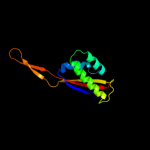
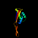
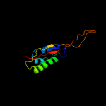
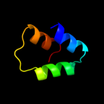
Top model
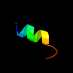
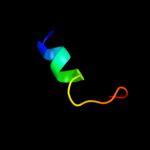
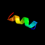
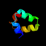
Image coloured by rainbow N → C terminus
Model (left) based on template d2zjrp1
Top template information
Fold:Ribosomal protein L22
Superfamily:Ribosomal protein L22
Family:Ribosomal protein L22
Confidence and coverage
Confidence:100.0% Coverage: 99%
109 residues ( 99% of your sequence) have been modelled with 100.0% confidence by the single highest scoring template.
3D viewing
Interactive 3D view in Jmol

Sequence analysis 

Secondary structure and disorder prediction 

   1........10.........20.........30.........40.........50.........60
Sequence  METIAKHRHARSSAQKVRLVADLIRGKKVSQALDILTYTNKKAAVLVKKVLESAIANAEH
Secondary structure 


SS confidence 


Disorder  ??


Disorder confidence 


   .........70.........80.........90.........100.........110
Sequence  NDGADIDDLKVTKIFVDEGPSMKRIMPRAKGRADRILKRTSHITVVVSDR
Secondary structure 


SS confidence 


Disorder  ????
?


?
?

??


??
Disorder confidence 


Confidence Key
High(9)                    Low (0)
?Disordered
Alpha helix
Beta strand

Domain analysis 

Hover over an aligned region to see model and summary info

Please note, only up to the top 20 hits are modelled to reduce computer load

RankAligned region

PDB 2zjr chain P domain 1

3D model

Region: 2 - 110
Aligned: 109
Modelled: 109
Confidence: 100.0%
Identity: 49%
Fold: Ribosomal protein L22
Superfamily: Ribosomal protein L22
Family: Ribosomal protein L22

Phyre2

PDB 1i4j chain A

3D model

Region: 1 - 110
Aligned: 107
Modelled: 110
Confidence: 100.0%
Identity: 57%
Fold: Ribosomal protein L22
Superfamily: Ribosomal protein L22
Family: Ribosomal protein L22

Phyre2

PDB 3bbo chain U

3D model

Region: 2 - 110
Aligned: 109
Modelled: 109
Confidence: 100.0%
Identity: 36%
PDB header:ribosome
Chain: U: PDB Molecule:ribosomal protein l22;
PDBTitle: homology model for the spinach chloroplast 50s subunit2 fitted to 9.4a cryo-em map of the 70s chlororibosome

Phyre2

PDB 1vqo chain R domain 1

3D model

Region: 2 - 109
Aligned: 107
Modelled: 108
Confidence: 100.0%
Identity: 24%
Fold: Ribosomal protein L22
Superfamily: Ribosomal protein L22
Family: Ribosomal protein L22

Phyre2

PDB 4a17 chain Q

3D model

Region: 2 - 110
Aligned: 108
Modelled: 109
Confidence: 100.0%
Identity: 22%
PDB header:ribosome
Chain: Q: PDB Molecule:rpl17;
PDBTitle: t.thermophila 60s ribosomal subunit in complex with2 initiation factor 6. this file contains 5s rrna,3 5.8s rrna and proteins of molecule 2.

Phyre2

PDB 3iz5 chain V

3D model

Region: 3 - 110
Aligned: 107
Modelled: 108
Confidence: 100.0%
Identity: 30%
PDB header:ribosome
Chain: V: PDB Molecule:60s ribosomal protein l17 (l22p);
PDBTitle: localization of the large subunit ribosomal proteins into a 5.5 a2 cryo-em map of triticum aestivum translating 80s ribosome

Phyre2

PDB 3jyw chain N

3D model

Region: 3 - 110
Aligned: 107
Modelled: 108
Confidence: 100.0%
Identity: 34%
PDB header:ribosome
Chain: N: PDB Molecule:60s ribosomal protein l17(a);
PDBTitle: structure of the 60s proteins for eukaryotic ribosome based on cryo-em2 map of thermomyces lanuginosus ribosome at 8.9a resolution

Phyre2

PDB 2gyc chain Q domain 1

3D model

Region: 5 - 110
Aligned: 106
Modelled: 106
Confidence: 100.0%
Identity: 100%
Fold: Ribosomal protein L22
Superfamily: Ribosomal protein L22
Family: Ribosomal protein L22

Phyre2

PDB 2ftc chain M

3D model

Region: 8 - 110
Aligned: 103
Modelled: 103
Confidence: 100.0%
Identity: 29%
PDB header:ribosome
Chain: M: PDB Molecule:mitochondrial ribosomal protein l22 isoform a;
PDBTitle: structural model for the large subunit of the mammalian mitochondrial2 ribosome

Phyre2

PDB 2zkr chain R

3D model

Region: 2 - 110
Aligned: 106
Modelled: 108
Confidence: 100.0%
Identity: 30%
PDB header:ribosomal protein/rna
Chain: R: PDB Molecule:rna expansion segment es39 part i;
PDBTitle: structure of a mammalian ribosomal 60s subunit within an2 80s complex obtained by docking homology models of the rna3 and proteins into an 8.7 a cryo-em map

Phyre2

PDB 1s1i chain N

3D model

Region: 2 - 109
Aligned: 107
Modelled: 108
Confidence: 100.0%
Identity: 34%
PDB header:ribosome
Chain: N: PDB Molecule:60s ribosomal protein l17-a;
PDBTitle: structure of the ribosomal 80s-eef2-sordarin complex from2 yeast obtained by docking atomic models for rna and protein3 components into a 11.7 a cryo-em map. this file, 1s1i,4 contains 60s subunit. the 40s ribosomal subunit is in file5 1s1h.

Phyre2

PDB 1r9p chain A

3D model

Region: 17 - 67
Aligned: 51
Modelled: 51
Confidence: 85.9%
Identity: 20%
Fold: SufE/NifU
Superfamily: SufE/NifU
Family: NifU/IscU domain

Phyre2

PDB 2z7e chain B

3D model

Region: 17 - 63
Aligned: 47
Modelled: 47
Confidence: 74.8%
Identity: 19%
PDB header:biosynthetic protein
Chain: B: PDB Molecule:nifu-like protein;
PDBTitle: crystal structure of aquifex aeolicus iscu with bound [2fe-2 2s] cluster

Phyre2

PDB 1wfz chain A

3D model

Region: 17 - 63
Aligned: 47
Modelled: 47
Confidence: 67.9%
Identity: 21%
Fold: SufE/NifU
Superfamily: SufE/NifU
Family: NifU/IscU domain

Phyre2

PDB 3ka5 chain A

3D model

Region: 24 - 39
Aligned: 16
Modelled: 14
Confidence: 32.6%
Identity: 13%
PDB header:chaperone
Chain: A: PDB Molecule:ribosome-associated protein y (psrp-1);
PDBTitle: crystal structure of ribosome-associated protein y (psrp-1)2 from clostridium acetobutylicum. northeast structural3 genomics consortium target id car123a

Phyre2

PDB 1fs1 chain B domain 1

3D model

Region: 17 - 31
Aligned: 15
Modelled: 15
Confidence: 29.6%
Identity: 33%
Fold: Skp1 dimerisation domain-like
Superfamily: Skp1 dimerisation domain-like
Family: Skp1 dimerisation domain-like

Phyre2

PDB 3lyv chain F

3D model

Region: 24 - 39
Aligned: 16
Modelled: 14
Confidence: 28.9%
Identity: 13%
PDB header:chaperone
Chain: F: PDB Molecule:ribosome-associated factor y;
PDBTitle: crystal structure of a domain of ribosome-associated factor y from2 streptococcus pyogenes serotype m6. northeast structural genomics3 consortium target id dr64a

Phyre2

PDB 2oux chain A domain 1

3D model

Region: 19 - 61
Aligned: 43
Modelled: 43
Confidence: 27.4%
Identity: 21%
Fold: alpha-alpha superhelix
Superfamily: MgtE N-terminal domain-like
Family: MgtE N-terminal domain-like

Phyre2

PDB 3k2t chain A

3D model

Region: 27 - 39
Aligned: 13
Modelled: 13
Confidence: 26.9%
Identity: 8%
PDB header:structural genomics, unknown function
Chain: A: PDB Molecule:lmo2511 protein;
PDBTitle: crystal structure of lmo2511 protein from listeria2 monocytogenes, northeast structural genomics consortium3 target lkr84a

Phyre2

PDB 1xjs chain A

3D model

Region: 17 - 57
Aligned: 41
Modelled: 41
Confidence: 26.3%
Identity: 34%
Fold: SufE/NifU
Superfamily: SufE/NifU
Family: NifU/IscU domain

Phyre2
1

d2zjrp1
2

d1i4ja_
3

c3bboU_
4

d1vqor1
5

c4a17Q_
6

c3iz5V_
7

c3jywN_
8

d2gycq1
9

c2ftcM_
10

c2zkrr_
11

c1s1iN_
12

d1r9pa_
13

c2z7eB_
14

d1wfza_
15

c3ka5A_
16

d1fs1b1
17

c3lyvF_
18

d2ouxa1
19

c3k2tA_
20

d1xjsa_
21


22


23


24


25


26


27


28


29


30


31


32


33


34


35


36


37


38


39


40


41


42


43


44


45


46


47


48


Detailed template information 

#
Template Alignment Coverage3D Model Confidence
% i.d. Template Information
1d2zjrp1


100.0 49 Fold:Ribosomal protein L22
Superfamily:Ribosomal protein L22
Family:Ribosomal protein L22
2d1i4ja_


100.0 57 Fold:Ribosomal protein L22
Superfamily:Ribosomal protein L22
Family:Ribosomal protein L22
3c3bboU_


100.0 36 PDB header:ribosome
Chain: U: PDB Molecule:ribosomal protein l22;
PDBTitle: homology model for the spinach chloroplast 50s subunit2 fitted to 9.4a cryo-em map of the 70s chlororibosome
4d1vqor1


100.0 24 Fold:Ribosomal protein L22
Superfamily:Ribosomal protein L22
Family:Ribosomal protein L22
5c4a17Q_


100.0 22 PDB header:ribosome
Chain: Q: PDB Molecule:rpl17;
PDBTitle: t.thermophila 60s ribosomal subunit in complex with2 initiation factor 6. this file contains 5s rrna,3 5.8s rrna and proteins of molecule 2.
6c3iz5V_


100.0 30 PDB header:ribosome
Chain: V: PDB Molecule:60s ribosomal protein l17 (l22p);
PDBTitle: localization of the large subunit ribosomal proteins into a 5.5 a2 cryo-em map of triticum aestivum translating 80s ribosome
7c3jywN_


100.0 34 PDB header:ribosome
Chain: N: PDB Molecule:60s ribosomal protein l17(a);
PDBTitle: structure of the 60s proteins for eukaryotic ribosome based on cryo-em2 map of thermomyces lanuginosus ribosome at 8.9a resolution
8d2gycq1


100.0 100 Fold:Ribosomal protein L22
Superfamily:Ribosomal protein L22
Family:Ribosomal protein L22
9c2ftcM_


100.0 29 PDB header:ribosome
Chain: M: PDB Molecule:mitochondrial ribosomal protein l22 isoform a;
PDBTitle: structural model for the large subunit of the mammalian mitochondrial2 ribosome
10c2zkrr_


100.0 30 PDB header:ribosomal protein/rna
Chain: R: PDB Molecule:rna expansion segment es39 part i;
PDBTitle: structure of a mammalian ribosomal 60s subunit within an2 80s complex obtained by docking homology models of the rna3 and proteins into an 8.7 a cryo-em map
11c1s1iN_


100.0 34 PDB header:ribosome
Chain: N: PDB Molecule:60s ribosomal protein l17-a;
PDBTitle: structure of the ribosomal 80s-eef2-sordarin complex from2 yeast obtained by docking atomic models for rna and protein3 components into a 11.7 a cryo-em map. this file, 1s1i,4 contains 60s subunit. the 40s ribosomal subunit is in file5 1s1h.
12d1r9pa_


85.9 20 Fold:SufE/NifU
Superfamily:SufE/NifU
Family:NifU/IscU domain
13c2z7eB_


74.8 19 PDB header:biosynthetic protein
Chain: B: PDB Molecule:nifu-like protein;
PDBTitle: crystal structure of aquifex aeolicus iscu with bound [2fe-2 2s] cluster
14d1wfza_


67.9 21 Fold:SufE/NifU
Superfamily:SufE/NifU
Family:NifU/IscU domain
15c3ka5A_


32.6 13 PDB header:chaperone
Chain: A: PDB Molecule:ribosome-associated protein y (psrp-1);
PDBTitle: crystal structure of ribosome-associated protein y (psrp-1)2 from clostridium acetobutylicum. northeast structural3 genomics consortium target id car123a
16d1fs1b1


29.6 33 Fold:Skp1 dimerisation domain-like
Superfamily:Skp1 dimerisation domain-like
Family:Skp1 dimerisation domain-like
17c3lyvF_


28.9 13 PDB header:chaperone
Chain: F: PDB Molecule:ribosome-associated factor y;
PDBTitle: crystal structure of a domain of ribosome-associated factor y from2 streptococcus pyogenes serotype m6. northeast structural genomics3 consortium target id dr64a
18d2ouxa1


27.4 21 Fold:alpha-alpha superhelix
Superfamily:MgtE N-terminal domain-like
Family:MgtE N-terminal domain-like
19c3k2tA_


26.9 8 PDB header:structural genomics, unknown function
Chain: A: PDB Molecule:lmo2511 protein;
PDBTitle: crystal structure of lmo2511 protein from listeria2 monocytogenes, northeast structural genomics consortium3 target lkr84a
20d1xjsa_


26.3 34 Fold:SufE/NifU
Superfamily:SufE/NifU
Family:NifU/IscU domain
21d1fs2b1


not modelled 23.6 33 Fold:Skp1 dimerisation domain-like
Superfamily:Skp1 dimerisation domain-like
Family:Skp1 dimerisation domain-like
22d1nexa1


not modelled 19.6 33 Fold:Skp1 dimerisation domain-like
Superfamily:Skp1 dimerisation domain-like
Family:Skp1 dimerisation domain-like
23d1dwka1


not modelled 19.4 16 Fold:lambda repressor-like DNA-binding domains
Superfamily:lambda repressor-like DNA-binding domains
Family:Cyanase N-terminal domain
24c2zqeA_


not modelled 14.6 36 PDB header:dna binding protein
Chain: A: PDB Molecule:muts2 protein;
PDBTitle: crystal structure of the smr domain of thermus thermophilus muts2
25d1pyya2


not modelled 13.6 13 Fold:Penicillin-binding protein 2x (pbp-2x), c-terminal domain
Superfamily:Penicillin-binding protein 2x (pbp-2x), c-terminal domain
Family:Penicillin-binding protein 2x (pbp-2x), c-terminal domain
26d1su0b_


not modelled 13.5 25 Fold:SufE/NifU
Superfamily:SufE/NifU
Family:NifU/IscU domain
27d1rp5a2


not modelled 13.1 14 Fold:Penicillin-binding protein 2x (pbp-2x), c-terminal domain
Superfamily:Penicillin-binding protein 2x (pbp-2x), c-terminal domain
Family:Penicillin-binding protein 2x (pbp-2x), c-terminal domain
28c1rh1A_


not modelled 12.9 21 PDB header:antibiotic
Chain: A: PDB Molecule:colicin b;
PDBTitle: crystal structure of the cytotoxic bacterial protein2 colicin b at 2.5 a resolution
29c3ouvA_


not modelled 12.3 21 PDB header:transferase
Chain: A: PDB Molecule:serine/threonine protein kinase;
PDBTitle: semet derivative of l512m mutant of pasta domain 3 of mycobacterium2 tuberculosis pknb
30d1r4va_


not modelled 11.9 29 Fold:Histone-fold
Superfamily:Histone-fold
Family:Bacterial histone-fold protein
31c2i88A_


not modelled 11.5 29 PDB header:membrane protein
Chain: A: PDB Molecule:colicin-e1;
PDBTitle: crystal structure of the channel-forming domain of colicin2 e1
32d1k25a2


not modelled 10.6 19 Fold:Penicillin-binding protein 2x (pbp-2x), c-terminal domain
Superfamily:Penicillin-binding protein 2x (pbp-2x), c-terminal domain
Family:Penicillin-binding protein 2x (pbp-2x), c-terminal domain
33c2qq4A_


not modelled 9.5 36 PDB header:metal binding protein
Chain: A: PDB Molecule:iron-sulfur cluster biosynthesis protein iscu;
PDBTitle: crystal structure of iron-sulfur cluster biosynthesis2 protein iscu (ttha1736) from thermus thermophilus hb8
34d1ciia1


not modelled 9.2 25 Fold:Toxins' membrane translocation domains
Superfamily:Colicin
Family:Colicin
35c3fewX_


not modelled 9.0 24 PDB header:immune system
Chain: X: PDB Molecule:colicin s4;
PDBTitle: structure and function of colicin s4, a colicin with a2 duplicated receptor binding domain
36c1xc0A_


not modelled 8.4 29 PDB header:signaling protein
Chain: A: PDB Molecule:pardaxin p-4;
PDBTitle: twenty lowest energy structures of pa4 by solution nmr
37d2ovra1


not modelled 8.1 25 Fold:Skp1 dimerisation domain-like
Superfamily:Skp1 dimerisation domain-like
Family:Skp1 dimerisation domain-like
38d1a87a_


not modelled 8.0 25 Fold:Toxins' membrane translocation domains
Superfamily:Colicin
Family:Colicin
39c1a87A_


not modelled 8.0 25 PDB header:bacteriocin
Chain: A: PDB Molecule:colicin n;
PDBTitle: colicin n
40c2p1nD_


not modelled 8.0 40 PDB header:signaling protein
Chain: D: PDB Molecule:skp1-like protein 1a;
PDBTitle: mechanism of auxin perception by the tir1 ubiqutin ligase
41d1pyya1


not modelled 8.0 7 Fold:Penicillin-binding protein 2x (pbp-2x), c-terminal domain
Superfamily:Penicillin-binding protein 2x (pbp-2x), c-terminal domain
Family:Penicillin-binding protein 2x (pbp-2x), c-terminal domain
42c2guzD_


not modelled 7.2 19 PDB header:chaperone, protein transport
Chain: D: PDB Molecule:mitochondrial import inner membrane translocase
PDBTitle: structure of the tim14-tim16 complex of the mitochondrial2 protein import motor
43d1k25a1


not modelled 7.2 14 Fold:Penicillin-binding protein 2x (pbp-2x), c-terminal domain
Superfamily:Penicillin-binding protein 2x (pbp-2x), c-terminal domain
Family:Penicillin-binding protein 2x (pbp-2x), c-terminal domain
44d1v95a_


not modelled 7.2 15 Fold:Anticodon-binding domain-like
Superfamily:Class II aaRS ABD-related
Family:Anticodon-binding domain of Class II aaRS
45c2qdoC_


not modelled 6.9 14 PDB header:photosynthesis
Chain: C: PDB Molecule:nbla protein;
PDBTitle: nbla protein from t. vulcanus
46d1wwia1


not modelled 6.8 35 Fold:Histone-fold
Superfamily:Histone-fold
Family:Bacterial histone-fold protein
47d1jiha2


not modelled 6.3 13 Fold:DNA/RNA polymerases
Superfamily:DNA/RNA polymerases
Family:Lesion bypass DNA polymerase (Y-family), catalytic domain
48d1rh1a2


not modelled 5.8 22 Fold:Toxins' membrane translocation domains
Superfamily:Colicin
Family:Colicin
Binding site prediction 
Due to computational demand, binding site predictions are not run for batch jobs

If you want to predict binding sites, please manually submit your model of choice to 3DLigandSite


Phyre is for academic use only

Please cite: Protein structure prediction on the web: a case study using the Phyre server
Kelley LA and Sternberg MJE. Nature Protocols 4, 363 - 371 (2009) [pdf] [Import into BibTeX]
 
If you use the binding site predictions from 3DLigandSite, please also cite:
3DLigandSite: predicting ligand-binding sites using similar structures.
Wass MN, Kelley LA and Sternberg MJ Nucleic Acids Research 38, W469-73 (2010) [PubMed]
 
© Structural Bioinformatics Group
Imperial College London
Lawrence Kelley, Benjamin Jefferys
Disclaimer
Terms and Conditions
Component software
Template detection: HHpred 1.51
Secondary structure prediction: Psi-pred 2.5
Disorder prediction: Disopred 2.4
Transmembrane prediction: Memsat_SVM
Multi-template modelling and ab initio: Poing 1.0