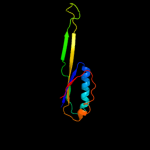
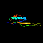
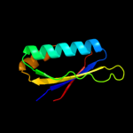
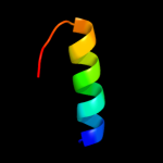
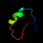
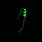
Region: 5 - 101 Aligned: 96 Modelled: 97 Confidence: 100.0% Identity: 27% PDB header:ribosomal protein/rna Chain: J: PDB Molecule: PDBTitle: structure of a mammalian ribosomal 40s subunit within an2 80s complex obtained by docking homology models of the rna3 and proteins into an 8.7 a cryo-em map
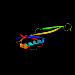
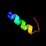
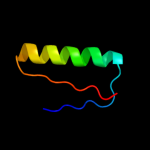
Region: 5 - 100 Aligned: 95 Modelled: 96 Confidence: 100.0% Identity: 25% PDB header:ribosome Chain: J: PDB Molecule:40s ribosomal protein s20; PDBTitle: structure of the ribosomal 80s-eef2-sordarin complex from2 yeast obtained by docking atomic models for rna and protein3 components into a 11.7 a cryo-em map. this file, 1s1h,4 contains 40s subunit. the 60s ribosomal subunit is in file5 1s1i.
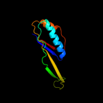
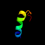
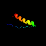
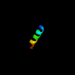
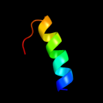
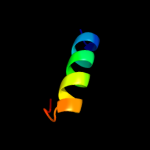
Region: 18 - 36 Aligned: 19 Modelled: 19 Confidence: 12.6% Identity: 37% PDB header:ribosome Chain: K: PDB Molecule:60s ribosomal protein l12; PDBTitle: structure of the ribosomal 80s-eef2-sordarin complex from2 yeast obtained by docking atomic models for rna and protein3 components into a 11.7 a cryo-em map. this file, 1s1i,4 contains 60s subunit. the 40s ribosomal subunit is in file5 1s1h.
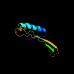
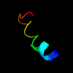
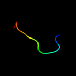
Region: 18 - 36 Aligned: 19 Modelled: 19 Confidence: 11.4% Identity: 26% PDB header:ribosome Chain: I: PDB Molecule:50s ribosomal protein l11p; PDBTitle: the structure of ccda-phe-cap-bio and the antibiotic sparsomycin bound2 to the large ribosomal subunit of haloarcula marismortui
Region: 18 - 36 Aligned: 19 Modelled: 19 Confidence: 9.8% Identity: 32% PDB header:ribosomal protein/rna Chain: I: PDB Molecule:rna expansion segment es15 part i; PDBTitle: structure of a mammalian ribosomal 60s subunit within an2 80s complex obtained by docking homology models of the rna3 and proteins into an 8.7 a cryo-em map
Region: 18 - 36 Aligned: 19 Modelled: 19 Confidence: 8.2% Identity: 37% PDB header:ribosome Chain: A: PDB Molecule:50s ribosomal protein l11; PDBTitle: fitting of l11 protein and elongation factor g (ef-g) in2 the cryo-em map of e. coli 70s ribosome bound with ef-g,3 gdp and fusidic acid
Phyre2
21
22
23
24
25
26
27
28
29
30
31
32
33
34
35
36
Detailed template information
Binding site prediction
Due to computational demand, binding site predictions are not run for batch jobs
If you want to predict binding sites, please manually submit your model of choice to 3DLigandSite
Phyre is for academic use only
Please cite: Protein structure prediction on
the web: a case study using the Phyre server
Kelley LA and Sternberg MJE. Nature Protocols
4, 363 - 371 (2009) [pdf] [Import into BibTeX]
If you use the binding site
predictions from 3DLigandSite, please also cite:
3DLigandSite: predicting ligand-binding sites using similar structures.
Wass MN, Kelley LA and Sternberg
MJ Nucleic Acids Research 38, W469-73 (2010) [PubMed]