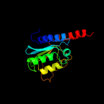
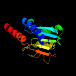
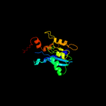
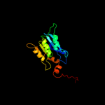
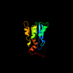
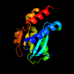
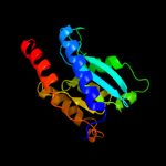
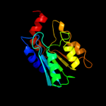
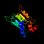
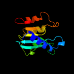
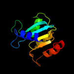
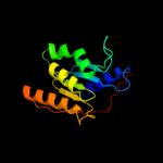
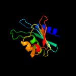
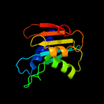
1 d1z3aa1
100.0
100
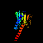
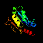
Fold: Cytidine deaminase-likeSuperfamily: Cytidine deaminase-likeFamily: Deoxycytidylate deaminase-like2 c2nx8A_
100.0
37
PDB header: hydrolaseChain: A: PDB Molecule: trna-specific adenosine deaminase;PDBTitle: the crystal structure of the trna-specific adenosine deaminase from2 streptococcus pyogenes
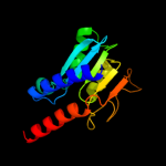
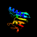
3 d2b3ja1
100.0
44
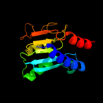
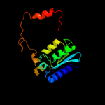
Fold: Cytidine deaminase-likeSuperfamily: Cytidine deaminase-likeFamily: Deoxycytidylate deaminase-like4 c3ocqA_
100.0
92
PDB header: hydrolaseChain: A: PDB Molecule: putative cytosine/adenosine deaminase;PDBTitle: crystal structure of trna-specific adenosine deaminase from salmonella2 enterica
5 d1wwra1
100.0
41
Fold: Cytidine deaminase-likeSuperfamily: Cytidine deaminase-likeFamily: Deoxycytidylate deaminase-like6 c2o7pA_
100.0
34
PDB header: hydrolase, oxidoreductaseChain: A: PDB Molecule: riboflavin biosynthesis protein ribd;PDBTitle: the crystal structure of ribd from escherichia coli in complex with2 the oxidised nadp+ cofactor in the active site of the reductase3 domain
7 c2d5nB_
100.0
38
PDB header: hydrolase, oxidoreductaseChain: B: PDB Molecule: riboflavin biosynthesis protein ribd;PDBTitle: crystal structure of a bifunctional deaminase and reductase2 involved in riboflavin biosynthesis
8 c2hxvA_
100.0
34
PDB header: biosynthetic proteinChain: A: PDB Molecule: diaminohydroxyphosphoribosylaminopyrimidine deaminase/ 5-PDBTitle: crystal structure of a diaminohydroxyphosphoribosylaminopyrimidine2 deaminase/ 5-amino-6-(5-phosphoribosylamino)uracil reductase (tm1828)3 from thermotoga maritima at 1.80 a resolution
9 d2g84a1
100.0
31
Fold: Cytidine deaminase-likeSuperfamily: Cytidine deaminase-likeFamily: Deoxycytidylate deaminase-like10 c3dh1D_
100.0
34
PDB header: hydrolaseChain: D: PDB Molecule: trna-specific adenosine deaminase 2;PDBTitle: crystal structure of human trna-specific adenosine-34 deaminase2 subunit adat2
11 d2a8na1
100.0
42
Fold: Cytidine deaminase-likeSuperfamily: Cytidine deaminase-likeFamily: Deoxycytidylate deaminase-like12 d1wkqa_
100.0
29
Fold: Cytidine deaminase-likeSuperfamily: Cytidine deaminase-likeFamily: Deoxycytidylate deaminase-like13 d1p6oa_
100.0
29
Fold: Cytidine deaminase-likeSuperfamily: Cytidine deaminase-likeFamily: Deoxycytidylate deaminase-like14 d2hxva2
100.0
26
Fold: Cytidine deaminase-likeSuperfamily: Cytidine deaminase-likeFamily: Deoxycytidylate deaminase-like15 d2b3za2
100.0
40
Fold: Cytidine deaminase-likeSuperfamily: Cytidine deaminase-likeFamily: Deoxycytidylate deaminase-like16 d1vq2a_
100.0
27
Fold: Cytidine deaminase-likeSuperfamily: Cytidine deaminase-likeFamily: Deoxycytidylate deaminase-like17 c2hvwC_
100.0
29
PDB header: hydrolaseChain: C: PDB Molecule: deoxycytidylate deaminase;PDBTitle: crystal structure of dcmp deaminase from streptococcus2 mutans
18 c2w4lC_
100.0
28
PDB header: hydrolaseChain: C: PDB Molecule: deoxycytidylate deaminase;PDBTitle: human dcmp deaminase
19 d1uwza_
98.3
16
Fold: Cytidine deaminase-likeSuperfamily: Cytidine deaminase-likeFamily: Cytidine deaminase20 d2d30a1
98.3
17
Fold: Cytidine deaminase-likeSuperfamily: Cytidine deaminase-likeFamily: Cytidine deaminase21 d1r5ta_
not modelled
98.2
18
Fold: Cytidine deaminase-likeSuperfamily: Cytidine deaminase-likeFamily: Cytidine deaminase22 d1mq0a_
not modelled
98.2
17
Fold: Cytidine deaminase-likeSuperfamily: Cytidine deaminase-likeFamily: Cytidine deaminase23 d2fr5a1
not modelled
98.2
18
Fold: Cytidine deaminase-likeSuperfamily: Cytidine deaminase-likeFamily: Cytidine deaminase24 c3dmoD_
not modelled
98.2
22
PDB header: hydrolaseChain: D: PDB Molecule: cytidine deaminase;PDBTitle: 1.6 a crystal structure of cytidine deaminase from2 burkholderia pseudomallei
25 c3ijfX_
not modelled
98.1
17
PDB header: hydrolaseChain: X: PDB Molecule: cytidine deaminase;PDBTitle: crystal structure of cytidine deaminase from mycobacterium2 tuberculosis
26 c3r2nC_
not modelled
98.0
15
PDB header: hydrolaseChain: C: PDB Molecule: cytidine deaminase;PDBTitle: crystal structure of cytidine deaminase from mycobacterium leprae
27 c3b8fB_
not modelled
98.0
12
PDB header: hydrolaseChain: B: PDB Molecule: putative blasticidin s deaminase;PDBTitle: crystal structure of the cytidine deaminase from bacillus anthracis
28 d1alna1
not modelled
97.8
23
Fold: Cytidine deaminase-likeSuperfamily: Cytidine deaminase-likeFamily: Cytidine deaminase29 d2z3ga1
not modelled
97.8
13
Fold: Cytidine deaminase-likeSuperfamily: Cytidine deaminase-likeFamily: Cytidine deaminase30 c3oj6C_
not modelled
97.7
10
PDB header: hydrolaseChain: C: PDB Molecule: blasticidin-s deaminase;PDBTitle: crystal structure of blasticidin s deaminase from coccidioides immitis
31 d1alna2
not modelled
97.6
15
Fold: Cytidine deaminase-likeSuperfamily: Cytidine deaminase-likeFamily: Cytidine deaminase32 c1alnA_
not modelled
97.5
22
PDB header: hydrolaseChain: A: PDB Molecule: cytidine deaminase;PDBTitle: crystal structure of cytidine deaminase complexed with 3-deazacytidine
33 c3g8qA_
not modelled
97.0
25
PDB header: rna binding proteinChain: A: PDB Molecule: predicted rna-binding protein, contains thumpPDBTitle: a cytidine deaminase edits c-to-u in transfer rnas in2 archaea
34 c2nytB_
not modelled
93.6
28
PDB header: hydrolaseChain: B: PDB Molecule: probable c->u-editing enzyme apobec-2;PDBTitle: the apobec2 crystal structure and functional implications2 for aid
35 c2kboA_
not modelled
92.0
25
PDB header: hydrolaseChain: A: PDB Molecule: dna dc->du-editing enzyme apobec-3g;PDBTitle: structure, interaction, and real-time monitoring of the2 enzymatic reaction of wild type apobec3g
36 c3gxgA_
not modelled
51.0
9
PDB header: hydrolaseChain: A: PDB Molecule: putative phosphatase (duf442);PDBTitle: crystal structure of putative phosphatase (duf442) (yp_001181608.1)2 from shewanella putrefaciens cn-32 at 1.60 a resolution
37 d1zbfa1
not modelled
49.7
21
Fold: Ribonuclease H-like motifSuperfamily: Ribonuclease H-likeFamily: Ribonuclease H38 c3e9jC_
not modelled
42.9
71
PDB header: oxidoreductaseChain: C: PDB Molecule: thiol/disulfide oxidoreductase dsbb;PDBTitle: structure of the charge-transfer intermediate of the2 transmembrane redox catalyst dsbb
39 d2hi7b1
not modelled
42.9
71
Fold: Bromodomain-likeSuperfamily: DsbB-likeFamily: DsbB-like40 d2b0va1
not modelled
30.6
14
Fold: NudixSuperfamily: NudixFamily: MutT-like41 c3dkuB_
not modelled
27.7
11
PDB header: hydrolaseChain: B: PDB Molecule: putative phosphohydrolase;PDBTitle: crystal structure of nudix hydrolase orf153, ymfb, from2 escherichia coli k-1
42 c3ggmB_
not modelled
24.8
13
PDB header: structural genomics, unknown functionChain: B: PDB Molecule: uncharacterized protein bt9727_2919;PDBTitle: crystal structure of bt9727_2919 from bacillus2 thuringiensis subsp. northeast structural genomics target3 bur228b
43 c2lk2A_
not modelled
20.9
17
PDB header: transcriptionChain: A: PDB Molecule: homeobox protein tgif1;PDBTitle: solution nmr structure of homeobox domain (171-248) of human homeobox2 protein tgif1, northeast structural genomics consortium target3 hr4411b
44 c3rh7A_
not modelled
15.7
32
PDB header: oxidoreductaseChain: A: PDB Molecule: hypothetical oxidoreductase;PDBTitle: crystal structure of a hypothetical oxidoreductase (sma0793) from2 sinorhizobium meliloti 1021 at 3.00 a resolution
45 c3o8sA_
not modelled
15.3
9
PDB header: hydrolaseChain: A: PDB Molecule: adp-ribose pyrophosphatase;PDBTitle: crystal structure of an adp-ribose pyrophosphatase (ssu98_1448) from2 streptococcus suis 89-1591 at 2.27 a resolution
46 c3nznA_
not modelled
14.6
20
PDB header: oxidoreductaseChain: A: PDB Molecule: glutaredoxin;PDBTitle: the crystal structure of the glutaredoxin from methanosarcina mazei2 go1
47 c3q4iA_
not modelled
13.7
18
PDB header: hydrolaseChain: A: PDB Molecule: phosphohydrolase (mutt/nudix family protein);PDBTitle: crystal structure of cdp-chase in complex with gd3+
48 c3d2oB_
not modelled
13.5
18
PDB header: hydrolase, biosynthetic proteinChain: B: PDB Molecule: upf0343 protein ngo0387;PDBTitle: crystal structure of manganese-metallated gtp cyclohydrolase2 type ib
49 d1j9ba_
not modelled
13.2
17
Fold: Thioredoxin foldSuperfamily: Thioredoxin-likeFamily: ArsC-like50 c2imrA_
not modelled
13.0
36
PDB header: structural genomics, unknown functionChain: A: PDB Molecule: hypothetical protein dr_0824;PDBTitle: crystal structure of amidohydrolase dr_0824 from2 deinococcus radiodurans
51 d1ttza_
not modelled
12.3
40
Fold: Thioredoxin foldSuperfamily: Thioredoxin-likeFamily: Thioltransferase52 d2fb5a1
not modelled
11.7
29
Fold: YojJ-likeSuperfamily: YojJ-likeFamily: YojJ-like53 c3ld0Q_
not modelled
11.6
63
PDB header: gene regulationChain: Q: PDB Molecule: inhibitor of trap, regulated by t-box (trp) sequence rtpa;PDBTitle: crystal structure of b.licheniformis anti-trap protein, an antagonist2 of trap-rna interactions
54 d1hr6a1
not modelled
11.4
13
Fold: LuxS/MPP-like metallohydrolaseSuperfamily: LuxS/MPP-like metallohydrolaseFamily: MPP-like55 c3ipzA_
not modelled
10.7
15
PDB header: electron transport, oxidoreductaseChain: A: PDB Molecule: monothiol glutaredoxin-s14, chloroplastic;PDBTitle: crystal structure of arabidopsis monothiol glutaredoxin atgrxcp
56 d1xdpa4
not modelled
10.6
16
Fold: Phospholipase D/nucleaseSuperfamily: Phospholipase D/nucleaseFamily: Polyphosphate kinase C-terminal domain57 d2o8ra4
not modelled
10.4
16
Fold: Phospholipase D/nucleaseSuperfamily: Phospholipase D/nucleaseFamily: Polyphosphate kinase C-terminal domain58 c4a1oB_
not modelled
9.7
21
PDB header: transferase-hydrolaseChain: B: PDB Molecule: bifunctional purine biosynthesis protein purh;PDBTitle: crystal structure of mycobacterium tuberculosis purh complexed with2 aicar and a novel nucleotide cfair, at 2.48 a resolution.
59 c2oodA_
not modelled
9.7
21
PDB header: hydrolaseChain: A: PDB Molecule: blr3880 protein;PDBTitle: crystal structure of guanine deaminase from bradyrhizobium japonicum
60 c2klxA_
not modelled
9.6
23
PDB header: oxidoreductaseChain: A: PDB Molecule: glutaredoxin;PDBTitle: solution structure of glutaredoxin from bartonella henselae str.2 houston
61 c2r5rA_
not modelled
9.3
18
PDB header: structural genomics, unknown functionChain: A: PDB Molecule: upf0343 protein ne1163;PDBTitle: the crystal structure of duf198 from nitrosomonas europaea2 atcc 19718
62 d1zcza2
not modelled
9.0
16
Fold: Cytidine deaminase-likeSuperfamily: Cytidine deaminase-likeFamily: AICAR transformylase domain of bifunctional purine biosynthesis enzyme ATIC63 c2dmnA_
not modelled
8.4
17
PDB header: transcriptionChain: A: PDB Molecule: homeobox protein tgif2lx;PDBTitle: the solution structure of the homeobox domain of human2 homeobox protein tgif2lx
64 d1nuia2
not modelled
8.4
27
Fold: Rubredoxin-likeSuperfamily: Zinc beta-ribbonFamily: DNA primase zinc finger65 c2pq1B_
not modelled
8.0
29
PDB header: hydrolaseChain: B: PDB Molecule: ap4a hydrolase;PDBTitle: crystal structure of ap4a hydrolase complexed with amp and2 atp (aq_158) from aquifex aeolicus vf5
66 c2kq2A_
not modelled
7.6
15
PDB header: hydrolaseChain: A: PDB Molecule: ribonuclease h-related protein;PDBTitle: solution nmr structure of the apo form of a ribonuclease h2 domain of protein dsy1790 from desulfitobacterium3 hafniense, northeast structural genomics target dhr1a
67 d2drpa2
not modelled
7.2
60
Fold: beta-beta-alpha zinc fingersSuperfamily: beta-beta-alpha zinc fingersFamily: Classic zinc finger, C2H268 c2khpA_
not modelled
7.0
32
PDB header: electron transportChain: A: PDB Molecule: glutaredoxin;PDBTitle: solution structure of glutaredoxin from brucella melitensis
69 c3hstD_
not modelled
6.9
15
PDB header: hydrolaseChain: D: PDB Molecule: protein rv2228c/mt2287;PDBTitle: n-terminal rnase h domain of rv2228c from mycobacterium tuberculosis2 as a fusion protein with maltose binding protein
70 d1i1qb_
not modelled
6.8
29
Fold: Flavodoxin-likeSuperfamily: Class I glutamine amidotransferase-likeFamily: Class I glutamine amidotransferases (GAT)71 d2akla2
not modelled
6.4
56
Fold: Rubredoxin-likeSuperfamily: Zinc beta-ribbonFamily: PhnA zinc-binding domain72 c2bx9J_
not modelled
5.7
57
PDB header: transcription regulationChain: J: PDB Molecule: tryptophan rna-binding attenuator protein-inhibitoryPDBTitle: crystal structure of b.subtilis anti-trap protein, an2 antagonist of trap-rna interactions
73 c2xzn5_
not modelled
5.7
70
PDB header: ribosomeChain: 5: PDB Molecule: ribosomal protein s26e containing protein;PDBTitle: crystal structure of the eukaryotic 40s ribosomal2 subunit in complex with initiation factor 1. this file3 contains the 40s subunit and initiation factor for4 molecule 2
74 d1vdda_
not modelled
5.5
21
Fold: Recombination protein RecRSuperfamily: Recombination protein RecRFamily: Recombination protein RecR75 d2r5yb1
not modelled
5.4
11
Fold: DNA/RNA-binding 3-helical bundleSuperfamily: Homeodomain-likeFamily: Homeodomain76 d2baia1
not modelled
5.3
44
Fold: Viral leader polypeptide zinc fingerSuperfamily: Viral leader polypeptide zinc fingerFamily: Viral leader polypeptide zinc finger77 d1k61a_
not modelled
5.3
15
Fold: DNA/RNA-binding 3-helical bundleSuperfamily: Homeodomain-likeFamily: Homeodomain78 d1du6a_
not modelled
5.2
9
Fold: DNA/RNA-binding 3-helical bundleSuperfamily: Homeodomain-likeFamily: Homeodomain79 c2o8rA_
not modelled
5.1
16
PDB header: transferaseChain: A: PDB Molecule: polyphosphate kinase;PDBTitle: crystal structure of polyphosphate kinase from2 porphyromonas gingivalis


