| 1 | c2rqlA_
|
|
 |
100.0 |
100 |
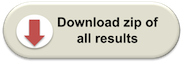
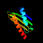
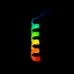
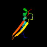
PDB header:translation
Chain: A: PDB Molecule:probable sigma-54 modulation protein;
PDBTitle: solution structure of the e. coli ribosome hibernation2 promoting factor hpf
|
| 2 | d1imua_
|
|
 |
100.0 |
34 |
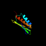
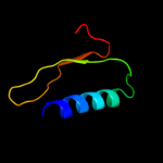
Fold:Ribosome binding protein Y (YfiA homologue)
Superfamily:Ribosome binding protein Y (YfiA homologue)
Family:Ribosome binding protein Y (YfiA homologue) |
| 3 | c3tqmD_
|
|
 |
99.9 |
33 |
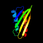
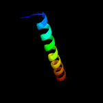
PDB header:protein binding
Chain: D: PDB Molecule:ribosome-associated factor y;
PDBTitle: structure of an ribosomal subunit interface protein from coxiella2 burnetii
|
| 4 | d1l4sa_
|
|
 |
99.9 |
38 |
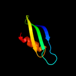
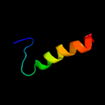
Fold:Ribosome binding protein Y (YfiA homologue)
Superfamily:Ribosome binding protein Y (YfiA homologue)
Family:Ribosome binding protein Y (YfiA homologue) |
| 5 | d2ywqa1
|
|
 |
99.9 |
33 |
Fold:Ribosome binding protein Y (YfiA homologue)
Superfamily:Ribosome binding protein Y (YfiA homologue)
Family:Ribosome binding protein Y (YfiA homologue) |
| 6 | d1sr9a3
|
|
 |
24.2 |
22 |
Fold:2-isopropylmalate synthase LeuA, allosteric (dimerisation) domain
Superfamily:2-isopropylmalate synthase LeuA, allosteric (dimerisation) domain
Family:2-isopropylmalate synthase LeuA, allosteric (dimerisation) domain |
| 7 | c3f6hA_
|
|
 |
23.6 |
21 |
PDB header:transferase
Chain: A: PDB Molecule:alpha-isopropylmalate synthase;
PDBTitle: crystal structure of the regulatory domain of licms in2 complexed with isoleucine - type iii
|
| 8 | d1uila_
|
|
 |
21.7 |
24 |
Fold:dsRBD-like
Superfamily:dsRNA-binding domain-like
Family:Double-stranded RNA-binding domain (dsRBD) |
| 9 | c3kxeD_
|
|
 |
19.9 |
18 |
PDB header:protein binding
Chain: D: PDB Molecule:antitoxin protein pard-1;
PDBTitle: a conserved mode of protein recognition and binding in a2 pard-pare toxin-antitoxin complex
|
| 10 | c1i7nA_
|
|
 |
19.7 |
13 |
PDB header:neuropeptide
Chain: A: PDB Molecule:synapsin ii;
PDBTitle: crystal structure analysis of the c domain of synapsin ii2 from rat brain
|
| 11 | c2kxoA_
|
|
 |
19.2 |
10 |
PDB header:cell cycle
Chain: A: PDB Molecule:cell division topological specificity factor;
PDBTitle: solution nmr structure of the cell division regulator mine protein2 from neisseria gonorrhoeae
|
| 12 | c1by0A_
|
|
 |
18.9 |
41 |
PDB header:rna binding protein
Chain: A: PDB Molecule:protein (hepatitis delta antigen);
PDBTitle: n-terminal leucine-repeat region of hepatitis delta antigen
|
| 13 | c1pk8D_
|
|
 |
18.0 |
9 |
PDB header:membrane protein
Chain: D: PDB Molecule:rat synapsin i;
PDBTitle: crystal structure of rat synapsin i c domain complexed to2 ca.atp
|
| 14 | c2yh5A_
|
|
 |
17.6 |
12 |
PDB header:lipid binding protein
Chain: A: PDB Molecule:dapx protein;
PDBTitle: structure of the c-terminal domain of bamc
|
| 15 | d2clyc1
|
|
 |
16.6 |
17 |
Fold:Mitochondrial ATP synthase coupling factor 6
Superfamily:Mitochondrial ATP synthase coupling factor 6
Family:Mitochondrial ATP synthase coupling factor 6 |
| 16 | d2r85a2
|
|
 |
12.3 |
8 |
Fold:ATP-grasp
Superfamily:Glutathione synthetase ATP-binding domain-like
Family:PurP ATP-binding domain-like |
| 17 | d2hkja2
|
|
 |
12.2 |
23 |
Fold:Ribosomal protein S5 domain 2-like
Superfamily:Ribosomal protein S5 domain 2-like
Family:DNA gyrase/MutL, second domain |
| 18 | d1vzsa_
|
|
 |
11.0 |
20 |
Fold:Mitochondrial ATP synthase coupling factor 6
Superfamily:Mitochondrial ATP synthase coupling factor 6
Family:Mitochondrial ATP synthase coupling factor 6 |
| 19 | c3r9jD_
|
|
 |
11.0 |
9 |
PDB header:cell cycle,hydrolase/cell cycle
Chain: D: PDB Molecule:cell division topological specificity factor;
PDBTitle: 4.3a resolution structure of a mind-mine(i24n) protein complex
|
| 20 | c1mx0D_
|
|
 |
10.8 |
23 |
PDB header:isomerase
Chain: D: PDB Molecule:type ii dna topoisomerase vi subunit b;
PDBTitle: structure of topoisomerase subunit
|
| 21 | c2p0aA_ |
|
not modelled |
10.2 |
17 |
PDB header:neuropeptide
Chain: A: PDB Molecule:synapsin-3;
PDBTitle: the crystal structure of human synapsin iii (syn3) in complex with2 amppnp
|
| 22 | d2e9xd1 |
|
not modelled |
9.8 |
20 |
Fold:GINS helical bundle-like
Superfamily:GINS helical bundle-like
Family:SLD5 N-terminal domain-like |
| 23 | c3r23B_ |
|
not modelled |
9.7 |
18 |
PDB header:ligase
Chain: B: PDB Molecule:d-alanine--d-alanine ligase;
PDBTitle: crystal structure of d-alanine--d-alanine ligase from bacillus2 anthracis
|
| 24 | d1hk8a_ |
|
not modelled |
9.6 |
16 |
Fold:PFL-like glycyl radical enzymes
Superfamily:PFL-like glycyl radical enzymes
Family:Class III anaerobic ribonucleotide reductase NRDD subunit |
| 25 | c1hk8A_ |
|
not modelled |
9.6 |
16 |
PDB header:oxidoreductase
Chain: A: PDB Molecule:anaerobic ribonucleotide-triphosphate reductase;
PDBTitle: structural basis for allosteric substrate specificity2 regulation in class iii ribonucleotide reductases:3 nrdd in complex with dgtp
|
| 26 | c3l2iB_ |
|
not modelled |
9.2 |
15 |
PDB header:lyase
Chain: B: PDB Molecule:3-dehydroquinate dehydratase;
PDBTitle: 1.85 angstrom crystal structure of the 3-dehydroquinate dehydratase2 (arod) from salmonella typhimurium lt2.
|
| 27 | d1o6da_ |
|
not modelled |
8.9 |
10 |
Fold:alpha/beta knot
Superfamily:alpha/beta knot
Family:YbeA-like |
| 28 | c3ehuA_ |
|
not modelled |
8.3 |
19 |
PDB header:membrane protein
Chain: A: PDB Molecule:fusion protein of crfr1 extracellular domain and mbp;
PDBTitle: crystal structure of the extracellular domain of human corticotropin2 releasing factor receptor type 1 (crfr1) in complex with crf
|
| 29 | d1sfla_ |
|
not modelled |
8.2 |
24 |
Fold:TIM beta/alpha-barrel
Superfamily:Aldolase
Family:Class I aldolase |
| 30 | d1gsoa3 |
|
not modelled |
8.2 |
5 |
Fold:ATP-grasp
Superfamily:Glutathione synthetase ATP-binding domain-like
Family:BC ATP-binding domain-like |
| 31 | d2i52a1 |
|
not modelled |
8.1 |
5 |
Fold:MK0786-like
Superfamily:MK0786-like
Family:MK0786-like |
| 32 | c3q2oB_ |
|
not modelled |
7.6 |
15 |
PDB header:lyase
Chain: B: PDB Molecule:phosphoribosylaminoimidazole carboxylase, atpase subunit;
PDBTitle: crystal structure of purk: n5-carboxyaminoimidazole ribonucleotide2 synthetase
|
| 33 | c3rd6A_ |
|
not modelled |
7.5 |
13 |
PDB header:structural genomics, unknown function
Chain: A: PDB Molecule:mll3558 protein;
PDBTitle: crystal structure of mll3558 protein from rhizobium loti. northeast2 structural genomics consortium target id mlr403
|
| 34 | c1hf9B_ |
|
not modelled |
7.4 |
31 |
PDB header:atpase inhibitor
Chain: B: PDB Molecule:atpase inhibitor (mitochondrial);
PDBTitle: c-terminal coiled-coil domain from bovine if1
|
| 35 | d1lr0a_ |
|
not modelled |
7.4 |
12 |
Fold:TolA/TonB C-terminal domain
Superfamily:TolA/TonB C-terminal domain
Family:TolA |
| 36 | c2yr1B_ |
|
not modelled |
6.7 |
21 |
PDB header:lyase
Chain: B: PDB Molecule:3-dehydroquinate dehydratase;
PDBTitle: crystal structure of 3-dehydroquinate dehydratase from geobacillus2 kaustophilus hta426
|
| 37 | d2r7ka2 |
|
not modelled |
6.5 |
12 |
Fold:ATP-grasp
Superfamily:Glutathione synthetase ATP-binding domain-like
Family:PurP ATP-binding domain-like |
| 38 | d1xppa_ |
|
not modelled |
6.1 |
13 |
Fold:DCoH-like
Superfamily:RBP11-like subunits of RNA polymerase
Family:RBP11/RpoL |
| 39 | c1q0vA_ |
|
not modelled |
6.0 |
19 |
PDB header:transport binding
Chain: A: PDB Molecule:hydrophilic protein; has cysteine rich putative
PDBTitle: solution structure of tandem uims of vps27
|
| 40 | d1ehia2 |
|
not modelled |
6.0 |
12 |
Fold:ATP-grasp
Superfamily:Glutathione synthetase ATP-binding domain-like
Family:ATP-binding domain of peptide synthetases |
| 41 | c3e5nA_ |
|
not modelled |
5.9 |
18 |
PDB header:ligase
Chain: A: PDB Molecule:d-alanine-d-alanine ligase a;
PDBTitle: crystal strucutre of d-alanine-d-alanine ligase from2 xanthomonas oryzae pv. oryzae kacc10331
|
| 42 | c2l5gB_ |
|
not modelled |
5.9 |
30 |
PDB header:transcription regulator
Chain: B: PDB Molecule:putative uncharacterized protein ncor2;
PDBTitle: co-ordinates and 1h, 13c and 15n chemical shift assignments for the2 complex of gps2 53-90 and smrt 167-207
|
| 43 | c1a92B_ |
|
not modelled |
5.9 |
35 |
PDB header:leucine zipper
Chain: B: PDB Molecule:delta antigen;
PDBTitle: oligomerization domain of hepatitis delta antigen
|
| 44 | c2l9pA_ |
|
not modelled |
5.7 |
11 |
PDB header:structural genomics, unknown function
Chain: A: PDB Molecule:uncharacterized protein;
PDBTitle: solution nmr structure of q5hli9 from staphylococcus epidermidis,2 northeast structural genomics consortium target ser147
|
| 45 | c1g6uB_ |
|
not modelled |
5.7 |
23 |
PDB header:de novo protein
Chain: B: PDB Molecule:domain swapped dimer;
PDBTitle: crystal structure of a domain swapped dimer
|
| 46 | c1ulzA_ |
|
not modelled |
5.7 |
10 |
PDB header:ligase
Chain: A: PDB Molecule:pyruvate carboxylase n-terminal domain;
PDBTitle: crystal structure of the biotin carboxylase subunit of pyruvate2 carboxylase
|
| 47 | d1ghha_ |
|
not modelled |
5.6 |
12 |
Fold:DNA damage-inducible protein DinI
Superfamily:DNA damage-inducible protein DinI
Family:DNA damage-inducible protein DinI |
| 48 | c3oqvA_ |
|
not modelled |
5.4 |
11 |
PDB header:protein binding
Chain: A: PDB Molecule:albc;
PDBTitle: albc, a cyclodipeptide synthase from streptomyces noursei
|
| 49 | c2q2eB_ |
|
not modelled |
5.3 |
27 |
PDB header:isomerase
Chain: B: PDB Molecule:type 2 dna topoisomerase 6 subunit b;
PDBTitle: crystal structure of the topoisomerase vi holoenzyme from2 methanosarcina mazei
|