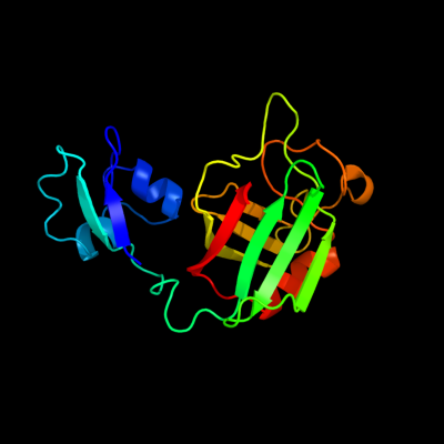
Phyre Home Retrieve Phyre Job Id
Emaill.a.kelley@imperial.ac.uk
DescriptionP39176
DateThu Jan 5 11:58:22 GMT 2012
Unique Job IDa3cabd4d51c330d5

Summary 

Top model
Image coloured by rainbow N → C terminus
Model (left) based on template c1y7mB_
Top template information
PDB header:structural genomics, unknown function
Chain: B: PDB Molecule:hypothetical protein bsu14040;
PDBTitle: crystal structure of the b. subtilis ykud protein at 2 a2 resolution
Confidence and coverage
Confidence:100.0% Coverage: 52%
161 residues ( 52% of your sequence) have been modelled with 100.0% confidence by the single highest scoring template.
You may wish to submit your sequence to Phyrealarm. This will automatically scan your sequence every week for new potential templates as they appear in the Phyre2 library.
3D viewing
Interactive 3D view in Jmol

Sequence analysis 

Secondary structure and disorder prediction 

   1........10.........20.........30.........40.........50.........60
Sequence  MRRVNILCSFALLFASHTSLAVTYPLPPEGSRLVGQSFTVTVPDHNTQPLETFAAQYGQG
Secondary structure 


SS confidence 


Disorder  ????


???????????

???????????????
??
?


Disorder confidence 


   .........70.........80.........90.........100.........110.........120
Sequence  LSNMLEANPGADVFLPKSGSQLTIPQQLILPDTVRKGIVVNVAEMRLYYYPPDSNTVEVF
Secondary structure 


SS confidence 


Disorder 


?


???????


Disorder confidence 


   .........130.........140.........150.........160.........170.........180
Sequence  PIGIGQAGRETPRNWVTTVERKQEAPTWTPTPNTRREYAKRGESLPAFVPAGPDNPMGLY
Secondary structure 


SS confidence 


Disorder 


?


?


Disorder confidence 


   .........190.........200.........210.........220.........230.........240
Sequence  AIYIGRLYAIHGTNANFGIGLRVSQGCIRLRNDDIKYLFDNVPVGTRVQIIDQPVKYTTE
Secondary structure 


SS confidence 


Disorder 


?


Disorder confidence 


   .........250.........260.........270.........280.........290.........300
Sequence  PDGSNWLEVHEPLSRNRAEYESDRKVPLPVTPSLRAFINGQEVDVNRANAALQRRSGMPV
Secondary structure 


SS confidence 


Disorder 


???????????????????


????????


??


Disorder confidence 


   .........310
Sequence  QISSGSRQMF
Secondary structure 


SS confidence 


Disorder 


???????
Disorder confidence 


Confidence Key
High(9)                    Low (0)
?Disordered
Alpha helix
Beta strand

Domain analysis 

Hover over an aligned region to see model and summary info

Please note, only up to the top 20 hits are modelled to reduce computer load

RankAligned region

PDB 1y7m chain B

3D model

Region: 38 - 231
Aligned: 161
Modelled: 167
Confidence: 100.0%
Identity: 32%
PDB header:structural genomics, unknown function
Chain: B: PDB Molecule:hypothetical protein bsu14040;
PDBTitle: crystal structure of the b. subtilis ykud protein at 2 a2 resolution

Phyre2

PDB 1y7m chain A domain 1

3D model

Region: 91 - 230
Aligned: 111
Modelled: 113
Confidence: 100.0%
Identity: 36%
Fold: L,D-transpeptidase catalytic domain-like
Superfamily: L,D-transpeptidase catalytic domain-like
Family: L,D-transpeptidase catalytic domain-like

Phyre2

PDB 1zat chain A domain 1

3D model

Region: 93 - 230
Aligned: 118
Modelled: 120
Confidence: 100.0%
Identity: 29%
Fold: L,D-transpeptidase catalytic domain-like
Superfamily: L,D-transpeptidase catalytic domain-like
Family: L,D-transpeptidase catalytic domain-like

Phyre2

PDB 2hkl chain B

3D model

Region: 91 - 230
Aligned: 120
Modelled: 122
Confidence: 100.0%
Identity: 28%
PDB header:transferase
Chain: B: PDB Molecule:l,d-transpeptidase;
PDBTitle: crystal structure of enterococcus faecium l,d-2 transpeptidase c442s mutant

Phyre2

PDB 2l9y chain A

3D model

Region: 35 - 87
Aligned: 51
Modelled: 53
Confidence: 98.3%
Identity: 24%
PDB header:sugar binding protein
Chain: A: PDB Molecule:cvnh-lysm lectin;
PDBTitle: solution structure of the mocvnh-lysm module from the rice blast2 fungus magnaporthe oryzae protein (mgg_03307)

Phyre2

PDB 1y7m chain A domain 2

3D model

Region: 39 - 86
Aligned: 45
Modelled: 48
Confidence: 98.0%
Identity: 29%
Fold: LysM domain
Superfamily: LysM domain
Family: LysM domain

Phyre2

PDB 2djp chain A

3D model

Region: 36 - 85
Aligned: 48
Modelled: 50
Confidence: 97.9%
Identity: 21%
PDB header:structural genomics, unknown function
Chain: A: PDB Molecule:hypothetical protein sb145;
PDBTitle: the solution structure of the lysm domain of human2 hypothetical protein sb145

Phyre2

PDB 1e0g chain A

3D model

Region: 39 - 87
Aligned: 45
Modelled: 49
Confidence: 97.8%
Identity: 20%
Fold: LysM domain
Superfamily: LysM domain
Family: LysM domain

Phyre2

PDB 2gu1 chain A

3D model

Region: 39 - 118
Aligned: 77
Modelled: 80
Confidence: 95.0%
Identity: 16%
PDB header:hydrolase
Chain: A: PDB Molecule:zinc peptidase;
PDBTitle: crystal structure of a zinc containing peptidase from2 vibrio cholerae

Phyre2

PDB 3mca chain B

3D model

Region: 206 - 229
Aligned: 24
Modelled: 24
Confidence: 51.1%
Identity: 25%
PDB header:translation regulation/hydrolase
Chain: B: PDB Molecule:protein dom34;
PDBTitle: structure of the dom34-hbs1 complex and implications for its role in2 no-go decay

Phyre2

PDB 1wjj chain A

3D model

Region: 206 - 249
Aligned: 39
Modelled: 42
Confidence: 46.7%
Identity: 23%
Fold: OB-fold
Superfamily: Nucleic acid-binding proteins
Family: Single strand DNA-binding domain, SSB

Phyre2

PDB 1h5n chain C

3D model

Region: 125 - 233
Aligned: 96
Modelled: 109
Confidence: 41.6%
Identity: 17%
PDB header:oxidoreductase
Chain: C: PDB Molecule:dmso reductase;
PDBTitle: dmso reductase modified by the presence of dms and air

Phyre2

PDB 2kke chain A

3D model

Region: 208 - 226
Aligned: 19
Modelled: 19
Confidence: 26.0%
Identity: 42%
PDB header:structural genomics, unknown function
Chain: A: PDB Molecule:uncharacterized protein;
PDBTitle: solution nmr structure of a dimeric protein of unknown2 function from methanobacterium thermoautotrophicum,3 northeast structural genomics consortium target tr5

Phyre2

PDB 1y5i chain A

3D model

Region: 125 - 233
Aligned: 98
Modelled: 109
Confidence: 17.0%
Identity: 15%
PDB header:oxidoreductase
Chain: A: PDB Molecule:respiratory nitrate reductase 1 alpha chain;
PDBTitle: the crystal structure of the narghi mutant nari-k86a

Phyre2

PDB 1ogy chain A domain 1

3D model

Region: 205 - 233
Aligned: 27
Modelled: 29
Confidence: 14.4%
Identity: 15%
Fold: Double psi beta-barrel
Superfamily: ADC-like
Family: Formate dehydrogenase/DMSO reductase, C-terminal domain

Phyre2

PDB 2k50 chain A

3D model

Region: 204 - 247
Aligned: 44
Modelled: 44
Confidence: 10.2%
Identity: 16%
PDB header:structural genomics, unknown function
Chain: A: PDB Molecule:replication factor a related protein;
PDBTitle: solution nmr structure of the replication factor a related2 protein from methanobacterium thermoautotrophicum.3 northeast structural genomics target tr91a.

Phyre2

PDB 2ken chain A

3D model

Region: 206 - 251
Aligned: 42
Modelled: 46
Confidence: 9.6%
Identity: 12%
PDB header:structural genomics, unknown function
Chain: A: PDB Molecule:conserved protein;
PDBTitle: solution nmr structure of the ob domain (residues 67-166)2 of mm0293 from methanosarcina mazei. northeast structural3 genomics consortium target mar214a.

Phyre2

PDB 3rf1 chain B

3D model

Region: 203 - 216
Aligned: 14
Modelled: 12
Confidence: 9.3%
Identity: 29%
PDB header:ligase
Chain: B: PDB Molecule:glycyl-trna synthetase alpha subunit;
PDBTitle: the crystal structure of glycyl-trna synthetase subunit alpha from2 campylobacter jejuni subsp. jejuni nctc 11168

Phyre2

PDB 2vw9 chain B

3D model

Region: 208 - 249
Aligned: 42
Modelled: 42
Confidence: 9.0%
Identity: 14%
PDB header:dna-binding protein
Chain: B: PDB Molecule:single-stranded dna binding protein;
PDBTitle: single stranded dna binding protein complex from2 helicobacter pylori

Phyre2

PDB 2ihe chain A

3D model

Region: 207 - 244
Aligned: 38
Modelled: 38
Confidence: 8.9%
Identity: 18%
PDB header:dna binding protein
Chain: A: PDB Molecule:single-stranded dna-binding protein;
PDBTitle: crystal structure of wild-type single-stranded dna binding protein2 from thermus aquaticus

Phyre2
1

c1y7mB_
2

d1y7ma1
3

d1zata1
4

c2hklB_
5

c2l9yA_
6

d1y7ma2
7

c2djpA_
8

d1e0ga_
9

c2gu1A_
10

c3mcaB_
11

d1wjja_
12

c1h5nC_
13

c2kkeA_
14

c1y5iA_
15

d1ogya1
16

c2k50A_
17

c2kenA_
18

c3rf1B_
19

c2vw9B_
20

c2iheA_
21


22


23


24


25


26


27


28


29


Detailed template information 

#
Template Alignment Coverage3D Model Confidence
% i.d. Template Information
1c1y7mB_


100.0 32 PDB header:structural genomics, unknown function
Chain: B: PDB Molecule:hypothetical protein bsu14040;
PDBTitle: crystal structure of the b. subtilis ykud protein at 2 a2 resolution
2d1y7ma1


100.0 36 Fold:L,D-transpeptidase catalytic domain-like
Superfamily:L,D-transpeptidase catalytic domain-like
Family:L,D-transpeptidase catalytic domain-like
3d1zata1


100.0 29 Fold:L,D-transpeptidase catalytic domain-like
Superfamily:L,D-transpeptidase catalytic domain-like
Family:L,D-transpeptidase catalytic domain-like
4c2hklB_


100.0 28 PDB header:transferase
Chain: B: PDB Molecule:l,d-transpeptidase;
PDBTitle: crystal structure of enterococcus faecium l,d-2 transpeptidase c442s mutant
5c2l9yA_


98.3 24 PDB header:sugar binding protein
Chain: A: PDB Molecule:cvnh-lysm lectin;
PDBTitle: solution structure of the mocvnh-lysm module from the rice blast2 fungus magnaporthe oryzae protein (mgg_03307)
6d1y7ma2


98.0 29 Fold:LysM domain
Superfamily:LysM domain
Family:LysM domain
7c2djpA_


97.9 21 PDB header:structural genomics, unknown function
Chain: A: PDB Molecule:hypothetical protein sb145;
PDBTitle: the solution structure of the lysm domain of human2 hypothetical protein sb145
8d1e0ga_


97.8 20 Fold:LysM domain
Superfamily:LysM domain
Family:LysM domain
9c2gu1A_


95.0 16 PDB header:hydrolase
Chain: A: PDB Molecule:zinc peptidase;
PDBTitle: crystal structure of a zinc containing peptidase from2 vibrio cholerae
10c3mcaB_


51.1 25 PDB header:translation regulation/hydrolase
Chain: B: PDB Molecule:protein dom34;
PDBTitle: structure of the dom34-hbs1 complex and implications for its role in2 no-go decay
11d1wjja_


46.7 23 Fold:OB-fold
Superfamily:Nucleic acid-binding proteins
Family:Single strand DNA-binding domain, SSB
12c1h5nC_


41.6 17 PDB header:oxidoreductase
Chain: C: PDB Molecule:dmso reductase;
PDBTitle: dmso reductase modified by the presence of dms and air
13c2kkeA_


26.0 42 PDB header:structural genomics, unknown function
Chain: A: PDB Molecule:uncharacterized protein;
PDBTitle: solution nmr structure of a dimeric protein of unknown2 function from methanobacterium thermoautotrophicum,3 northeast structural genomics consortium target tr5
14c1y5iA_


17.0 15 PDB header:oxidoreductase
Chain: A: PDB Molecule:respiratory nitrate reductase 1 alpha chain;
PDBTitle: the crystal structure of the narghi mutant nari-k86a
15d1ogya1


14.4 15 Fold:Double psi beta-barrel
Superfamily:ADC-like
Family:Formate dehydrogenase/DMSO reductase, C-terminal domain
16c2k50A_


10.2 16 PDB header:structural genomics, unknown function
Chain: A: PDB Molecule:replication factor a related protein;
PDBTitle: solution nmr structure of the replication factor a related2 protein from methanobacterium thermoautotrophicum.3 northeast structural genomics target tr91a.
17c2kenA_


9.6 12 PDB header:structural genomics, unknown function
Chain: A: PDB Molecule:conserved protein;
PDBTitle: solution nmr structure of the ob domain (residues 67-166)2 of mm0293 from methanosarcina mazei. northeast structural3 genomics consortium target mar214a.
18c3rf1B_


9.3 29 PDB header:ligase
Chain: B: PDB Molecule:glycyl-trna synthetase alpha subunit;
PDBTitle: the crystal structure of glycyl-trna synthetase subunit alpha from2 campylobacter jejuni subsp. jejuni nctc 11168
19c2vw9B_


9.0 14 PDB header:dna-binding protein
Chain: B: PDB Molecule:single-stranded dna binding protein;
PDBTitle: single stranded dna binding protein complex from2 helicobacter pylori
20c2iheA_


8.9 18 PDB header:dna binding protein
Chain: A: PDB Molecule:single-stranded dna-binding protein;
PDBTitle: crystal structure of wild-type single-stranded dna binding protein2 from thermus aquaticus
21d2hthb1


not modelled 7.3 27 Fold:PH domain-like barrel
Superfamily:PH domain-like
Family:VPS36 N-terminal domain-like
22c1r1gA_


not modelled 7.2 36 PDB header:toxin
Chain: A: PDB Molecule:neurotoxin bmk37;
PDBTitle: crystal structure of the scorpion toxin bmbkttx1
23d1r1ga_


not modelled 7.2 36 Fold:Knottins (small inhibitors, toxins, lectins)
Superfamily:Scorpion toxin-like
Family:Short-chain scorpion toxins
24c1eqqD_


not modelled 7.2 15 PDB header:replication/rna
Chain: D: PDB Molecule:single stranded dna binding protein;
PDBTitle: single stranded dna binding protein and ssdna complex
25c1r1gB_


not modelled 6.9 36 PDB header:toxin
Chain: B: PDB Molecule:neurotoxin bmk37;
PDBTitle: crystal structure of the scorpion toxin bmbkttx1
26d1y0pa3


not modelled 6.4 17 Fold:Succinate dehydrogenase/fumarate reductase flavoprotein, catalytic domain
Superfamily:Succinate dehydrogenase/fumarate reductase flavoprotein, catalytic domain
Family:Succinate dehydrogenase/fumarate reductase flavoprotein, catalytic domain
27d2c42a3


not modelled 6.1 25 Fold:TK C-terminal domain-like
Superfamily:TK C-terminal domain-like
Family:Pyruvate-ferredoxin oxidoreductase, PFOR, domain II
28d2hh8a1


not modelled 5.6 33 Fold:YdfO-like
Superfamily:YdfO-like
Family:YdfO-like
29c1eu1A_


not modelled 5.5 13 PDB header:oxidoreductase
Chain: A: PDB Molecule:dimethyl sulfoxide reductase;
PDBTitle: the crystal structure of rhodobacter sphaeroides dimethylsulfoxide2 reductase reveals two distinct molybdenum coordination environments.

Binding site prediction 

Due to computational demand, binding site predictions are not run for batch jobs

If you want to predict binding sites, please manually submit your model of choice to 3DLigandSite


Phyre is for academic use only

Please cite: Protein structure prediction on the web: a case study using the Phyre server
Kelley LA and Sternberg MJE. Nature Protocols 4, 363 - 371 (2009) [pdf] [Import into BibTeX]
 
If you use the binding site predictions from 3DLigandSite, please also cite:
3DLigandSite: predicting ligand-binding sites using similar structures.
Wass MN, Kelley LA and Sternberg MJ Nucleic Acids Research 38, W469-73 (2010) [PubMed]
 
© Structural Bioinformatics Group
Imperial College London
Lawrence Kelley, Benjamin Jefferys
Disclaimer
Terms and Conditions
Component software
Template detection: HHpred 1.51
Secondary structure prediction: Psi-pred 2.5
Disorder prediction: Disopred 2.4
Transmembrane prediction: Memsat_SVM
Multi-template modelling and ab initio: Poing 1.0