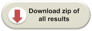
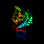
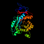
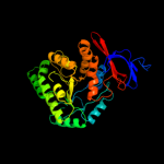
1 c3nqbB_
100.0
20
PDB header: hydrolaseChain: B: PDB Molecule: adenine deaminase 2;PDBTitle: crystal structure of adenine deaminase from agrobacterium tumefaciens2 (str. c 58)
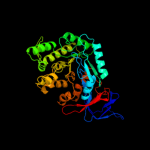
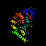
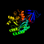
2 c1nfgA_
100.0
18
PDB header: hydrolaseChain: A: PDB Molecule: d-hydantoinase;PDBTitle: structure of d-hydantoinase
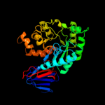
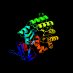
3 c1gkpD_
100.0
17
PDB header: hydrolaseChain: D: PDB Molecule: hydantoinase;PDBTitle: d-hydantoinase (dihydropyrimidinase) from thermus sp. in2 space group c2221
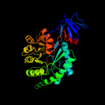
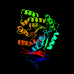
4 c2pajA_
100.0
14
PDB header: hydrolaseChain: A: PDB Molecule: putative cytosine/guanine deaminase;PDBTitle: crystal structure of an amidohydrolase from an environmental sample of2 sargasso sea
5 c3be7B_
100.0
14
PDB header: hydrolaseChain: B: PDB Molecule: zn-dependent arginine carboxypeptidase;PDBTitle: crystal structure of zn-dependent arginine carboxypeptidase
6 c2vr2A_
100.0
19
PDB header: hydrolaseChain: A: PDB Molecule: dihydropyrimidinase;PDBTitle: human dihydropyrimidinase
7 c2gseC_
100.0
16
PDB header: hydrolaseChain: C: PDB Molecule: dihydropyrimidinase-related protein 2;PDBTitle: crystal structure of human dihydropyrimidinease-like 2
8 c2ftwA_
100.0
16
PDB header: hydrolaseChain: A: PDB Molecule: dihydropyrimidine amidohydrolase;PDBTitle: crystal structure of dihydropyrimidinase from dictyostelium discoideum
9 c3hpaB_
100.0
18
PDB header: hydrolaseChain: B: PDB Molecule: amidohydrolase;PDBTitle: crystal structure of an amidohydrolase gi:44264246 from an2 evironmental sample of sargasso sea
10 c2bb0A_
100.0
18
PDB header: hydrolaseChain: A: PDB Molecule: imidazolonepropionase;PDBTitle: structure of imidazolonepropionase from bacillus subtilis
11 c1k1dF_
100.0
18
PDB header: hydrolaseChain: F: PDB Molecule: d-hydantoinase;PDBTitle: crystal structure of d-hydantoinase
12 c3ooqC_
100.0
19
PDB header: hydrolaseChain: C: PDB Molecule: amidohydrolase;PDBTitle: crystal structure of amidohydrolase from thermotoga maritima msb8
13 c3gnhA_
100.0
15
PDB header: hydrolaseChain: A: PDB Molecule: l-lysine, l-arginine carboxypeptidase cc2672;PDBTitle: crystal structure of l-lysine, l-arginine carboxypeptidase cc2672 from2 caulobacter crescentus cb15 complexed with n-methyl phosphonate3 derivative of l-arginine.
14 c3hm7A_
100.0
17
PDB header: hydrolaseChain: A: PDB Molecule: allantoinase;PDBTitle: crystal structure of allantoinase from bacillus halodurans c-125
15 c2fvmA_
100.0
18
PDB header: hydrolaseChain: A: PDB Molecule: dihydropyrimidinase;PDBTitle: crystal structure of dihydropyrimidinase from saccharomyces kluyveri2 in complex with the reaction product n-carbamyl-beta-alanine
16 c3dc8B_
100.0
16
PDB header: hydrolaseChain: B: PDB Molecule: dihydropyrimidinase;PDBTitle: crystal structure of dihydropyrimidinase from sinorhizobium meliloti
17 c1gkrA_
100.0
16
PDB header: hydrolaseChain: A: PDB Molecule: non-atp dependent l-selective hydantoinase;PDBTitle: l-hydantoinase (dihydropyrimidinase) from arthrobacter2 aurescens
18 c3lsbA_
100.0
16
PDB header: hydrolaseChain: A: PDB Molecule: triazine hydrolase;PDBTitle: crystal structure of the mutant e241q of atrazine chlorohydrolase trzn2 from arthrobacter aurescens tc1 complexed with zinc and ametrin
19 c3e74D_
100.0
19
PDB header: hydrolaseChain: D: PDB Molecule: allantoinase;PDBTitle: crystal structure of e. coli allantoinase with iron ions at2 the metal center
20 c2i9uA_
100.0
15
PDB header: hydrolaseChain: A: PDB Molecule: cytosine/guanine deaminase related protein;PDBTitle: crystal structure of guanine deaminase from c. acetobutylicum with2 bound guanine in the active site
21 c3griB_
not modelled
100.0
15
PDB header: hydrolaseChain: B: PDB Molecule: dihydroorotase;PDBTitle: the crystal structure of a dihydroorotase from staphylococcus aureus
22 c2q09A_
not modelled
100.0
15
PDB header: hydrolaseChain: A: PDB Molecule: imidazolonepropionase;PDBTitle: crystal structure of imidazolonepropionase from environmental sample2 with bound inhibitor 3-(2,5-dioxo-imidazolidin-4-yl)-propionic acid
23 c3d6nA_
not modelled
100.0
16
PDB header: hydrolase/transferaseChain: A: PDB Molecule: dihydroorotase;PDBTitle: crystal structure of aquifex dihydroorotase activated by aspartate2 transcarbamoylase
24 c2gwnA_
not modelled
100.0
16
PDB header: structural genomics, unknown functionChain: A: PDB Molecule: dihydroorotase;PDBTitle: the structure of putative dihydroorotase from porphyromonas2 gingivalis.
25 c3lnpA_
not modelled
100.0
15
PDB header: hydrolaseChain: A: PDB Molecule: amidohydrolase family protein olei01672_1_465;PDBTitle: crystal structure of amidohydrolase family protein2 olei01672_1_465 from oleispira antarctica
26 c1p1mA_
not modelled
100.0
18
PDB header: structural genomics, unknown functionChain: A: PDB Molecule: hypothetical protein tm0936;PDBTitle: structure of thermotoga maritima amidohydrolase tm09362 bound to ni and methionine
27 c2gokA_
not modelled
100.0
16
PDB header: hydrolaseChain: A: PDB Molecule: imidazolonepropionase;PDBTitle: crystal structure of the imidazolonepropionase from agrobacterium2 tumefaciens at 1.87 a resolution
28 c1xrfA_
not modelled
100.0
16
PDB header: hydrolaseChain: A: PDB Molecule: dihydroorotase;PDBTitle: the crystal structure of a novel, latent dihydroorotase from aquifex2 aeolicus at 1.7 a resolution
29 c2r8cB_
not modelled
100.0
17
PDB header: structural genomics, unknown functionChain: B: PDB Molecule: putative amidohydrolase;PDBTitle: crystal structure of uncharacterized protein eaj56179
30 c2vhlB_
not modelled
100.0
16
PDB header: hydrolaseChain: B: PDB Molecule: n-acetylglucosamine-6-phosphate deacetylase;PDBTitle: the three-dimensional structure of the n-acetylglucosamine-2 6-phosphate deacetylase from bacillus subtilis
31 c3mpgB_
not modelled
100.0
17
PDB header: hydrolaseChain: B: PDB Molecule: dihydroorotase;PDBTitle: dihydroorotase from bacillus anthracis
32 c2aqoB_
not modelled
100.0
17
PDB header: hydrolaseChain: B: PDB Molecule: isoaspartyl dipeptidase;PDBTitle: crystal structure of e. coli isoaspartyl dipeptidase mutant e77q
33 c2z00A_
not modelled
100.0
17
PDB header: hydrolaseChain: A: PDB Molecule: dihydroorotase;PDBTitle: crystal structure of dihydroorotase from thermus thermophilus
34 c3e0lB_
not modelled
100.0
18
PDB header: hydrolaseChain: B: PDB Molecule: guanine deaminase;PDBTitle: computationally designed ammelide deaminase
35 c2qs8A_
not modelled
100.0
14
PDB header: hydrolaseChain: A: PDB Molecule: xaa-pro dipeptidase;PDBTitle: crystal structure of a xaa-pro dipeptidase with bound2 methionine in the active site
36 c3v7pA_
not modelled
100.0
14
PDB header: hydrolaseChain: A: PDB Molecule: amidohydrolase family protein;PDBTitle: crystal structure of amidohydrolase nis_0429 (target efi-500396) from2 nitratiruptor sp. sb155-2
37 c2p50C_
not modelled
100.0
13
PDB header: hydrolaseChain: C: PDB Molecule: n-acetylglucosamine-6-phosphate deacetylase;PDBTitle: crystal structure of n-acetyl-d-glucosamine-6-phosphate deacetylase2 liganded with zn
38 c3feqB_
not modelled
100.0
17
PDB header: structural genomics, unknown functionChain: B: PDB Molecule: putative amidohydrolase;PDBTitle: crystal structure of uncharacterized protein eah89906
39 c1r9yA_
not modelled
100.0
18
PDB header: hydrolaseChain: A: PDB Molecule: cytosine deaminase;PDBTitle: bacterial cytosine deaminase d314a mutant.
40 c2ubpC_
not modelled
100.0
18
PDB header: hydrolaseChain: C: PDB Molecule: protein (urease alpha subunit);PDBTitle: structure of native urease from bacillus pasteurii
41 c2qt3A_
not modelled
100.0
17
PDB header: hydrolaseChain: A: PDB Molecule: n-isopropylammelide isopropyl amidohydrolase;PDBTitle: crystal structure of n-isopropylammelide isopropylaminohydrolase atzc2 from pseudomonas sp. strain adp complexed with zn
42 c1e9yB_
not modelled
100.0
17
PDB header: hydrolaseChain: B: PDB Molecule: urease subunit beta;PDBTitle: crystal structure of helicobacter pylori urease in complex with2 acetohydroxamic acid
43 c2p9bA_
not modelled
100.0
17
PDB header: hydrolaseChain: A: PDB Molecule: possible prolidase;PDBTitle: crystal structure of putative prolidase from2 bifidobacterium longum
44 c3gipB_
not modelled
100.0
18
PDB header: hydrolaseChain: B: PDB Molecule: n-acyl-d-glutamate deacylase;PDBTitle: crystal structure of n-acyl-d-glutamate deacylase from2 bordetella bronchiseptica complexed with zinc, acetate and3 formate ions.
45 c2oodA_
not modelled
100.0
16
PDB header: hydrolaseChain: A: PDB Molecule: blr3880 protein;PDBTitle: crystal structure of guanine deaminase from bradyrhizobium japonicum
46 c2vunC_
not modelled
100.0
17
PDB header: hydrolaseChain: C: PDB Molecule: enamidase;PDBTitle: the crystal structure of enamidase at 1.9 a resolution - a2 new member of the amidohydrolase superfamily
47 c3egjA_
not modelled
100.0
15
PDB header: hydrolaseChain: A: PDB Molecule: n-acetylglucosamine-6-phosphate deacetylase;PDBTitle: n-acetylglucosamine-6-phosphate deacetylase from vibrio cholerae.
48 c1o12B_
not modelled
100.0
19
PDB header: hydrolaseChain: B: PDB Molecule: n-acetylglucosamine-6-phosphate deacetylase;PDBTitle: crystal structure of n-acetylglucosamine-6-phosphate2 deacetylase (tm0814) from thermotoga maritima at 2.5 a3 resolution
49 c1fwcC_
not modelled
100.0
15
PDB header: hydrolaseChain: C: PDB Molecule: urease;PDBTitle: klebsiella aerogenes urease, c319a variant at ph 8.5
50 c3la4A_
not modelled
100.0
18
PDB header: hydrolaseChain: A: PDB Molecule: urease;PDBTitle: crystal structure of the first plant urease from jack bean (canavalia2 ensiformis)
51 c2icsA_
not modelled
100.0
19
PDB header: hydrolaseChain: A: PDB Molecule: adenine deaminase;PDBTitle: crystal structure of an adenine deaminase
52 c1rjqA_
not modelled
100.0
16
PDB header: hydrolaseChain: A: PDB Molecule: d-aminoacylase;PDBTitle: the crystal structure of the d-aminoacylase mutant d366a
53 c3mduA_
not modelled
100.0
13
PDB header: hydrolaseChain: A: PDB Molecule: n-formimino-l-glutamate iminohydrolase;PDBTitle: the structure of n-formimino-l-glutamate iminohydrolase from2 pseudomonas aeruginosa complexed with n-guanidino-l-glutamate
54 c2ogjB_
not modelled
100.0
19
PDB header: hydrolaseChain: B: PDB Molecule: dihydroorotase;PDBTitle: crystal structure of a dihydroorotase
55 c3etkA_
not modelled
100.0
11
PDB header: hydrolaseChain: A: PDB Molecule: uncharacterized metal-dependent hydrolase;PDBTitle: crystal structure of an uncharacterized metal-dependent2 hydrolase from pyrococcus furiosus
56 c3ighX_
not modelled
100.0
12
PDB header: hydrolaseChain: X: PDB Molecule: uncharacterized metal-dependent hydrolase;PDBTitle: crystal structure of an uncharacterized metal-dependent2 hydrolase from pyrococcus horikoshii ot3
57 c2imrA_
not modelled
100.0
13
PDB header: structural genomics, unknown functionChain: A: PDB Molecule: hypothetical protein dr_0824;PDBTitle: crystal structure of amidohydrolase dr_0824 from2 deinococcus radiodurans
58 d2uz9a2
not modelled
99.8
15
Fold: TIM beta/alpha-barrelSuperfamily: Metallo-dependent hydrolasesFamily: SAH/MTA deaminase-like59 c3msrA_
not modelled
99.8
13
PDB header: hydrolaseChain: A: PDB Molecule: amidohydrolases;PDBTitle: the crystal structure of an amidohydrolase from mycoplasma synoviae
60 d1gkpa2
not modelled
99.7
13
Fold: TIM beta/alpha-barrelSuperfamily: Metallo-dependent hydrolasesFamily: Hydantoinase (dihydropyrimidinase), catalytic domain61 d3be7a2
not modelled
99.7
13
Fold: TIM beta/alpha-barrelSuperfamily: Metallo-dependent hydrolasesFamily: Zn-dependent arginine carboxypeptidase-like62 d2paja2
not modelled
99.7
13
Fold: TIM beta/alpha-barrelSuperfamily: Metallo-dependent hydrolasesFamily: SAH/MTA deaminase-like63 d2i9ua2
not modelled
99.7
15
Fold: TIM beta/alpha-barrelSuperfamily: Metallo-dependent hydrolasesFamily: SAH/MTA deaminase-like64 d4ubpc2
not modelled
99.7
17
Fold: TIM beta/alpha-barrelSuperfamily: Metallo-dependent hydrolasesFamily: alpha-subunit of urease, catalytic domain65 d2r8ca2
not modelled
99.7
16
Fold: TIM beta/alpha-barrelSuperfamily: Metallo-dependent hydrolasesFamily: Zn-dependent arginine carboxypeptidase-like66 d2ooda2
not modelled
99.7
14
Fold: TIM beta/alpha-barrelSuperfamily: Metallo-dependent hydrolasesFamily: SAH/MTA deaminase-like67 d2qs8a2
not modelled
99.7
12
Fold: TIM beta/alpha-barrelSuperfamily: Metallo-dependent hydrolasesFamily: Zn-dependent arginine carboxypeptidase-like68 d2bb0a2
not modelled
99.7
13
Fold: TIM beta/alpha-barrelSuperfamily: Metallo-dependent hydrolasesFamily: Imidazolonepropionase-like69 d1ra0a2
not modelled
99.6
20
Fold: TIM beta/alpha-barrelSuperfamily: Metallo-dependent hydrolasesFamily: Cytosine deaminase catalytic domain70 d1kcxa2
not modelled
99.6
13
Fold: TIM beta/alpha-barrelSuperfamily: Metallo-dependent hydrolasesFamily: Hydantoinase (dihydropyrimidinase), catalytic domain71 d2puza2
not modelled
99.6
19
Fold: TIM beta/alpha-barrelSuperfamily: Metallo-dependent hydrolasesFamily: Imidazolonepropionase-like72 c3pnuA_
not modelled
99.6
12
PDB header: hydrolaseChain: A: PDB Molecule: dihydroorotase;PDBTitle: 2.4 angstrom crystal structure of dihydroorotase (pyrc) from2 campylobacter jejuni.
73 d2ftwa2
not modelled
99.6
13
Fold: TIM beta/alpha-barrelSuperfamily: Metallo-dependent hydrolasesFamily: Hydantoinase (dihydropyrimidinase), catalytic domain74 d2imra2
not modelled
99.6
11
Fold: TIM beta/alpha-barrelSuperfamily: Metallo-dependent hydrolasesFamily: DR0824-like75 d2q09a2
not modelled
99.6
16
Fold: TIM beta/alpha-barrelSuperfamily: Metallo-dependent hydrolasesFamily: Imidazolonepropionase-like76 d2p9ba2
not modelled
99.6
11
Fold: TIM beta/alpha-barrelSuperfamily: Metallo-dependent hydrolasesFamily: Imidazolonepropionase-like77 d1p1ma2
not modelled
99.6
15
Fold: TIM beta/alpha-barrelSuperfamily: Metallo-dependent hydrolasesFamily: SAH/MTA deaminase-like78 d1un7a2
not modelled
99.6
15
Fold: TIM beta/alpha-barrelSuperfamily: Metallo-dependent hydrolasesFamily: N-acetylglucosamine-6-phosphate deacetylase, NagA, catalytic domain79 d1ynya2
not modelled
99.6
13
Fold: TIM beta/alpha-barrelSuperfamily: Metallo-dependent hydrolasesFamily: Hydantoinase (dihydropyrimidinase), catalytic domain80 d1ejxc1
not modelled
99.5
21
Fold: Composite domain of metallo-dependent hydrolasesSuperfamily: Composite domain of metallo-dependent hydrolasesFamily: alpha-Subunit of urease81 c3jzeC_
not modelled
99.5
11
PDB header: hydrolaseChain: C: PDB Molecule: dihydroorotase;PDBTitle: 1.8 angstrom resolution crystal structure of dihydroorotase (pyrc)2 from salmonella enterica subsp. enterica serovar typhimurium str. lt2
82 d2fvka1
not modelled
99.5
26
Fold: Composite domain of metallo-dependent hydrolasesSuperfamily: Composite domain of metallo-dependent hydrolasesFamily: Hydantoinase (dihydropyrimidinase)83 d1nfga2
not modelled
99.5
13
Fold: TIM beta/alpha-barrelSuperfamily: Metallo-dependent hydrolasesFamily: Hydantoinase (dihydropyrimidinase), catalytic domain84 d2eg6a1
not modelled
99.5
10
Fold: TIM beta/alpha-barrelSuperfamily: Metallo-dependent hydrolasesFamily: Dihydroorotase85 d1e9yb1
not modelled
99.5
22
Fold: Composite domain of metallo-dependent hydrolasesSuperfamily: Composite domain of metallo-dependent hydrolasesFamily: alpha-Subunit of urease86 d1xrta2
not modelled
99.5
15
Fold: TIM beta/alpha-barrelSuperfamily: Metallo-dependent hydrolasesFamily: Hydantoinase (dihydropyrimidinase), catalytic domain87 d1yrra2
not modelled
99.5
15
Fold: TIM beta/alpha-barrelSuperfamily: Metallo-dependent hydrolasesFamily: N-acetylglucosamine-6-phosphate deacetylase, NagA, catalytic domain88 d1onwa2
not modelled
99.5
14
Fold: TIM beta/alpha-barrelSuperfamily: Metallo-dependent hydrolasesFamily: Isoaspartyl dipeptidase, catalytic domain89 d1k1da2
not modelled
99.5
14
Fold: TIM beta/alpha-barrelSuperfamily: Metallo-dependent hydrolasesFamily: Hydantoinase (dihydropyrimidinase), catalytic domain90 d1onwa1
not modelled
99.5
19
Fold: Composite domain of metallo-dependent hydrolasesSuperfamily: Composite domain of metallo-dependent hydrolasesFamily: Isoaspartyl dipeptidase91 d1gkra2
not modelled
99.5
16
Fold: TIM beta/alpha-barrelSuperfamily: Metallo-dependent hydrolasesFamily: Hydantoinase (dihydropyrimidinase), catalytic domain92 c3ggmB_
not modelled
99.4
23
PDB header: structural genomics, unknown functionChain: B: PDB Molecule: uncharacterized protein bt9727_2919;PDBTitle: crystal structure of bt9727_2919 from bacillus2 thuringiensis subsp. northeast structural genomics target3 bur228b
93 d1i0da_
not modelled
99.4
15
Fold: TIM beta/alpha-barrelSuperfamily: Metallo-dependent hydrolasesFamily: Phosphotriesterase-like94 d2p9ba1
not modelled
99.4
14
Fold: Composite domain of metallo-dependent hydrolasesSuperfamily: Composite domain of metallo-dependent hydrolasesFamily: Imidazolonepropionase-like95 d1yrra1
not modelled
99.4
17
Fold: Composite domain of metallo-dependent hydrolasesSuperfamily: Composite domain of metallo-dependent hydrolasesFamily: N-acetylglucosamine-6-phosphate deacetylase, NagA96 d2r8ca1
not modelled
99.4
21
Fold: Composite domain of metallo-dependent hydrolasesSuperfamily: Composite domain of metallo-dependent hydrolasesFamily: Zn-dependent arginine carboxypeptidase-like97 d2icsa2
not modelled
99.4
17
Fold: TIM beta/alpha-barrelSuperfamily: Metallo-dependent hydrolasesFamily: Adenine deaminase-like98 d2fvka2
not modelled
99.4
15
Fold: TIM beta/alpha-barrelSuperfamily: Metallo-dependent hydrolasesFamily: Hydantoinase (dihydropyrimidinase), catalytic domain99 d2d2ja1
not modelled
99.3
17
Fold: TIM beta/alpha-barrelSuperfamily: Metallo-dependent hydrolasesFamily: Phosphotriesterase-like100 d1k1da1
not modelled
99.3
21
Fold: Composite domain of metallo-dependent hydrolasesSuperfamily: Composite domain of metallo-dependent hydrolasesFamily: Hydantoinase (dihydropyrimidinase)101 c1pscA_
not modelled
99.2
13
PDB header: hydrolaseChain: A: PDB Molecule: phosphotriesterase;PDBTitle: phosphotriesterase from pseudomonas diminuta
102 d2ftwa1
not modelled
99.2
17
Fold: Composite domain of metallo-dependent hydrolasesSuperfamily: Composite domain of metallo-dependent hydrolasesFamily: Hydantoinase (dihydropyrimidinase)103 d1ynya1
not modelled
99.2
15
Fold: Composite domain of metallo-dependent hydrolasesSuperfamily: Composite domain of metallo-dependent hydrolasesFamily: Hydantoinase (dihydropyrimidinase)104 d1o12a2
not modelled
99.2
14
Fold: TIM beta/alpha-barrelSuperfamily: Metallo-dependent hydrolasesFamily: N-acetylglucosamine-6-phosphate deacetylase, NagA, catalytic domain105 d1gkpa1
not modelled
99.1
15
Fold: Composite domain of metallo-dependent hydrolasesSuperfamily: Composite domain of metallo-dependent hydrolasesFamily: Hydantoinase (dihydropyrimidinase)106 c3pnzD_
not modelled
99.0
14
PDB header: hydrolaseChain: D: PDB Molecule: phosphotriesterase family protein;PDBTitle: crystal structure of the lactonase lmo2620 from listeria monocytogenes
107 c2zc1A_
not modelled
99.0
15
PDB header: hydrolaseChain: A: PDB Molecule: phosphotriesterase;PDBTitle: organophosphorus hydrolase from deinococcus radiodurans
108 c3f4cA_
not modelled
99.0
12
PDB header: hydrolaseChain: A: PDB Molecule: organophosphorus hydrolase;PDBTitle: crystal structure of organophosphorus hydrolase from geobacillus2 stearothermophilus strain 10, with glycerol bound
109 d1kcxa1
not modelled
99.0
11
Fold: Composite domain of metallo-dependent hydrolasesSuperfamily: Composite domain of metallo-dependent hydrolasesFamily: Hydantoinase (dihydropyrimidinase)110 d1bf6a_
not modelled
99.0
11
Fold: TIM beta/alpha-barrelSuperfamily: Metallo-dependent hydrolasesFamily: Phosphotriesterase-like111 c3ou8B_
not modelled
98.9
12
PDB header: hydrolaseChain: B: PDB Molecule: adenosine deaminase;PDBTitle: the crystal structure of adenosine deaminase from pseudomonas2 aeruginosa
112 d1m7ja1
not modelled
98.9
18
Fold: Composite domain of metallo-dependent hydrolasesSuperfamily: Composite domain of metallo-dependent hydrolasesFamily: D-aminoacylase113 c3ou8A_
not modelled
98.8
10
PDB header: hydrolaseChain: A: PDB Molecule: adenosine deaminase;PDBTitle: the crystal structure of adenosine deaminase from pseudomonas2 aeruginosa
114 d1m7ja3
not modelled
98.8
16
Fold: TIM beta/alpha-barrelSuperfamily: Metallo-dependent hydrolasesFamily: D-aminoacylase, catalytic domain115 c2vc7A_
not modelled
98.7
12
PDB header: hydrolaseChain: A: PDB Molecule: aryldialkylphosphatase;PDBTitle: structural basis for natural lactonase and promiscuous2 phosphotriesterase activities
116 d3be7a1
not modelled
98.7
20
Fold: Composite domain of metallo-dependent hydrolasesSuperfamily: Composite domain of metallo-dependent hydrolasesFamily: Zn-dependent arginine carboxypeptidase-like117 d2paja1
not modelled
98.6
15
Fold: Composite domain of metallo-dependent hydrolasesSuperfamily: Composite domain of metallo-dependent hydrolasesFamily: SAH/MTA deaminase-like118 d2bb0a1
not modelled
98.6
20
Fold: Composite domain of metallo-dependent hydrolasesSuperfamily: Composite domain of metallo-dependent hydrolasesFamily: Imidazolonepropionase-like119 d1nfga1
not modelled
98.4
21
Fold: Composite domain of metallo-dependent hydrolasesSuperfamily: Composite domain of metallo-dependent hydrolasesFamily: Hydantoinase (dihydropyrimidinase)120 d1p1ma1
not modelled
98.2
23
Fold: Composite domain of metallo-dependent hydrolasesSuperfamily: Composite domain of metallo-dependent hydrolasesFamily: SAH/MTA deaminase-like