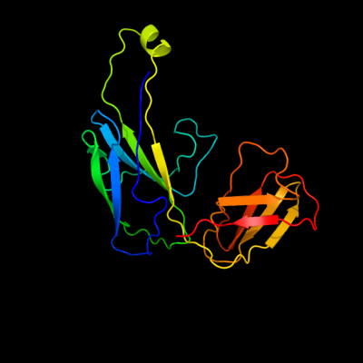
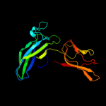
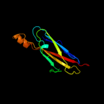
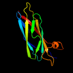
1 c1z9sA_
100.0
16
PDB header: chaperone/immune systemChain: A: PDB Molecule: chaperone protein caf1m;PDBTitle: crystal structure of the native chaperone:subunit:subunit2 caf1m:caf1:caf1 complex
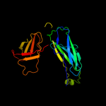
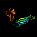
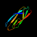
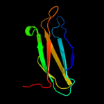
2 c2co7B_
100.0
11
PDB header: fibril proteinChain: B: PDB Molecule: putative fimbriae assembly chaperone;PDBTitle: salmonella enterica safa pilin in complex with the safb2 chaperone (type ii)
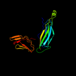
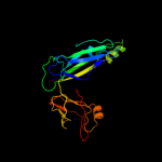
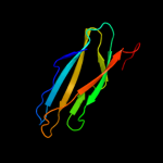
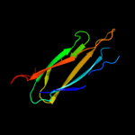
3 c3q48B_
100.0
13
PDB header: chaperoneChain: B: PDB Molecule: chaperone cupb2;PDBTitle: crystal structure of pseudomonas aeruginosa cupb2 chaperone
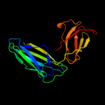
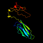
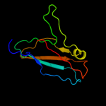
4 c1qunA_
100.0
14
PDB header: chaperone/structural proteinChain: A: PDB Molecule: papd-like chaperone fimc;PDBTitle: x-ray structure of the fimc-fimh chaperone adhesin complex2 from uropathogenic e.coli
5 c1l4iA_
100.0
12
PDB header: chaperoneChain: A: PDB Molecule: sfae protein;PDBTitle: crystal structure of the periplasmic chaperone sfae
6 c1qpxA_
100.0
11
PDB header: chaperoneChain: A: PDB Molecule: papd chaperone;PDBTitle: crystal structures of self-capping papd chaperone homodimers
7 c3f6iB_
99.9
14
PDB header: chaperoneChain: B: PDB Molecule: chaperone protein faee;PDBTitle: structure of the semet labeled f4 fibrial chaperone faee
8 d1p5va1
99.9
20
Fold: Immunoglobulin-like beta-sandwichSuperfamily: PapD-likeFamily: Pilus chaperone9 d2co7b1
99.9
12
Fold: Immunoglobulin-like beta-sandwichSuperfamily: PapD-likeFamily: Pilus chaperone10 d2j2za1
99.9
11
Fold: Immunoglobulin-like beta-sandwichSuperfamily: PapD-likeFamily: Pilus chaperone11 d3bwuc1
99.9
13
Fold: Immunoglobulin-like beta-sandwichSuperfamily: PapD-likeFamily: Pilus chaperone12 d1l4ia1
99.9
10
Fold: Immunoglobulin-like beta-sandwichSuperfamily: PapD-likeFamily: Pilus chaperone13 d1m1sa_
97.9
11
Fold: Immunoglobulin-like beta-sandwichSuperfamily: PapD-likeFamily: MSP-like14 d1rowa_
97.7
12
Fold: Immunoglobulin-like beta-sandwichSuperfamily: PapD-likeFamily: MSP-like15 c2qsvA_
97.6
16
PDB header: structural genomics, unknown functionChain: A: PDB Molecule: uncharacterized protein;PDBTitle: crystal structure of protein of unknown function from porphyromonas2 gingivalis w83
16 d1grwa_
97.2
14
Fold: Immunoglobulin-like beta-sandwichSuperfamily: PapD-likeFamily: MSP-like17 d3bwuc2
96.7
14
Fold: C2 domain-likeSuperfamily: Periplasmic chaperone C-domainFamily: Periplasmic chaperone C-domain18 c1z9oB_
96.6
13
PDB header: protein binding/lipid binding proteinChain: B: PDB Molecule: vesicle-associated membrane protein-associated protein a;PDBTitle: 1.9 angstrom crystal structure of the rat vap-a msp homology domain in2 complex with the rat orp1 ffat motif
19 d1mspa_
95.9
15
Fold: Immunoglobulin-like beta-sandwichSuperfamily: PapD-likeFamily: MSP-like20 c2e6jA_
95.5
10
PDB header: structural genomics, unknown functionChain: A: PDB Molecule: hydin protein;PDBTitle: solution structure of the c-terminal papd-like domain from2 human hydin protein
21 c2ys4A_
not modelled
95.3
13
PDB header: structural genomics, unknown functionChain: A: PDB Molecule: hydrocephalus-inducing protein homolog;PDBTitle: solution structure of the n-terminal papd-like domain of2 hydin protein from human
22 d1wica_
not modelled
94.3
16
Fold: Immunoglobulin-like beta-sandwichSuperfamily: PapD-likeFamily: MSP-like23 d1w8oa1
not modelled
94.2
14
Fold: Immunoglobulin-like beta-sandwichSuperfamily: E set domainsFamily: E-set domains of sugar-utilizing enzymes24 d2j2za2
not modelled
94.0
12
Fold: C2 domain-likeSuperfamily: Periplasmic chaperone C-domainFamily: Periplasmic chaperone C-domain25 c3o0lB_
not modelled
94.0
9
PDB header: structural genomics, unknown functionChain: B: PDB Molecule: uncharacterized protein;PDBTitle: crystal structure of a pfam duf1425 family member (shew_1734) from2 shewanella sp. pv-4 at 1.81 a resolution
26 d1p5va2
not modelled
94.0
10
Fold: C2 domain-likeSuperfamily: Periplasmic chaperone C-domainFamily: Periplasmic chaperone C-domain27 d2co7b2
not modelled
93.4
8
Fold: C2 domain-likeSuperfamily: Periplasmic chaperone C-domainFamily: Periplasmic chaperone C-domain28 c3qbtH_
not modelled
91.7
17
PDB header: protein transport/hydrolaseChain: H: PDB Molecule: inositol polyphosphate 5-phosphatase ocrl-1;PDBTitle: crystal structure of ocrl1 540-678 in complex with rab8a:gppnhp
29 d1l4ia2
not modelled
88.4
14
Fold: C2 domain-likeSuperfamily: Periplasmic chaperone C-domainFamily: Periplasmic chaperone C-domain30 c3qisA_
not modelled
88.3
17
PDB header: hydrolase/protein bindingChain: A: PDB Molecule: inositol polyphosphate 5-phosphatase ocrl-1;PDBTitle: recognition of the f&h motif by the lowe syndrome protein ocrl
31 c2jwyA_
not modelled
84.7
17
PDB header: lipoproteinChain: A: PDB Molecule: uncharacterized lipoprotein yaji;PDBTitle: solution nmr structure of uncharacterized lipoprotein yaji from2 escherichia coli. northeast structural genomics target er540
32 c3ac0B_
not modelled
80.8
14
PDB header: hydrolaseChain: B: PDB Molecule: beta-glucosidase i;PDBTitle: crystal structure of beta-glucosidase from kluyveromyces marxianus in2 complex with glucose
33 d2vzsa2
not modelled
75.7
11
Fold: Immunoglobulin-like beta-sandwichSuperfamily: beta-Galactosidase/glucuronidase domainFamily: beta-Galactosidase/glucuronidase domain34 c2x41A_
not modelled
71.2
13
PDB header: hydrolaseChain: A: PDB Molecule: beta-glucosidase;PDBTitle: structure of beta-glucosidase 3b from thermotoga neapolitana2 in complex with glucose
35 c1l9mB_
not modelled
65.9
13
PDB header: transferaseChain: B: PDB Molecule: protein-glutamine glutamyltransferase e3;PDBTitle: three-dimensional structure of the human transglutaminase 32 enzyme: binding of calcium ions change structure for3 activation
36 c3isyA_
not modelled
59.2
10
PDB header: protein bindingChain: A: PDB Molecule: intracellular proteinase inhibitor;PDBTitle: crystal structure of an intracellular proteinase inhibitor (ipi,2 bsu11130) from bacillus subtilis at 2.61 a resolution
37 c2r39A_
not modelled
51.3
12
PDB header: structural genomics, unknown functionChain: A: PDB Molecule: fixg-related protein;PDBTitle: crystal structure of fixg-related protein from vibrio parahaemolyticus
38 c3cfuA_
not modelled
47.2
16
PDB header: lipoproteinChain: A: PDB Molecule: uncharacterized lipoprotein yjha;PDBTitle: crystal structure of the yjha protein from bacillus2 subtilis. northeast structural genomics consortium target3 sr562
39 d1exha_
not modelled
47.1
14
Fold: Common fold of diphtheria toxin/transcription factors/cytochrome fSuperfamily: Carbohydrate-binding domainFamily: Cellulose-binding domain family II40 d1yq2a1
not modelled
45.7
7
Fold: Immunoglobulin-like beta-sandwichSuperfamily: beta-Galactosidase/glucuronidase domainFamily: beta-Galactosidase/glucuronidase domain41 d1ufga_
not modelled
45.7
14
Fold: Immunoglobulin-like beta-sandwichSuperfamily: Lamin A/C globular tail domainFamily: Lamin A/C globular tail domain42 c2lllA_
not modelled
44.8
9
PDB header: structural proteinChain: A: PDB Molecule: lamin-b2;PDBTitle: solution nmr structure of c-terminal globular domain of human lamin-2 b2, northeast structural genomics consortium target hr8546a
43 d4ubpb_
not modelled
44.1
11
Fold: beta-clipSuperfamily: Urease, beta-subunitFamily: Urease, beta-subunit44 d1e9ya1
not modelled
43.6
15
Fold: beta-clipSuperfamily: Urease, beta-subunitFamily: Urease, beta-subunit45 c3qgaD_
not modelled
38.0
11
PDB header: hydrolaseChain: D: PDB Molecule: fusion of urease beta and gamma subunits;PDBTitle: 3.0 a model of iron containing urease urea2b2 from helicobacter2 mustelae
46 c3jt0B_
not modelled
37.8
13
PDB header: structural proteinChain: B: PDB Molecule: lamin-b1;PDBTitle: crystal structure of the c-terminal fragment (426-558)2 lamin-b1 from homo sapiens, northeast structural genomics3 consortium target hr5546a
47 c2kl6A_
not modelled
37.1
15
PDB header: structural genomics, unknown functionChain: A: PDB Molecule: uncharacterized protein;PDBTitle: solution nmr structure of the cardb domain of pf1109 from2 pyrococcus furiosus. northeast structural genomics3 consortium target pfr193a
48 d1ejxb_
not modelled
32.1
17
Fold: beta-clipSuperfamily: Urease, beta-subunitFamily: Urease, beta-subunit49 d1ifra_
not modelled
31.9
12
Fold: Immunoglobulin-like beta-sandwichSuperfamily: Lamin A/C globular tail domainFamily: Lamin A/C globular tail domain50 c1yewI_
not modelled
26.9
17
PDB header: oxidoreductase, membrane proteinChain: I: PDB Molecule: particulate methane monooxygenase, b subunit;PDBTitle: crystal structure of particulate methane monooxygenase
51 d1hmja_
not modelled
26.7
26
Fold: RPB5-like RNA polymerase subunitSuperfamily: RPB5-like RNA polymerase subunitFamily: RPB552 c3rgbA_
not modelled
26.0
17
PDB header: oxidoreductaseChain: A: PDB Molecule: methane monooxygenase subunit b2;PDBTitle: crystal structure of particulate methane monooxygenase from2 methylococcus capsulatus (bath)
53 d2uubs1
not modelled
23.7
29
Fold: Ribosomal protein S19Superfamily: Ribosomal protein S19Family: Ribosomal protein S1954 d2gy9s1
not modelled
23.3
29
Fold: Ribosomal protein S19Superfamily: Ribosomal protein S19Family: Ribosomal protein S1955 d1ivta_
not modelled
20.2
11
Fold: Immunoglobulin-like beta-sandwichSuperfamily: Lamin A/C globular tail domainFamily: Lamin A/C globular tail domain56 d1vjja2
not modelled
19.4
12
Fold: Immunoglobulin-like beta-sandwichSuperfamily: Transglutaminase, two C-terminal domainsFamily: Transglutaminase, two C-terminal domains57 c3bbnS_
not modelled
18.4
23
PDB header: ribosomeChain: S: PDB Molecule: ribosomal protein s19;PDBTitle: homology model for the spinach chloroplast 30s subunit2 fitted to 9.4a cryo-em map of the 70s chlororibosome.
58 d1kyaa2
not modelled
18.3
11
Fold: Cupredoxin-likeSuperfamily: CupredoxinsFamily: Multidomain cupredoxins59 c1s1hS_
not modelled
17.8
29
PDB header: ribosomeChain: S: PDB Molecule: 40s ribosomal protein s15;PDBTitle: structure of the ribosomal 80s-eef2-sordarin complex from2 yeast obtained by docking atomic models for rna and protein3 components into a 11.7 a cryo-em map. this file, 1s1h,4 contains 40s subunit. the 60s ribosomal subunit is in file5 1s1i.
60 d2cj3a1
not modelled
16.0
18
Fold: Cupredoxin-likeSuperfamily: CupredoxinsFamily: Plastocyanin/azurin-like61 c3rfrI_
not modelled
15.6
15
PDB header: oxidoreductaseChain: I: PDB Molecule: pmob;PDBTitle: crystal structure of particulate methane monooxygenase (pmmo) from2 methylocystis sp. strain m
62 c2zkqs_
not modelled
15.4
29
PDB header: ribosomal protein/rnaChain: S: PDB Molecule: PDBTitle: structure of a mammalian ribosomal 40s subunit within an2 80s complex obtained by docking homology models of the rna3 and proteins into an 8.7 a cryo-em map
63 c3ndyG_
not modelled
15.4
17
PDB header: hydrolaseChain: G: PDB Molecule: endoglucanase d;PDBTitle: the structure of the catalytic and carbohydrate binding domain of2 endoglucanase d from clostridium cellulovorans
64 c2xzmS_
not modelled
14.5
29
PDB header: ribosomeChain: S: PDB Molecule: rps15e;PDBTitle: crystal structure of the eukaryotic 40s ribosomal2 subunit in complex with initiation factor 1. this file3 contains the 40s subunit and initiation factor for4 molecule 1
65 c1e9zA_
not modelled
14.3
25
PDB header: hydrolaseChain: A: PDB Molecule: urease subunit alpha;PDBTitle: crystal structure of helicobacter pylori urease
66 c2je8B_
not modelled
12.9
10
PDB header: hydrolaseChain: B: PDB Molecule: beta-mannosidase;PDBTitle: structure of a beta-mannosidase from bacteroides2 thetaiotaomicron
67 d1plaa_
not modelled
12.7
25
Fold: Cupredoxin-likeSuperfamily: CupredoxinsFamily: Plastocyanin/azurin-like68 d1eika_
not modelled
12.6
24
Fold: RPB5-like RNA polymerase subunitSuperfamily: RPB5-like RNA polymerase subunitFamily: RPB569 d1bxua_
not modelled
12.1
11
Fold: Cupredoxin-likeSuperfamily: CupredoxinsFamily: Plastocyanin/azurin-like70 d2q5ba1
not modelled
12.0
11
Fold: Cupredoxin-likeSuperfamily: CupredoxinsFamily: Plastocyanin/azurin-like71 d1pcsa_
not modelled
11.4
14
Fold: Cupredoxin-likeSuperfamily: CupredoxinsFamily: Plastocyanin/azurin-like72 c3mdjB_
not modelled
11.3
10
PDB header: hydrolase/hydrolase inhibitorChain: B: PDB Molecule: endoplasmic reticulum aminopeptidase 1;PDBTitle: er aminopeptidase, erap1, bound to the zinc aminopeptidase inhibitor,2 bestatin
73 d1gyca2
not modelled
11.1
19
Fold: Cupredoxin-likeSuperfamily: CupredoxinsFamily: Multidomain cupredoxins74 d1bqka_
not modelled
10.9
22
Fold: Cupredoxin-likeSuperfamily: CupredoxinsFamily: Plastocyanin/azurin-like75 d9pcya_
not modelled
10.8
21
Fold: Cupredoxin-likeSuperfamily: CupredoxinsFamily: Plastocyanin/azurin-like76 c2f1eA_
not modelled
10.0
12
PDB header: structural genomics, unknown functionChain: A: PDB Molecule: protein apag;PDBTitle: solution structure of apag protein
77 d1dzfa2
not modelled
9.7
25
Fold: RPB5-like RNA polymerase subunitSuperfamily: RPB5-like RNA polymerase subunitFamily: RPB578 d1ag6a_
not modelled
9.5
18
Fold: Cupredoxin-likeSuperfamily: CupredoxinsFamily: Plastocyanin/azurin-like79 d1g0da2
not modelled
9.4
9
Fold: Immunoglobulin-like beta-sandwichSuperfamily: Transglutaminase, two C-terminal domainsFamily: Transglutaminase, two C-terminal domains80 d1hfua2
not modelled
9.3
5
Fold: Cupredoxin-likeSuperfamily: CupredoxinsFamily: Multidomain cupredoxins81 d1plca_
not modelled
9.1
18
Fold: Cupredoxin-likeSuperfamily: CupredoxinsFamily: Plastocyanin/azurin-like82 c2kutA_
not modelled
9.0
7
PDB header: structural genomics, unknown functionChain: A: PDB Molecule: uncharacterized protein;PDBTitle: solution structure of gmr58a from geobacter metallireducens.2 northeast structural genomics consortium target gmr58a
83 d1aoza2
not modelled
8.8
10
Fold: Cupredoxin-likeSuperfamily: CupredoxinsFamily: Multidomain cupredoxins84 d1hc1a3
not modelled
8.7
19
Fold: Immunoglobulin-like beta-sandwichSuperfamily: E set domainsFamily: Arthropod hemocyanin, C-terminal domain85 d1v7wa2
not modelled
8.6
16
Fold: SupersandwichSuperfamily: Galactose mutarotase-likeFamily: Glycosyltransferase family 36 N-terminal domain86 d7pcya_
not modelled
8.3
21
Fold: Cupredoxin-likeSuperfamily: CupredoxinsFamily: Plastocyanin/azurin-like87 c2pmzV_
not modelled
8.1
25
PDB header: translation, transferaseChain: V: PDB Molecule: dna-directed rna polymerase subunit h;PDBTitle: archaeal rna polymerase from sulfolobus solfataricus
88 c1pzdA_
not modelled
7.6
15
PDB header: endocytosis/exocytosisChain: A: PDB Molecule: coatomer gamma subunit;PDBTitle: structural identification of a conserved appendage domain2 in the carboxyl-terminus of the copi gamma-subunit.
89 d3pmga4
not modelled
7.6
11
Fold: TBP-likeSuperfamily: Phosphoglucomutase, C-terminal domainFamily: Phosphoglucomutase, C-terminal domain90 d2jxma1
not modelled
7.4
18
Fold: Cupredoxin-likeSuperfamily: CupredoxinsFamily: Plastocyanin/azurin-like91 c3qnfA_
not modelled
7.4
12
PDB header: hydrolaseChain: A: PDB Molecule: endoplasmic reticulum aminopeptidase 1;PDBTitle: crystal structure of the open state of human endoplasmic reticulum2 aminopeptidase 1 erap1
92 d1ex0a2
not modelled
7.2
10
Fold: Immunoglobulin-like beta-sandwichSuperfamily: Transglutaminase, two C-terminal domainsFamily: Transglutaminase, two C-terminal domains93 d1v10a2
not modelled
6.9
10
Fold: Cupredoxin-likeSuperfamily: CupredoxinsFamily: Multidomain cupredoxins94 d1pmya_
not modelled
6.6
19
Fold: Cupredoxin-likeSuperfamily: CupredoxinsFamily: Plastocyanin/azurin-like95 d1adwa_
not modelled
6.6
18
Fold: Cupredoxin-likeSuperfamily: CupredoxinsFamily: Plastocyanin/azurin-like96 d1bypa_
not modelled
6.5
21
Fold: Cupredoxin-likeSuperfamily: CupredoxinsFamily: Plastocyanin/azurin-like97 d2plta_
not modelled
6.5
18
Fold: Cupredoxin-likeSuperfamily: CupredoxinsFamily: Plastocyanin/azurin-like98 d1nc7a_
not modelled
5.9
21
Fold: Hypothetical protein TM1070Superfamily: Hypothetical protein TM1070Family: Hypothetical protein TM107099 c3izbR_
not modelled
5.6
31
PDB header: ribosomeChain: R: PDB Molecule: 40s ribosomal protein rps15 (s19p);PDBTitle: localization of the small subunit ribosomal proteins into a 6.1 a2 cryo-em map of saccharomyces cerevisiae translating 80s ribosome