| Secondary structure and disorder prediction | |
| | |
1 | . | . | . | . | . | . | . | . | 10 | . | . | . | . | . | . | . | . | . | 20 | . | . | . | . | . | . | . | . | . | 30 | . | . | . | . | . | . | . | . | . | 40 | . | . | . | . | . | . | . | . | . | 50 | . | . | . | . | . | . | . | . | . | 60 |
| Sequence | |
M | T | T | Q | V | R | K | N | V | M | D | M | F | I | D | G | A | R | R | G | F | T | I | A | T | T | N | L | L | P | N | V | V | M | A | F | V | I | I | Q | A | L | K | I | T | G | L | L | D | W | V | G | H | I | C | E | P | V | M | A |
| Secondary structure | |
|
|
|
|
|
|
|
|  | | | | | | | | | | | | | | | | | | | | | | | | | | | | | | | | | | | |
|
| | | | | | | | | | | | | | |
| SS confidence | |
|
|
|
|
|
|
|
|
|
|
|
|
|
|
|
|
|
|
|
|
|
|
|
|
|
|
|
|
|
|
|
|
|
|
|
|
|
|
|
|
|
|
|
|
|
|
|
|
|
|
|
|
|
|
|
|
|
|
|
|
| Disorder | |
? | ? | ? | ? | ? | ? | ? |
|
|
|
|
|
|
|
|
|
|
|
|
|
|
|
|
|
|
|
|
|
|
|
|
|
|
|
|
|
|
|
|
|
|
|
|
|
|
|
|
|
|
|
|
|
|
|
|
|
|
|
|
|
| Disorder confidence | |
|
|
|
|
|
|
|
|
|
|
|
|
|
|
|
|
|
|
|
|
|
|
|
|
|
|
|
|
|
|
|
|
|
|
|
|
|
|
|
|
|
|
|
|
|
|
|
|
|
|
|
|
|
|
|
|
|
|
|
|
| |
| | |
. | . | . | . | . | . | . | . | . | 70 | . | . | . | . | . | . | . | . | . | 80 | . | . | . | . | . | . | . | . | . | 90 | . | . | . | . | . | . | . | . | . | 100 | . | . | . | . | . | . | . | . | . | 110 | . | . | . | . | . | . | . | . | . | 120 |
| Sequence | |
L | W | G | L | P | G | E | A | A | T | V | L | L | A | A | L | M | S | M | G | G | A | V | G | V | A | A | S | L | A | T | A | G | A | L | T | G | H | D | V | T | V | L | L | P | A | M | Y | L | M | G | N | P | V | Q | N | V | G | R | C |
| Secondary structure | |
| |
|
|
| | | | | | | | | | | |
|
|
| | | | | | | | | | | | |
|
|
|
|
| | | | | | | | | | | | | | | | | | | | | | | | |
| SS confidence | |
|
|
|
|
|
|
|
|
|
|
|
|
|
|
|
|
|
|
|
|
|
|
|
|
|
|
|
|
|
|
|
|
|
|
|
|
|
|
|
|
|
|
|
|
|
|
|
|
|
|
|
|
|
|
|
|
|
|
|
|
| Disorder | |
|
|
|
|
|
|
|
|
|
|
|
|
|
|
|
| ? | ? |
|
| ? |
|
|
|
|
|
|
|
|
|
|
| ? |
|
|
|
|
|
|
|
|
|
|
|
|
|
|
|
|
|
|
|
|
|
|
|
|
|
|
|
| Disorder confidence | |
|
|
|
|
|
|
|
|
|
|
|
|
|
|
|
|
|
|
|
|
|
|
|
|
|
|
|
|
|
|
|
|
|
|
|
|
|
|
|
|
|
|
|
|
|
|
|
|
|
|
|
|
|
|
|
|
|
|
|
|
| |
| | |
. | . | . | . | . | . | . | . | . | 130 | . | . | . | . | . | . | . | . | . | 140 | . | . | . | . | . | . | . | . | . | 150 | . | . | . |
| Sequence | |
L | G | T | A | E | V | N | A | K | Y | Y | P | H | I | I | T | V | C | V | I | N | A | L | L | S | I | W | V | M | Q | L | I | V |
| Secondary structure | |
| | | | |
|
|
| | | | | | | | | | | | | | | | | | | | | | | | |
|
| SS confidence | |
|
|
|
|
|
|
|
|
|
|
|
|
|
|
|
|
|
|
|
|
|
|
|
|
|
|
|
|
|
|
|
|
|
| Disorder | |
|
|
|
|
|
|
| ? |
|
|
|
|
|
|
|
|
|
|
|
|
|
|
|
|
|
|
|
|
|
|
| ? | ? |
| Disorder confidence | |
|
|
|
|
|
|
|
|
|
|
|
|
|
|
|
|
|
|
|
|
|
|
|
|
|
|
|
|
|
|
|
|
|
| |
| Confidence Key |
| High(9) | |
|
|
|
|
|
|
|
|
|
Low (0) |
| ? | Disordered |
| Alpha helix |
  | Beta strand |
Hover over an aligned region to see model and summary info
Please note, only up to the top 20 hits are modelled to reduce computer load
|
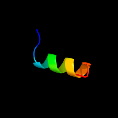
| 1 |
|
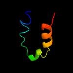
PDB 3bux chain B domain 3
Region: 6 - 21
Aligned: 16
Modelled: 16
Confidence: 65.0%
Identity: 44%
Fold: SH2-like
Superfamily: SH2 domain
Family: SH2 domain
Phyre2
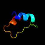
| 2 |
|
PDB 1fbv chain A
Region: 6 - 21
Aligned: 16
Modelled: 16
Confidence: 19.9%
Identity: 44%
PDB header:ligase
Chain: A: PDB Molecule:signal transduction protein cbl;
PDBTitle: structure of a cbl-ubch7 complex: ring domain function in2 ubiquitin-protein ligases
Phyre2
| 3 |
|
PDB 2zif chain B
Region: 52 - 88
Aligned: 33
Modelled: 37
Confidence: 7.0%
Identity: 15%
PDB header:transferase
Chain: B: PDB Molecule:putative modification methylase;
PDBTitle: crystal structure of ttha0409, putative dna modification2 methylase from thermus thermophilus hb8- complexed with s-3 adenosyl-l-methionine
Phyre2
| 4 |
|
PDB 2xpn chain A
Region: 25 - 58
Aligned: 33
Modelled: 34
Confidence: 6.8%
Identity: 18%
PDB header:transcription
Chain: A: PDB Molecule:iws1;
PDBTitle: crystal structure of a spt6-iws1(spn1) complex from2 encephalitozoon cuniculi, form i
Phyre2
| 5 |
|
PDB 1nw6 chain A
Region: 52 - 88
Aligned: 33
Modelled: 37
Confidence: 5.7%
Identity: 15%
PDB header:transferase
Chain: A: PDB Molecule:modification methylase rsri;
PDBTitle: structure of the beta class n6-adenine dna methyltransferase rsri2 bound to sinefungin
Phyre2
|
| Detailed template information | |
Due to computational demand, binding site predictions are not run for batch jobs
If you want to predict binding sites, please manually submit your model of choice to 3DLigandSite
Phyre is for academic use only
| Please cite: Protein structure prediction on
the web: a case study using the Phyre server |
| Kelley LA and Sternberg MJE. Nature Protocols
4, 363 - 371 (2009) [pdf] [Import into BibTeX] |
| |
| If you use the binding site
predictions from 3DLigandSite, please also cite: |
| 3DLigandSite: predicting ligand-binding sites using similar structures. |
| Wass MN, Kelley LA and Sternberg
MJ Nucleic Acids Research 38, W469-73 (2010) [PubMed] |
| |
|
|
|
|