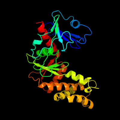
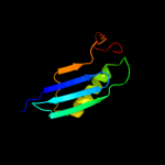
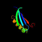
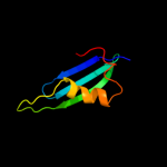
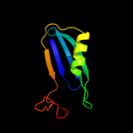
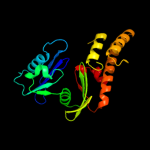
| 1 | c3nuwC_
|
|
 |
100.0 |
80 |
PDB header:transferase
Chain: C: PDB Molecule:2-oxo-3-deoxygalactonate kinase;
PDBTitle: crystal structure of 2-oxo-3-deoxygalactonate kinase from klebsiella2 pneumoniae
|
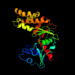
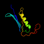
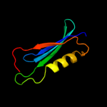
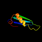
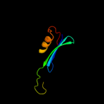
| 2 | c3t69A_
|
|
 |
100.0 |
34 |
PDB header:transferase
Chain: A: PDB Molecule:putative 2-dehydro-3-deoxygalactonokinase;
PDBTitle: crystal structure of a putative 2-dehydro-3-deoxygalactonokinase2 protein from sinorhizobium meliloti
|
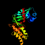
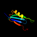
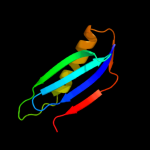
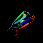
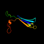
| 3 | c3gg4B_
|
|
 |
96.1 |
12 |
PDB header:transferase
Chain: B: PDB Molecule:glycerol kinase;
PDBTitle: the crystal structure of glycerol kinase from yersinia2 pseudotuberculosis
|
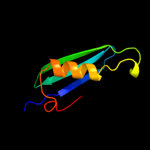
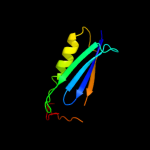
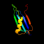
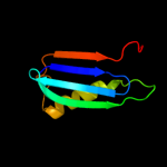
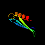
| 4 | c3i8bA_
|
|
 |
95.9 |
7 |
PDB header:transferase
Chain: A: PDB Molecule:xylulose kinase;
PDBTitle: the crystal structure of xylulose kinase from2 bifidobacterium adolescentis
|
| 5 | c3hz6A_
|
|
 |
95.8 |
14 |
PDB header:transferase
Chain: A: PDB Molecule:xylulokinase;
PDBTitle: crystal structure of xylulokinase from chromobacterium violaceum
|
| 6 | c2zf5O_
|
|
 |
95.6 |
15 |
PDB header:transferase
Chain: O: PDB Molecule:glycerol kinase;
PDBTitle: crystal structure of highly thermostable glycerol kinase from a2 hyperthermophilic archaeon
|
| 7 | c3gbtA_
|
|
 |
95.3 |
15 |
PDB header:transferase
Chain: A: PDB Molecule:gluconate kinase;
PDBTitle: crystal structure of gluconate kinase from lactobacillus acidophilus
|
| 8 | d2ch5a2
|
|
 |
95.2 |
14 |
Fold:Ribonuclease H-like motif
Superfamily:Actin-like ATPase domain
Family:BadF/BadG/BcrA/BcrD-like |
| 9 | c3ezwD_
|
|
 |
94.6 |
18 |
PDB header:transferase
Chain: D: PDB Molecule:glycerol kinase;
PDBTitle: crystal structure of a hyperactive escherichia coli glycerol kinase2 mutant gly230 --> asp obtained using microfluidic crystallization3 devices
|
| 10 | c2w40C_
|
|
 |
94.6 |
14 |
PDB header:transferase
Chain: C: PDB Molecule:glycerol kinase, putative;
PDBTitle: crystal structure of plasmodium falciparum glycerol kinase2 with bound glycerol
|
| 11 | d2p3ra1
|
|
 |
94.6 |
16 |
Fold:Ribonuclease H-like motif
Superfamily:Actin-like ATPase domain
Family:Glycerol kinase |
| 12 | c3g25B_
|
|
 |
94.3 |
25 |
PDB header:transferase
Chain: B: PDB Molecule:glycerol kinase;
PDBTitle: 1.9 angstrom crystal structure of glycerol kinase (glpk) from2 staphylococcus aureus in complex with glycerol.
|
| 13 | c3flcX_
|
|
 |
94.1 |
22 |
PDB header:transferase
Chain: X: PDB Molecule:glycerol kinase;
PDBTitle: crystal structure of the his-tagged h232r mutant of glycerol kinase2 from enterococcus casseliflavus with glycerol
|
| 14 | c1glbG_
|
|
 |
94.0 |
17 |
PDB header:phosphotransferase
Chain: G: PDB Molecule:glycerol kinase;
PDBTitle: structure of the regulatory complex of escherichia coli iiiglc with2 glycerol kinase
|
| 15 | c2d4wA_
|
|
 |
93.4 |
17 |
PDB header:transferase
Chain: A: PDB Molecule:glycerol kinase;
PDBTitle: crystal structure of glycerol kinase from cellulomonas sp.2 nt3060
|
| 16 | c2nlxA_
|
|
 |
93.0 |
15 |
PDB header:transferase
Chain: A: PDB Molecule:xylulose kinase;
PDBTitle: crystal structure of the apo e. coli xylulose kinase
|
| 17 | c2dpnB_
|
|
 |
92.0 |
23 |
PDB header:transferase
Chain: B: PDB Molecule:glycerol kinase;
PDBTitle: crystal structure of the glycerol kinase from thermus2 thermophilus hb8
|
| 18 | c3ifrB_
|
|
 |
91.1 |
18 |
PDB header:transferase
Chain: B: PDB Molecule:carbohydrate kinase, fggy;
PDBTitle: the crystal structure of xylulose kinase from rhodospirillum rubrum
|
| 19 | d1zc6a1
|
|
 |
90.8 |
15 |
Fold:Ribonuclease H-like motif
Superfamily:Actin-like ATPase domain
Family:BadF/BadG/BcrA/BcrD-like |
| 20 | d1huxa_
|
|
 |
90.1 |
16 |
Fold:Ribonuclease H-like motif
Superfamily:Actin-like ATPase domain
Family:BadF/BadG/BcrA/BcrD-like |
| 21 | c3h6eB_ |
|
not modelled |
89.8 |
23 |
PDB header:transferase
Chain: B: PDB Molecule:carbohydrate kinase, fggy;
PDBTitle: the crystal structure of a carbohydrate kinase from novosphingobium2 aromaticivorans
|
| 22 | d1r59o1 |
|
not modelled |
89.1 |
24 |
Fold:Ribonuclease H-like motif
Superfamily:Actin-like ATPase domain
Family:Glycerol kinase |
| 23 | c1zc6A_ |
|
not modelled |
89.0 |
17 |
PDB header:structural genomics, unknown function
Chain: A: PDB Molecule:probable n-acetylglucosamine kinase;
PDBTitle: crystal structure of putative n-acetylglucosamine kinase from2 chromobacterium violaceum. northeast structural genomics target3 cvr23.
|
| 24 | c1xupO_ |
|
not modelled |
88.7 |
22 |
PDB header:transferase
Chain: O: PDB Molecule:glycerol kinase;
PDBTitle: enterococcus casseliflavus glycerol kinase complexed with glycerol
|
| 25 | c3bf1C_ |
|
not modelled |
88.5 |
13 |
PDB header:transferase
Chain: C: PDB Molecule:type iii pantothenate kinase;
PDBTitle: type iii pantothenate kinase from thermotoga maritima2 complexed with pantothenate and adp
|
| 26 | c2cgkB_ |
|
not modelled |
87.3 |
13 |
PDB header:transferase
Chain: B: PDB Molecule:l-rhamnulose kinase;
PDBTitle: crystal structure of l-rhamnulose kinase from escherichia2 coli in an open uncomplexed conformation.
|
| 27 | c3jvpA_ |
|
not modelled |
86.5 |
26 |
PDB header:transferase
Chain: A: PDB Molecule:ribulokinase;
PDBTitle: crystal structure of ribulokinase from bacillus halodurans
|
| 28 | c1zxoB_ |
|
not modelled |
80.5 |
14 |
PDB header:unknown function
Chain: B: PDB Molecule:conserved hypothetical protein q8a1p1;
PDBTitle: x-ray crystal structure of protein q8a1p1 from bacteroides2 thetaiotaomicron. northeast structural genomics consortium3 target btr25.
|
| 29 | d1q18a1 |
|
not modelled |
78.7 |
22 |
Fold:Ribonuclease H-like motif
Superfamily:Actin-like ATPase domain
Family:Glucokinase |
| 30 | d1bdga1 |
|
not modelled |
78.6 |
15 |
Fold:Ribonuclease H-like motif
Superfamily:Actin-like ATPase domain
Family:Hexokinase |
| 31 | d1bg3a3 |
|
not modelled |
78.3 |
17 |
Fold:Ribonuclease H-like motif
Superfamily:Actin-like ATPase domain
Family:Hexokinase |
| 32 | c2v7yA_ |
|
not modelled |
75.3 |
33 |
PDB header:chaperone
Chain: A: PDB Molecule:chaperone protein dnak;
PDBTitle: crystal structure of the molecular chaperone dnak from2 geobacillus kaustophilus hta426 in post-atp hydrolysis3 state
|
| 33 | c3hm8D_ |
|
not modelled |
74.6 |
17 |
PDB header:transferase
Chain: D: PDB Molecule:hexokinase-3;
PDBTitle: crystal structure of the c-terminal hexokinase domain of human hk3
|
| 34 | d2ap1a2 |
|
not modelled |
73.1 |
9 |
Fold:Ribonuclease H-like motif
Superfamily:Actin-like ATPase domain
Family:ROK |
| 35 | d1ig8a1 |
|
not modelled |
69.7 |
31 |
Fold:Ribonuclease H-like motif
Superfamily:Actin-like ATPase domain
Family:Hexokinase |
| 36 | c2qm1D_ |
|
not modelled |
69.5 |
19 |
PDB header:transferase
Chain: D: PDB Molecule:glucokinase;
PDBTitle: crystal structure of glucokinase from enterococcus faecalis
|
| 37 | c1zbsA_ |
|
not modelled |
68.6 |
12 |
PDB header:structural genomics, unknown function
Chain: A: PDB Molecule:hypothetical protein pg1100;
PDBTitle: crystal structure of the putative n-acetylglucosamine kinase (pg1100)2 from porphyromonas gingivalis, northeast structural genomics target3 pgr18
|
| 38 | d1czan3 |
|
not modelled |
67.9 |
17 |
Fold:Ribonuclease H-like motif
Superfamily:Actin-like ATPase domain
Family:Hexokinase |
| 39 | d2e8aa1 |
|
not modelled |
67.8 |
29 |
Fold:Ribonuclease H-like motif
Superfamily:Actin-like ATPase domain
Family:Actin/HSP70 |
| 40 | c1bdgA_ |
|
not modelled |
66.8 |
15 |
PDB header:hexokinase
Chain: A: PDB Molecule:hexokinase;
PDBTitle: hexokinase from schistosoma mansoni complexed with glucose
|
| 41 | d1jcea1 |
|
not modelled |
64.2 |
29 |
Fold:Ribonuclease H-like motif
Superfamily:Actin-like ATPase domain
Family:Actin/HSP70 |
| 42 | d1bg3a1 |
|
not modelled |
62.5 |
17 |
Fold:Ribonuclease H-like motif
Superfamily:Actin-like ATPase domain
Family:Hexokinase |
| 43 | d3bexa1 |
|
not modelled |
61.6 |
17 |
Fold:Ribonuclease H-like motif
Superfamily:Actin-like ATPase domain
Family:CoaX-like |
| 44 | c2v7zA_ |
|
not modelled |
59.0 |
21 |
PDB header:chaperone
Chain: A: PDB Molecule:heat shock cognate 71 kda protein;
PDBTitle: crystal structure of the 70-kda heat shock cognate protein2 from rattus norvegicus in post-atp hydrolysis state
|
| 45 | c3iucC_ |
|
not modelled |
59.0 |
17 |
PDB header:chaperone
Chain: C: PDB Molecule:heat shock 70kda protein 5 (glucose-regulated
PDBTitle: crystal structure of the human 70kda heat shock protein 52 (bip/grp78) atpase domain in complex with adp
|
| 46 | c1ig8A_ |
|
not modelled |
58.1 |
31 |
PDB header:transferase
Chain: A: PDB Molecule:hexokinase pii;
PDBTitle: crystal structure of yeast hexokinase pii with the correct2 amino acid sequence
|
| 47 | d1v4sa1 |
|
not modelled |
57.1 |
17 |
Fold:Ribonuclease H-like motif
Superfamily:Actin-like ATPase domain
Family:Hexokinase |
| 48 | d1czan1 |
|
not modelled |
56.5 |
13 |
Fold:Ribonuclease H-like motif
Superfamily:Actin-like ATPase domain
Family:Hexokinase |
| 49 | c3h1qB_ |
|
not modelled |
55.1 |
9 |
PDB header:structural protein
Chain: B: PDB Molecule:ethanolamine utilization protein eutj;
PDBTitle: crystal structure of ethanolamine utilization protein eutj from2 carboxydothermus hydrogenoformans
|
| 50 | c2ychA_ |
|
not modelled |
54.8 |
22 |
PDB header:cell cycle
Chain: A: PDB Molecule:competence protein pilm;
PDBTitle: pilm-piln type iv pilus biogenesis complex
|
| 51 | d1bupa1 |
|
not modelled |
54.5 |
30 |
Fold:Ribonuclease H-like motif
Superfamily:Actin-like ATPase domain
Family:Actin/HSP70 |
| 52 | c3d2fC_ |
|
not modelled |
51.6 |
15 |
PDB header:chaperone
Chain: C: PDB Molecule:heat shock protein homolog sse1;
PDBTitle: crystal structure of a complex of sse1p and hsp70
|
| 53 | c2khoA_ |
|
not modelled |
51.4 |
35 |
PDB header:chaperone
Chain: A: PDB Molecule:heat shock protein 70;
PDBTitle: nmr-rdc / xray structure of e. coli hsp70 (dnak) chaperone2 (1-605) complexed with adp and substrate
|
| 54 | c1dkgD_ |
|
not modelled |
49.9 |
32 |
PDB header:complex (hsp24/hsp70)
Chain: D: PDB Molecule:molecular chaperone dnak;
PDBTitle: crystal structure of the nucleotide exchange factor grpe2 bound to the atpase domain of the molecular chaperone dnak
|
| 55 | d1sz2a1 |
|
not modelled |
49.0 |
21 |
Fold:Ribonuclease H-like motif
Superfamily:Actin-like ATPase domain
Family:Glucokinase |
| 56 | c2aa4B_ |
|
not modelled |
48.4 |
23 |
PDB header:transferase
Chain: B: PDB Molecule:putative n-acetylmannosamine kinase;
PDBTitle: crystal structure of escherichia coli putative n-2 acetylmannosamine kinase, new york structural genomics3 consortium
|
| 57 | d1dkgd1 |
|
not modelled |
47.3 |
32 |
Fold:Ribonuclease H-like motif
Superfamily:Actin-like ATPase domain
Family:Actin/HSP70 |
| 58 | c3gjbA_ |
|
not modelled |
45.9 |
22 |
PDB header:biosynthetic protein
Chain: A: PDB Molecule:cytc3;
PDBTitle: cytc3 with fe(ii) and alpha-ketoglutarate
|
| 59 | c1hpmA_ |
|
not modelled |
43.9 |
23 |
PDB header:hydrolase (acting on acid anhydrides)
Chain: A: PDB Molecule:44k atpase fragment (n-terminal) of 7o kd heat-
PDBTitle: how potassium affects the activity of the molecular2 chaperone hsc70. ii. potassium binds specifically in the3 atpase active site
|
| 60 | c3htvA_ |
|
not modelled |
42.6 |
12 |
PDB header:transferase
Chain: A: PDB Molecule:d-allose kinase;
PDBTitle: crystal structure of d-allose kinase (np_418508.1) from escherichia2 coli k12 at 1.95 a resolution
|
| 61 | c1xc3A_ |
|
not modelled |
40.4 |
8 |
PDB header:transferase
Chain: A: PDB Molecule:putative fructokinase;
PDBTitle: structure of a putative fructokinase from bacillus subtilis
|
| 62 | c1v4sA_ |
|
not modelled |
40.3 |
17 |
PDB header:transferase
Chain: A: PDB Molecule:glucokinase isoform 2;
PDBTitle: crystal structure of human glucokinase
|
| 63 | c1jcgA_ |
|
not modelled |
39.7 |
27 |
PDB header:structural protein
Chain: A: PDB Molecule:rod shape-determining protein mreb;
PDBTitle: mreb from thermotoga maritima, amppnp
|
| 64 | d1vhxa_ |
|
not modelled |
39.5 |
16 |
Fold:Ribonuclease H-like motif
Superfamily:Ribonuclease H-like
Family:Putative Holliday junction resolvase RuvX |
| 65 | c1qhaA_ |
|
not modelled |
37.5 |
17 |
PDB header:transferase
Chain: A: PDB Molecule:protein (hexokinase);
PDBTitle: human hexokinase type i complexed with atp analogue amp-pnp
|
| 66 | d1z6ra2 |
|
not modelled |
37.0 |
5 |
Fold:Ribonuclease H-like motif
Superfamily:Actin-like ATPase domain
Family:ROK |
| 67 | d2ewsa1 |
|
not modelled |
36.6 |
16 |
Fold:Ribonuclease H-like motif
Superfamily:Actin-like ATPase domain
Family:Fumble-like |
| 68 | c1zcjA_ |
|
not modelled |
36.4 |
15 |
PDB header:oxidoreductase
Chain: A: PDB Molecule:peroxisomal bifunctional enzyme;
PDBTitle: crystal structure of 3-hydroxyacyl-coa dehydrogenase
|
| 69 | c2ap1A_ |
|
not modelled |
35.5 |
12 |
PDB header:transferase
Chain: A: PDB Molecule:putative regulator protein;
PDBTitle: crystal structure of the putative regulatory protein
|
| 70 | c3mcpA_ |
|
not modelled |
35.2 |
15 |
PDB header:transferase
Chain: A: PDB Molecule:glucokinase;
PDBTitle: crystal structure of glucokinase (bdi_1628) from parabacteroides2 distasonis atcc 8503 at 3.00 a resolution
|
| 71 | c3r8eA_ |
|
not modelled |
34.1 |
15 |
PDB header:transferase
Chain: A: PDB Molecule:hypothetical sugar kinase;
PDBTitle: crystal structure of a hypothetical sugar kinase (chu_1875) from2 cytophaga hutchinsonii atcc 33406 at 1.65 a resolution
|
| 72 | d1q1ra2 |
|
not modelled |
33.6 |
9 |
Fold:FAD/NAD(P)-binding domain
Superfamily:FAD/NAD(P)-binding domain
Family:FAD/NAD-linked reductases, N-terminal and central domains |
| 73 | c2gupA_ |
|
not modelled |
32.3 |
14 |
PDB header:transferase
Chain: A: PDB Molecule:rok family protein;
PDBTitle: structural genomics, the crystal structure of a rok family protein2 from streptococcus pneumoniae tigr4 in complex with sucrose
|
| 74 | c3k6jA_ |
|
not modelled |
32.2 |
8 |
PDB header:oxidoreductase
Chain: A: PDB Molecule:protein f01g10.3, confirmed by transcript evidence;
PDBTitle: crystal structure of the dehydrogenase part of multifuctional enzyme 12 from c.elegans
|
| 75 | c1e4gT_ |
|
not modelled |
32.1 |
26 |
PDB header:bacterial cell division
Chain: T: PDB Molecule:cell division protein ftsa;
PDBTitle: ftsa (atp-bound form) from thermotoga maritima
|
| 76 | d2pv7a2 |
|
not modelled |
31.6 |
19 |
Fold:NAD(P)-binding Rossmann-fold domains
Superfamily:NAD(P)-binding Rossmann-fold domains
Family:6-phosphogluconate dehydrogenase-like, N-terminal domain |
| 77 | c3zqoK_ |
|
not modelled |
30.6 |
29 |
PDB header:dna-binding protein
Chain: K: PDB Molecule:terminase small subunit;
PDBTitle: crystal structure of the small terminase oligomerization2 core domain from a spp1-like bacteriophage (crystal form 3)
|
| 78 | c2dwcB_ |
|
not modelled |
30.4 |
24 |
PDB header:transferase
Chain: B: PDB Molecule:433aa long hypothetical phosphoribosylglycinamide formyl
PDBTitle: crystal structure of probable phosphoribosylglycinamide formyl2 transferase from pyrococcus horikoshii ot3 complexed with adp
|
| 79 | c3aapA_ |
|
not modelled |
30.4 |
21 |
PDB header:hydrolase
Chain: A: PDB Molecule:ectonucleoside triphosphate diphosphohydrolase i;
PDBTitle: crystal structure of lp1ntpdase from legionella pneumophila
|
| 80 | d2aa4a1 |
|
not modelled |
29.5 |
21 |
Fold:Ribonuclease H-like motif
Superfamily:Actin-like ATPase domain
Family:ROK |
| 81 | c1dxyA_ |
|
not modelled |
29.2 |
20 |
PDB header:oxidoreductase
Chain: A: PDB Molecule:d-2-hydroxyisocaproate dehydrogenase;
PDBTitle: structure of d-2-hydroxyisocaproate dehydrogenase
|
| 82 | d1z05a3 |
|
not modelled |
28.8 |
11 |
Fold:Ribonuclease H-like motif
Superfamily:Actin-like ATPase domain
Family:ROK |
| 83 | c2ch5D_ |
|
not modelled |
28.8 |
13 |
PDB header:transferase
Chain: D: PDB Molecule:nagk protein;
PDBTitle: crystal structure of human n-acetylglucosamine kinase in2 complex with n-acetylglucosamine
|
| 84 | c3bazA_ |
|
not modelled |
28.6 |
12 |
PDB header:oxidoreductase
Chain: A: PDB Molecule:hydroxyphenylpyruvate reductase;
PDBTitle: structure of hydroxyphenylpyruvate reductase from coleus blumei in2 complex with nadp+
|
| 85 | d2gupa1 |
|
not modelled |
28.6 |
17 |
Fold:Ribonuclease H-like motif
Superfamily:Actin-like ATPase domain
Family:ROK |
| 86 | d1kjqa2 |
|
not modelled |
28.3 |
21 |
Fold:PreATP-grasp domain
Superfamily:PreATP-grasp domain
Family:BC N-terminal domain-like |
| 87 | c1mwmA_ |
|
not modelled |
26.6 |
26 |
PDB header:structural protein
Chain: A: PDB Molecule:parm;
PDBTitle: parm from plasmid r1 adp form
|
| 88 | c2d3tB_ |
|
not modelled |
26.5 |
7 |
PDB header:lyase, oxidoreductase/transferase
Chain: B: PDB Molecule:fatty oxidation complex alpha subunit;
PDBTitle: fatty acid beta-oxidation multienzyme complex from2 pseudomonas fragi, form v
|
| 89 | c2g1uA_ |
|
not modelled |
25.3 |
15 |
PDB header:transport protein
Chain: A: PDB Molecule:hypothetical protein tm1088a;
PDBTitle: crystal structure of a putative transport protein (tm1088a) from2 thermotoga maritima at 1.50 a resolution
|
| 90 | d2hmva1 |
|
not modelled |
24.1 |
14 |
Fold:NAD(P)-binding Rossmann-fold domains
Superfamily:NAD(P)-binding Rossmann-fold domains
Family:Potassium channel NAD-binding domain |
| 91 | d1d7ya2 |
|
not modelled |
23.9 |
14 |
Fold:FAD/NAD(P)-binding domain
Superfamily:FAD/NAD(P)-binding domain
Family:FAD/NAD-linked reductases, N-terminal and central domains |
| 92 | c2j6iC_ |
|
not modelled |
23.4 |
16 |
PDB header:oxidoreductase
Chain: C: PDB Molecule:formate dehydrogenase;
PDBTitle: candida boidinii formate dehydrogenase (fdh) c-terminal2 mutant
|
| 93 | d2hoea3 |
|
not modelled |
22.0 |
11 |
Fold:Ribonuclease H-like motif
Superfamily:Actin-like ATPase domain
Family:ROK |
| 94 | d1kyqa1 |
|
not modelled |
22.0 |
23 |
Fold:NAD(P)-binding Rossmann-fold domains
Superfamily:NAD(P)-binding Rossmann-fold domains
Family:Siroheme synthase N-terminal domain-like |
| 95 | c3vgkB_ |
|
not modelled |
20.2 |
13 |
PDB header:transferase
Chain: B: PDB Molecule:glucokinase;
PDBTitle: crystal structure of a rok family glucokinase from streptomyces2 griseus
|
| 96 | c2wtbA_ |
|
not modelled |
20.1 |
13 |
PDB header:oxidoreductase
Chain: A: PDB Molecule:fatty acid multifunctional protein (atmfp2);
PDBTitle: arabidopsis thaliana multifuctional protein, mfp2
|
| 97 | d2d1pa1 |
|
not modelled |
20.1 |
4 |
Fold:DsrEFH-like
Superfamily:DsrEFH-like
Family:DsrEF-like |
| 98 | c2x58B_ |
|
not modelled |
20.0 |
17 |
PDB header:oxidoreductase
Chain: B: PDB Molecule:peroxisomal bifunctional enzyme;
PDBTitle: the crystal structure of mfe1 liganded with coa
|
| 99 | c1z05A_ |
|
not modelled |
19.6 |
13 |
PDB header:transcription
Chain: A: PDB Molecule:transcriptional regulator, rok family;
PDBTitle: crystal structure of the rok family transcriptional regulator, homolog2 of e.coli mlc protein.
|