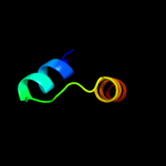
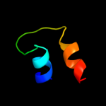
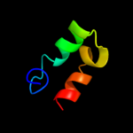
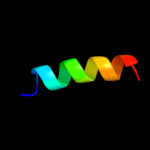
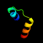
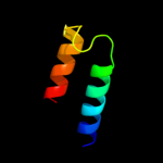
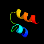
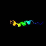
1 c3a1yF_
50.6
37
PDB header: ribosomal proteinChain: F: PDB Molecule: 50s ribosomal protein p1 (l12p);PDBTitle: the structure of protein complex
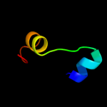
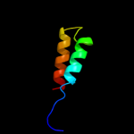
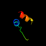
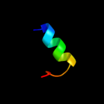
2 c3trkA_
46.5
24
PDB header: hydrolaseChain: A: PDB Molecule: nonstructural polyprotein;PDBTitle: structure of the chikungunya virus nsp2 protease
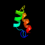
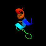
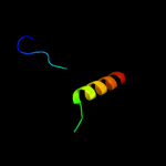
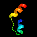
3 d1m6ya1
41.3
18
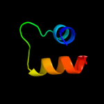
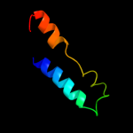
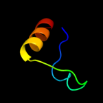
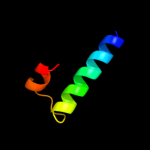
Fold: SAM domain-likeSuperfamily: Putative methyltransferase TM0872, insert domainFamily: Putative methyltransferase TM0872, insert domain4 d1sgla_
40.3
11
Fold: Ribonuclease Rh-likeSuperfamily: Ribonuclease Rh-likeFamily: Ribonuclease Rh-like5 d1iooa_
39.4
7
Fold: Ribonuclease Rh-likeSuperfamily: Ribonuclease Rh-likeFamily: Ribonuclease Rh-like6 d1or4a_
36.5
18
Fold: Globin-likeSuperfamily: Globin-likeFamily: Globins7 d1bola_
33.6
7
Fold: Ribonuclease Rh-likeSuperfamily: Ribonuclease Rh-likeFamily: Ribonuclease Rh-like8 c2jeuA_
27.4
27
PDB header: transcriptionChain: A: PDB Molecule: regulatory protein e2;PDBTitle: transcription activator structure reveals redox control of2 a replication initiation reaction
9 d1wg8a1
26.7
18
Fold: SAM domain-likeSuperfamily: Putative methyltransferase TM0872, insert domainFamily: Putative methyltransferase TM0872, insert domain10 c3d3zA_
25.7
15
PDB header: hydrolaseChain: A: PDB Molecule: actibind;PDBTitle: crystal structure of actibind a t2 rnase
11 d1k32a4
24.5
38
Fold: ClpP/crotonaseSuperfamily: ClpP/crotonaseFamily: Tail specific protease, catalytic domain12 c2w1oA_
21.4
11
PDB header: translationChain: A: PDB Molecule: 60s acidic ribosomal protein p2;PDBTitle: nmr structure of dimerization domain of human ribosomal2 protein p2
13 d1ev0a_
20.6
19
Fold: Cell division protein MinE topological specificity domainSuperfamily: Cell division protein MinE topological specificity domainFamily: Cell division protein MinE topological specificity domain14 c2q0oC_
20.4
38
PDB header: transcriptionChain: C: PDB Molecule: probable transcriptional repressor tram;PDBTitle: crystal structure of an anti-activation complex in bacterial quorum2 sensing
15 c3iz5w_
19.9
22
PDB header: ribosomeChain: W: PDB Molecule: 60s ribosomal protein l22 (l22e);PDBTitle: localization of the large subunit ribosomal proteins into a 5.5 a2 cryo-em map of triticum aestivum translating 80s ribosome
16 c2rchA_
19.0
26
PDB header: lyaseChain: A: PDB Molecule: cytochrome p450 74a;PDBTitle: crystal structure of arabidopsis thaliana allene oxide synthase (aos,2 cytochrome p450 74a, cyp74a) complexed with 13(s)-hod at 1.85 a3 resolution
17 c3izcw_
17.8
22
PDB header: ribosomeChain: W: PDB Molecule: 60s ribosomal protein rpl22 (l22e);PDBTitle: localization of the large subunit ribosomal proteins into a 6.1 a2 cryo-em map of saccharomyces cerevisiae translating 80s ribosome
18 d1r6na_
17.5
18
Fold: E2 regulatory, transactivation domainSuperfamily: E2 regulatory, transactivation domainFamily: E2 regulatory, transactivation domain19 d1dixa_
17.4
11
Fold: Ribonuclease Rh-likeSuperfamily: Ribonuclease Rh-likeFamily: Ribonuclease Rh-like20 c2kxoA_
17.1
19
PDB header: cell cycleChain: A: PDB Molecule: cell division topological specificity factor;PDBTitle: solution nmr structure of the cell division regulator mine protein2 from neisseria gonorrhoeae
21 c2w31A_
not modelled
16.7
15
PDB header: oxygen transportChain: A: PDB Molecule: globin;PDBTitle: globin domain of geobacter sulfurreducens globin-coupled2 sensor
22 d1jy5a_
not modelled
16.2
14
Fold: Ribonuclease Rh-likeSuperfamily: Ribonuclease Rh-likeFamily: Ribonuclease Rh-like23 c1n6dE_
not modelled
15.5
38
PDB header: hydrolaseChain: E: PDB Molecule: tricorn protease;PDBTitle: tricorn protease in complex with tetrapeptide chloromethyl2 ketone derivative
24 c1k32E_
not modelled
15.5
38
PDB header: hydrolaseChain: E: PDB Molecule: tricorn protease;PDBTitle: crystal structure of the tricorn protease
25 c1tueG_
not modelled
15.4
15
PDB header: replicationChain: G: PDB Molecule: regulatory protein e2;PDBTitle: the x-ray structure of the papillomavirus helicase in2 complex with its molecular matchmaker e2
26 d1tueb_
not modelled
15.3
15
Fold: E2 regulatory, transactivation domainSuperfamily: E2 regulatory, transactivation domainFamily: E2 regulatory, transactivation domain27 d2nnua1
not modelled
15.0
15
Fold: E2 regulatory, transactivation domainSuperfamily: E2 regulatory, transactivation domainFamily: E2 regulatory, transactivation domain28 d1iyba_
not modelled
14.4
14
Fold: Ribonuclease Rh-likeSuperfamily: Ribonuclease Rh-likeFamily: Ribonuclease Rh-like29 c1vd3A_
not modelled
12.3
15
PDB header: hydrolaseChain: A: PDB Molecule: rnase ngr3;PDBTitle: ribonuclease nt in complex with 2'-ump
30 d1faoa_
not modelled
11.8
15
Fold: PH domain-like barrelSuperfamily: PH domain-likeFamily: Pleckstrin-homology domain (PH domain)31 c1faoA_
not modelled
11.8
15
PDB header: signaling proteinChain: A: PDB Molecule: dual adaptor of phosphotyrosine and 3-PDBTitle: structure of the pleckstrin homology domain from2 dapp1/phish in complex with inositol 1,3,4,5-3 tetrakisphosphate
32 c1m6yA_
not modelled
11.4
17
PDB header: transferaseChain: A: PDB Molecule: s-adenosyl-methyltransferase mraw;PDBTitle: crystal structure analysis of tm0872, a putative sam-2 dependent methyltransferase, complexed with sah
33 d2p5ka1
not modelled
11.4
29
Fold: DNA/RNA-binding 3-helical bundleSuperfamily: "Winged helix" DNA-binding domainFamily: Arginine repressor (ArgR), N-terminal DNA-binding domain34 c3uo9B_
not modelled
10.9
15
PDB header: hydrolase/hydrolase inhibitorChain: B: PDB Molecule: glutaminase kidney isoform, mitochondrial;PDBTitle: crystal structure of human gac in complex with glutamate and bptes
35 d1wsca1
not modelled
10.6
18
Fold: AMMECR1-likeSuperfamily: AMMECR1-likeFamily: AMMECR1-like36 c3ayhA_
not modelled
10.5
13
PDB header: transcriptionChain: A: PDB Molecule: dna-directed rna polymerase iii subunit rpc9;PDBTitle: crystal structure of the c17/25 subcomplex from s. pombe rna2 polymerase iii
37 d1aoya_
not modelled
10.3
23
Fold: DNA/RNA-binding 3-helical bundleSuperfamily: "Winged helix" DNA-binding domainFamily: Arginine repressor (ArgR), N-terminal DNA-binding domain38 c3b73A_
not modelled
10.3
8
PDB header: structural genomics, unknown functionChain: A: PDB Molecule: phih1 repressor-like protein;PDBTitle: crystal structure of the phih1 repressor-like protein from2 haloarcula marismortui
39 c1b4aA_
not modelled
10.2
29
PDB header: repressorChain: A: PDB Molecule: arginine repressor;PDBTitle: structure of the arginine repressor from bacillus stearothermophilus
40 d1xdza_
not modelled
10.1
24
Fold: S-adenosyl-L-methionine-dependent methyltransferasesSuperfamily: S-adenosyl-L-methionine-dependent methyltransferasesFamily: Glucose-inhibited division protein B (GidB)41 d1f9na1
not modelled
10.1
29
Fold: DNA/RNA-binding 3-helical bundleSuperfamily: "Winged helix" DNA-binding domainFamily: Arginine repressor (ArgR), N-terminal DNA-binding domain42 c2p0vA_
not modelled
10.0
24
PDB header: structural genomics, unknown functionChain: A: PDB Molecule: hypothetical protein bt3781;PDBTitle: crystal structure of bt3781 protein from bacteroides2 thetaiotaomicron, northeast structural genomics target3 btr58
43 d2p0va1
not modelled
10.0
24
Fold: alpha/alpha toroidSuperfamily: Six-hairpin glycosidasesFamily: CPF0428-like44 d1v9va1
not modelled
9.7
19
Fold: Bromodomain-likeSuperfamily: MAST3 pre-PK domain-likeFamily: MAST3 pre-PK domain-like45 d2nvpa1
not modelled
9.3
33
Fold: alpha/alpha toroidSuperfamily: Six-hairpin glycosidasesFamily: CPF0428-like46 d1b4aa1
not modelled
9.2
29
Fold: DNA/RNA-binding 3-helical bundleSuperfamily: "Winged helix" DNA-binding domainFamily: Arginine repressor (ArgR), N-terminal DNA-binding domain47 c3ereD_
not modelled
9.1
36
PDB header: dna binding protein/dnaChain: D: PDB Molecule: arginine repressor;PDBTitle: crystal structure of the arginine repressor protein from mycobacterium2 tuberculosis in complex with the dna operator
48 d2nn5a1
not modelled
8.6
24
Fold: TBP-likeSuperfamily: Bet v1-likeFamily: AHSA1 domain49 c2kxwB_
not modelled
8.3
50
PDB header: calcium-binding protein/metal transportChain: B: PDB Molecule: sodium channel protein type 2 subunit alpha;PDBTitle: structure of the c-domain fragment of apo calmodulin bound to the iq2 motif of nav1.2
50 d1eo0a_
not modelled
7.8
21
Fold: N-cbl likeSuperfamily: Conserved domain common to transcription factors TFIIS, elongin A, CRSP70Family: Conserved domain common to transcription factors TFIIS, elongin A, CRSP7051 c2dymE_
not modelled
7.8
8
PDB header: protein turnover/protein turnoverChain: E: PDB Molecule: autophagy protein 5;PDBTitle: the crystal structure of saccharomyces cerevisiae atg5-2 atg16(1-46) complex
52 c3ivuB_
not modelled
7.6
18
PDB header: transferaseChain: B: PDB Molecule: homocitrate synthase, mitochondrial;PDBTitle: homocitrate synthase lys4 bound to 2-og
53 c3r9jD_
not modelled
7.6
19
PDB header: cell cycle,hydrolase/cell cycleChain: D: PDB Molecule: cell division topological specificity factor;PDBTitle: 4.3a resolution structure of a mind-mine(i24n) protein complex
54 d2c35a1
not modelled
7.4
17
Fold: SAM domain-likeSuperfamily: HRDC-likeFamily: RNA polymerase II subunit RBP4 (RpoF)55 c3ss4C_
not modelled
7.4
15
PDB header: hydrolaseChain: C: PDB Molecule: glutaminase c;PDBTitle: crystal structure of mouse glutaminase c, phosphate-bound form
56 c3htuE_
not modelled
7.2
20
PDB header: protein transportChain: E: PDB Molecule: vacuolar protein-sorting-associated protein 25;PDBTitle: crystal structure of the human vps25-vps20 subcomplex
57 d2etha1
not modelled
6.7
7
Fold: DNA/RNA-binding 3-helical bundleSuperfamily: "Winged helix" DNA-binding domainFamily: MarR-like transcriptional regulators58 c2q9rA_
not modelled
6.6
29
PDB header: unknown functionChain: A: PDB Molecule: protein of unknown function;PDBTitle: crystal structure of a duf416 family protein (sbal_3149) from2 shewanella baltica os155 at 1.91 a resolution
59 c3izct_
not modelled
6.5
25
PDB header: ribosomeChain: T: PDB Molecule: 60s ribosomal protein rpl19 (l19e);PDBTitle: localization of the large subunit ribosomal proteins into a 6.1 a2 cryo-em map of saccharomyces cerevisiae translating 80s ribosome
60 c3lvuB_
not modelled
6.5
13
PDB header: transport proteinChain: B: PDB Molecule: abc transporter, periplasmic substrate-binding protein;PDBTitle: crystal structure of abc transporter, periplasmic substrate-binding2 protein spo2066 from silicibacter pomeroyi
61 d1wjta_
not modelled
6.4
23
Fold: N-cbl likeSuperfamily: Conserved domain common to transcription factors TFIIS, elongin A, CRSP70Family: Conserved domain common to transcription factors TFIIS, elongin A, CRSP7062 d2c0sa1
not modelled
6.4
38
Fold: ROP-likeSuperfamily: BAS1536-likeFamily: BAS1536-like63 c3v4gA_
not modelled
6.3
23
PDB header: dna binding proteinChain: A: PDB Molecule: arginine repressor;PDBTitle: 1.60 angstrom resolution crystal structure of an arginine repressor2 from vibrio vulnificus cmcp6
64 c2kwuA_
not modelled
6.3
38
PDB header: protein binding/signaling proteinChain: A: PDB Molecule: dna polymerase iota;PDBTitle: solution structure of ubm2 of murine polymerase iota in complex with2 ubiquitin
65 d1zq7a1
not modelled
6.3
19
Fold: AMMECR1-likeSuperfamily: AMMECR1-likeFamily: AMMECR1-like66 d2ea9a1
not modelled
6.3
42
Fold: Profilin-likeSuperfamily: YeeU-likeFamily: YagB/YeeU/YfjZ-like67 d1wfya_
not modelled
6.0
39
Fold: beta-Grasp (ubiquitin-like)Superfamily: Ubiquitin-likeFamily: Ras-binding domain, RBD68 d2h28a1
not modelled
6.0
39
Fold: Profilin-likeSuperfamily: YeeU-likeFamily: YagB/YeeU/YfjZ-like69 c2wqmA_
not modelled
5.8
14
PDB header: transferaseChain: A: PDB Molecule: serine/threonine-protein kinase nek7;PDBTitle: structure of apo human nek7
70 d2dy1a4
not modelled
5.8
31
Fold: Ferredoxin-likeSuperfamily: EF-G C-terminal domain-likeFamily: EF-G/eEF-2 domains III and V71 c1wlzD_
not modelled
5.6
20
PDB header: unknown functionChain: D: PDB Molecule: cap-binding protein complex interacting proteinPDBTitle: crystal structure of djbp fragment which was obtained by2 limited proteolysis
72 d1wlza1
not modelled
5.6
20
Fold: EF Hand-likeSuperfamily: EF-handFamily: EF-hand modules in multidomain proteins73 d1gh6a_
not modelled
5.6
15
Fold: Long alpha-hairpinSuperfamily: Chaperone J-domainFamily: Chaperone J-domain74 d2fcea1
not modelled
5.6
25
Fold: EF Hand-likeSuperfamily: EF-handFamily: Calmodulin-like75 d1iira_
not modelled
5.6
26
Fold: UDP-Glycosyltransferase/glycogen phosphorylaseSuperfamily: UDP-Glycosyltransferase/glycogen phosphorylaseFamily: Gtf glycosyltransferase76 d1mw7a_
not modelled
5.6
23
Fold: YebC-likeSuperfamily: YebC-likeFamily: YebC-like77 d1ucda_
not modelled
5.5
11
Fold: Ribonuclease Rh-likeSuperfamily: Ribonuclease Rh-likeFamily: Ribonuclease Rh-like78 d2inwa1
not modelled
5.5
44
Fold: Profilin-likeSuperfamily: YeeU-likeFamily: YagB/YeeU/YfjZ-like79 c2ckzC_
not modelled
5.3
21
PDB header: transferaseChain: C: PDB Molecule: dna-directed rna polymerase iii 18 kdPDBTitle: x-ray structure of rna polymerase iii subcomplex c17-c25.
80 c2k42A_
not modelled
5.3
11
PDB header: signaling proteinChain: A: PDB Molecule: wiskott-aldrich syndrome protein;PDBTitle: solution structure of the gtpase binding domain of wasp in2 complex with espfu, an ehec effector
81 d2nxpa1
not modelled
5.2
10
Fold: Taf5 N-terminal domain-likeSuperfamily: Taf5 N-terminal domain-likeFamily: Taf5 N-terminal domain-like82 c2p6pB_
not modelled
5.2
15
PDB header: transferaseChain: B: PDB Molecule: glycosyl transferase;PDBTitle: x-ray crystal structure of c-c bond-forming dtdp-d-olivose-transferase2 urdgt2
83 c1wg8B_
not modelled
5.1
18
PDB header: transferaseChain: B: PDB Molecule: predicted s-adenosylmethionine-dependentPDBTitle: crystal structure of a predicted s-adenosylmethionine-2 dependent methyltransferase tt1512 from thermus3 thermophilus hb8.


