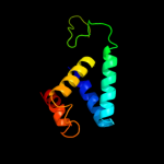
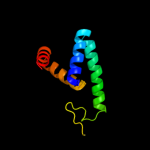
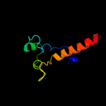
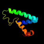
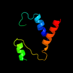
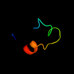
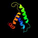
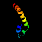
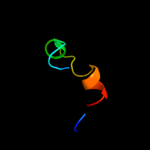
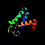
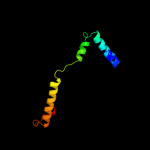
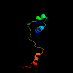
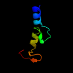
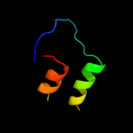
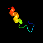
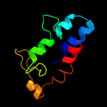
1 c3b7uX_
92.2
19
PDB header: hydrolaseChain: X: PDB Molecule: leukotriene a-4 hydrolase;PDBTitle: leukotriene a4 hydrolase complexed with kelatorphan
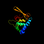
2 c3ciaA_
89.9
15
PDB header: hydrolaseChain: A: PDB Molecule: cold-active aminopeptidase;PDBTitle: crystal structure of cold-aminopeptidase from colwellia2 psychrerythraea
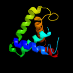
3 c3iukB_
89.7
25
PDB header: structural genomics, unknown functionChain: B: PDB Molecule: uncharacterized protein;PDBTitle: crystal structure of putative bacterial protein of unknown function2 (duf885, pf05960.1, ) from arthrobacter aurescens tc1, reveals fold3 similar to that of m32 carboxypeptidases
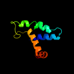
4 c2xpyA_
84.1
20
PDB header: hydrolaseChain: A: PDB Molecule: leukotriene a-4 hydrolase;PDBTitle: structure of native leukotriene a4 hydrolase from saccharomyces2 cerevisiae
5 c3o0yC_
83.6
24
PDB header: lipid binding proteinChain: C: PDB Molecule: lipoprotein;PDBTitle: the crystal structure of the putative lipoprotein from colwellia2 psychrerythraea
6 c3se6A_
79.7
16
PDB header: hydrolaseChain: A: PDB Molecule: endoplasmic reticulum aminopeptidase 2;PDBTitle: crystal structure of the human endoplasmic reticulum aminopeptidase 2
7 c3qnfA_
65.7
13
PDB header: hydrolaseChain: A: PDB Molecule: endoplasmic reticulum aminopeptidase 1;PDBTitle: crystal structure of the open state of human endoplasmic reticulum2 aminopeptidase 1 erap1
8 c1z5hB_
65.4
21
PDB header: hydrolaseChain: B: PDB Molecule: tricorn protease interacting factor f3;PDBTitle: crystal structures of the tricorn interacting factor f32 from thermoplasma acidophilum
9 c1yewC_
50.4
43
PDB header: oxidoreductase, membrane proteinChain: C: PDB Molecule: particulate methane monooxygenase subunit c2;PDBTitle: crystal structure of particulate methane monooxygenase
10 c3mdjB_
50.1
14
PDB header: hydrolase/hydrolase inhibitorChain: B: PDB Molecule: endoplasmic reticulum aminopeptidase 1;PDBTitle: er aminopeptidase, erap1, bound to the zinc aminopeptidase inhibitor,2 bestatin
11 d1q8ca_
35.9
23
Fold: NusB-likeSuperfamily: NusB-likeFamily: Hypothetical protein MG02712 d3b7sa3
34.3
19
Fold: Zincin-likeSuperfamily: Metalloproteases ("zincins"), catalytic domainFamily: Leukotriene A4 hydrolase catalytic domain13 c3chxG_
33.0
43
PDB header: membrane proteinChain: G: PDB Molecule: pmoc;PDBTitle: crystal structure of methylosinus trichosporium ob3b2 particulate methane monooxygenase (pmmo)
14 c2xdtA_
31.0
18
PDB header: hydrolaseChain: A: PDB Molecule: endoplasmic reticulum aminopeptidase 1;PDBTitle: crystal structure of the soluble domain of human2 endoplasmic reticulum aminopeptidase 1 erap1
15 d1wpba_
29.9
9
Fold: YfbU-likeSuperfamily: YfbU-likeFamily: YfbU-like16 c2knaA_
29.7
18
PDB header: apoptosisChain: A: PDB Molecule: baculoviral iap repeat-containing protein 4;PDBTitle: solution structure of uba domain of xiap
17 c2qffA_
28.5
19
PDB header: hydrolase inhibitorChain: A: PDB Molecule: hypothetical protein;PDBTitle: crystal structure of staphylococcal complement inhibitor
18 c2da4A_
27.8
26
PDB header: structural genomics, unknown functionChain: A: PDB Molecule: hypothetical protein dkfzp686k21156;PDBTitle: solution structure of the homeobox domain of the2 hypothetical protein, dkfzp686k21156
19 c1ceuA_
24.6
22
PDB header: viral proteinChain: A: PDB Molecule: protein (hiv-1 regulatory protein n-terminalPDBTitle: nmr structure of the (1-51) n-terminal domain of the hiv-12 regulatory protein
20 d1dl2a_
23.7
17
Fold: alpha/alpha toroidSuperfamily: Seven-hairpin glycosidasesFamily: Class I alpha-1;2-mannosidase, catalytic domain21 c3t4aG_
not modelled
23.4
25
PDB header: immune systemChain: G: PDB Molecule: fibrinogen-binding protein;PDBTitle: structure of a truncated form of staphylococcal complement inhibitor b2 bound to human c3c at 3.4 angstrom resolution
22 d1x9da1
not modelled
23.3
20
Fold: alpha/alpha toroidSuperfamily: Seven-hairpin glycosidasesFamily: Class I alpha-1;2-mannosidase, catalytic domain23 c1x9dA_
not modelled
23.3
20
PDB header: hydrolaseChain: A: PDB Molecule: endoplasmic reticulum mannosyl-oligosaccharide 1,PDBTitle: crystal structure of human class i alpha-1,2-mannosidase in2 complex with thio-disaccharide substrate analogue
24 d2zdra2
not modelled
23.1
16
Fold: TIM beta/alpha-barrelSuperfamily: AldolaseFamily: NeuB-like25 c2kfvA_
not modelled
22.1
13
PDB header: isomeraseChain: A: PDB Molecule: fk506-binding protein 3;PDBTitle: structure of the amino-terminal domain of human fk506-2 binding protein 3 / northeast structural genomics3 consortium target ht99a
26 c2vo9C_
not modelled
21.0
16
PDB header: hydrolaseChain: C: PDB Molecule: l-alanyl-d-glutamate peptidase;PDBTitle: crystal structure of the enzymatically active domain of the2 listeria monocytogenes bacteriophage 500 endolysin ply500
27 c1g6iA_
not modelled
20.6
17
PDB header: hydrolaseChain: A: PDB Molecule: class i alpha-1,2-mannosidase;PDBTitle: crystal structure of the yeast alpha-1,2-mannosidase with bound 1-2 deoxymannojirimycin at 1.59 a resolution
28 c2gtqA_
not modelled
18.4
12
PDB header: hydrolaseChain: A: PDB Molecule: aminopeptidase n;PDBTitle: crystal structure of aminopeptidase n from human pathogen neisseria2 meningitidis
29 d1lvfa_
not modelled
18.2
22
Fold: STAT-likeSuperfamily: t-snare proteinsFamily: t-snare proteins30 d1x2na1
not modelled
17.7
18
Fold: DNA/RNA-binding 3-helical bundleSuperfamily: Homeodomain-likeFamily: Homeodomain31 d1hcua_
not modelled
17.2
11
Fold: alpha/alpha toroidSuperfamily: Seven-hairpin glycosidasesFamily: Class I alpha-1;2-mannosidase, catalytic domain32 d1qd1a2
not modelled
17.0
20
Fold: Ferredoxin-likeSuperfamily: Formiminotransferase domain of formiminotransferase-cyclodeaminase.Family: Formiminotransferase domain of formiminotransferase-cyclodeaminase.33 d1nxca_
not modelled
16.8
16
Fold: alpha/alpha toroidSuperfamily: Seven-hairpin glycosidasesFamily: Class I alpha-1;2-mannosidase, catalytic domain34 d1m15a1
not modelled
16.7
19
Fold: Guanido kinase N-terminal domainSuperfamily: Guanido kinase N-terminal domainFamily: Guanido kinase N-terminal domain35 d2vo9a1
not modelled
16.6
16
Fold: Hedgehog/DD-peptidaseSuperfamily: Hedgehog/DD-peptidaseFamily: VanY-like36 c3bpqC_
not modelled
16.3
26
PDB header: toxinChain: C: PDB Molecule: antitoxin relb3;PDBTitle: crystal structure of relb-rele antitoxin-toxin complex from2 methanococcus jannaschii
37 d2oc6a1
not modelled
15.8
23
Fold: Secretion chaperone-likeSuperfamily: YdhG-likeFamily: YdhG-like38 c3fpvC_
not modelled
15.7
10
PDB header: heme binding proteinChain: C: PDB Molecule: extracellular haem-binding protein;PDBTitle: crystal structure of hbps
39 c2kvrA_
not modelled
15.6
4
PDB header: protein bindingChain: A: PDB Molecule: ubiquitin carboxyl-terminal hydrolase 7;PDBTitle: solution nmr structure of human ubiquitin specific protease2 usp7 ubl domain (residues 537-664). nesg target hr4395c/3 sgc-toronto
40 d1du6a_
not modelled
15.4
14
Fold: DNA/RNA-binding 3-helical bundleSuperfamily: Homeodomain-likeFamily: Homeodomain41 d1ndba1
not modelled
15.3
23
Fold: CoA-dependent acyltransferasesSuperfamily: CoA-dependent acyltransferasesFamily: Choline/Carnitine O-acyltransferase42 c2nrzB_
not modelled
14.9
19
PDB header: hydrolaseChain: B: PDB Molecule: uvrabc system protein c;PDBTitle: crystal structure of the c-terminal half of uvrc bound to2 its catalytic divalent cation
43 c3ayhA_
not modelled
14.7
22
PDB header: transcriptionChain: A: PDB Molecule: dna-directed rna polymerase iii subunit rpc9;PDBTitle: crystal structure of the c17/25 subcomplex from s. pombe rna2 polymerase iii
44 d2ri9a1
not modelled
14.3
13
Fold: alpha/alpha toroidSuperfamily: Seven-hairpin glycosidasesFamily: Class I alpha-1;2-mannosidase, catalytic domain45 d3cx5i1
not modelled
13.9
6
Fold: Single transmembrane helixSuperfamily: Subunit X (non-heme 7 kDa protein) of cytochrome bc1 complex (Ubiquinol-cytochrome c reductase)Family: Subunit X (non-heme 7 kDa protein) of cytochrome bc1 complex (Ubiquinol-cytochrome c reductase)46 c1krfA_
not modelled
13.8
13
PDB header: hydrolaseChain: A: PDB Molecule: mannosyl-oligosaccharide alpha-1,2-mannosidase;PDBTitle: structure of p. citrinum alpha 1,2-mannosidase reveals the basis for2 differences in specificity of the er and golgi class i enzymes
47 c2xr9A_
not modelled
13.8
26
PDB header: hydrolaseChain: A: PDB Molecule: ectonucleotide pyrophosphatase/phosphodiesterase familyPDBTitle: crystal structure of autotaxin (enpp2)
48 c3izbU_
not modelled
12.9
20
PDB header: ribosomeChain: U: PDB Molecule: 40s ribosomal protein s24;PDBTitle: localization of the small subunit ribosomal proteins into a 6.1 a2 cryo-em map of saccharomyces cerevisiae translating 80s ribosome
49 d1mv8a1
not modelled
12.8
28
Fold: 6-phosphogluconate dehydrogenase C-terminal domain-likeSuperfamily: 6-phosphogluconate dehydrogenase C-terminal domain-likeFamily: UDP-glucose/GDP-mannose dehydrogenase dimerisation domain50 d1miua2
not modelled
12.8
8
Fold: BRCA2 tower domainSuperfamily: BRCA2 tower domainFamily: BRCA2 tower domain51 d1ftra2
not modelled
12.7
86
Fold: Ferredoxin-likeSuperfamily: Formylmethanofuran:tetrahydromethanopterin formyltransferaseFamily: Formylmethanofuran:tetrahydromethanopterin formyltransferase52 c3mlcC_
not modelled
12.6
20
PDB header: isomeraseChain: C: PDB Molecule: fg41 malonate semialdehyde decarboxylase;PDBTitle: crystal structure of fg41msad inactivated by 3-chloropropiolate
53 d2g1da1
not modelled
12.2
9
Fold: Ribosomal proteins S24e, L23 and L15eSuperfamily: Ribosomal proteins S24e, L23 and L15eFamily: Ribosomal protein S24e54 d1m5sa2
not modelled
12.1
86
Fold: Ferredoxin-likeSuperfamily: Formylmethanofuran:tetrahydromethanopterin formyltransferaseFamily: Formylmethanofuran:tetrahydromethanopterin formyltransferase55 d1mylb_
not modelled
12.1
62
Fold: Ribbon-helix-helixSuperfamily: Ribbon-helix-helixFamily: Arc/Mnt-like phage repressors56 d1m5ha2
not modelled
12.0
86
Fold: Ferredoxin-likeSuperfamily: Formylmethanofuran:tetrahydromethanopterin formyltransferaseFamily: Formylmethanofuran:tetrahydromethanopterin formyltransferase57 c3ezkB_
not modelled
12.0
29
PDB header: hydrolaseChain: B: PDB Molecule: dna packaging protein gp17;PDBTitle: bacteriophage t4 gp17 motor assembly based on crystal2 structures and cryo-em reconstructions
58 d1eg1a_
not modelled
12.0
26
Fold: Concanavalin A-like lectins/glucanasesSuperfamily: Concanavalin A-like lectins/glucanasesFamily: Glycosyl hydrolase family 7 catalytic core59 c2vzaD_
not modelled
11.9
29
PDB header: cell adhesionChain: D: PDB Molecule: cell filamentation protein;PDBTitle: type iv secretion system effector protein bepa
60 c3pbpL_
not modelled
11.7
80
PDB header: transport protein,structural proteinChain: L: PDB Molecule: nucleoporin nup159;PDBTitle: structure of the yeast heterotrimeric nup82-nup159-nup116 nucleoporin2 complex
61 d1x6ma_
not modelled
11.5
18
Fold: Mss4-likeSuperfamily: Mss4-likeFamily: Glutathione-dependent formaldehyde-activating enzyme, Gfa62 c3pg8B_
not modelled
11.5
13
PDB header: transferaseChain: B: PDB Molecule: phospho-2-dehydro-3-deoxyheptonate aldolase;PDBTitle: truncated form of 3-deoxy-d-arabino-heptulosonate 7-phosphate synthase2 from thermotoga maritima
63 d1zxoa1
not modelled
11.2
57
Fold: Ribonuclease H-like motifSuperfamily: Actin-like ATPase domainFamily: BadF/BadG/BcrA/BcrD-like64 c2xrgA_
not modelled
11.2
26
PDB header: hydrolaseChain: A: PDB Molecule: ectonucleotide pyrophosphatase/phosphodiesterase familyPDBTitle: crystal structure of autotaxin (enpp2) in complex with the2 ha155 boronic acid inhibitor
65 c2dmnA_
not modelled
11.1
24
PDB header: transcriptionChain: A: PDB Molecule: homeobox protein tgif2lx;PDBTitle: the solution structure of the homeobox domain of human2 homeobox protein tgif2lx
66 d2a2la1
not modelled
11.1
10
Fold: Profilin-likeSuperfamily: GlcG-likeFamily: GlcG-like67 d1crka1
not modelled
10.8
19
Fold: Guanido kinase N-terminal domainSuperfamily: Guanido kinase N-terminal domainFamily: Guanido kinase N-terminal domain68 c3bbnR_
not modelled
10.8
38
PDB header: ribosomeChain: R: PDB Molecule: ribosomal protein s18;PDBTitle: homology model for the spinach chloroplast 30s subunit2 fitted to 9.4a cryo-em map of the 70s chlororibosome.
69 c3e38A_
not modelled
10.6
29
PDB header: hydrolaseChain: A: PDB Molecule: two-domain protein containing predicted php-like metal-PDBTitle: crystal structure of a two-domain protein containing predicted php-2 like metal-dependent phosphoesterase (bvu_3505) from bacteroides3 vulgatus atcc 8482 at 2.20 a resolution
70 d1myla_
not modelled
10.6
62
Fold: Ribbon-helix-helixSuperfamily: Ribbon-helix-helixFamily: Arc/Mnt-like phage repressors71 c3mgwA_
not modelled
10.5
25
PDB header: hydrolaseChain: A: PDB Molecule: lysozyme g;PDBTitle: thermodynamics and structure of a salmon cold-active goose-type2 lysozyme
72 c3cezA_
not modelled
10.4
29
PDB header: oxidoreductaseChain: A: PDB Molecule: methionine-r-sulfoxide reductase;PDBTitle: crystal structure of methionine-r-sulfoxide reductase from2 burkholderia pseudomallei
73 c1w9iA_
not modelled
10.3
13
PDB header: myosinChain: A: PDB Molecule: myosin ii heavy chain;PDBTitle: myosin ii dictyostelium discoideum motor domain s456y bound2 with mgadp-befx
74 d1t1ua1
not modelled
10.2
23
Fold: CoA-dependent acyltransferasesSuperfamily: CoA-dependent acyltransferasesFamily: Choline/Carnitine O-acyltransferase75 c2y8pA_
not modelled
10.2
8
PDB header: lyaseChain: A: PDB Molecule: endo-type membrane-bound lytic murein transglycosylase a;PDBTitle: crystal structure of an outer membrane-anchored endolytic2 peptidoglycan lytic transglycosylase (mlte) from3 escherichia coli
76 c2ktrA_
not modelled
10.2
7
PDB header: signaling protein, transport proteinChain: A: PDB Molecule: sequestosome-1;PDBTitle: nmr structure of p62 pb1 dimer determined based on pcs
77 d1bazb_
not modelled
10.1
62
Fold: Ribbon-helix-helixSuperfamily: Ribbon-helix-helixFamily: Arc/Mnt-like phage repressors78 c2nrrA_
not modelled
10.1
25
PDB header: hydrolaseChain: A: PDB Molecule: uvrabc system protein c;PDBTitle: crystal structure of the c-terminal rnaseh endonuclase2 domain of uvrc
79 c2l1uA_
not modelled
10.0
50
PDB header: oxidoreductaseChain: A: PDB Molecule: methionine-r-sulfoxide reductase b2, mitochondrial;PDBTitle: structure-functional analysis of mammalian msrb2 protein
80 c3c65A_
not modelled
10.0
12
PDB header: hydrolaseChain: A: PDB Molecule: uvrabc system protein c;PDBTitle: crystal structure of bacillus stearothermophilus uvrc 5'2 endonuclease domain
81 c3mnwP_
not modelled
9.9
30
PDB header: immune systemChain: P: PDB Molecule: gp41;PDBTitle: crystal structure of the non-neutralizing hiv antibody 13h11 fab2 fragment with a gp41 mper-derived peptide in a helical conformation
82 d1xrsb2
not modelled
9.9
16
Fold: Dodecin subunit-likeSuperfamily: D-lysine 5,6-aminomutase beta subunit KamE, N-terminal domainFamily: D-lysine 5,6-aminomutase beta subunit KamE, N-terminal domain83 d1atia2
not modelled
9.9
10
Fold: Class II aaRS and biotin synthetasesSuperfamily: Class II aaRS and biotin synthetasesFamily: Class II aminoacyl-tRNA synthetase (aaRS)-like, catalytic domain84 d1b28a_
not modelled
9.7
62
Fold: Ribbon-helix-helixSuperfamily: Ribbon-helix-helixFamily: Arc/Mnt-like phage repressors85 d1qh4a1
not modelled
9.6
13
Fold: Guanido kinase N-terminal domainSuperfamily: Guanido kinase N-terminal domainFamily: Guanido kinase N-terminal domain86 c2p58C_
not modelled
9.5
26
PDB header: transport protein/chaperoneChain: C: PDB Molecule: putative type iii secretion protein yscg;PDBTitle: structure of the yersinia pestis type iii secretion system2 needle protein yscf in complex with its chaperones3 ysce/yscg
87 d1jjcb2
not modelled
9.4
22
Fold: Putative DNA-binding domainSuperfamily: Putative DNA-binding domainFamily: Domains B1 and B5 of PheRS-beta, PheT88 d2ctda1
not modelled
9.4
44
Fold: beta-beta-alpha zinc fingersSuperfamily: beta-beta-alpha zinc fingersFamily: Classic zinc finger, C2H289 d1xm0a1
not modelled
9.4
50
Fold: Mss4-likeSuperfamily: Mss4-likeFamily: SelR domain90 d2qkwa1
not modelled
9.3
32
Fold: immunoglobulin/albumin-binding domain-likeSuperfamily: Avirulence protein AvrPtoFamily: Avirulence protein AvrPto91 c2qkwA_
not modelled
9.3
32
PDB header: transferaseChain: A: PDB Molecule: avirulence protein;PDBTitle: structural basis for activation of plant immunity by2 bacterial effector protein avrpto
92 c3kztB_
not modelled
9.3
12
PDB header: structural genomics, unknown functionChain: B: PDB Molecule: uncharacterized protein;PDBTitle: crystal structure of protein of unknown function (np_812423.1) from2 bacteroides thetaiotaomicron vpi-5482 at 2.10 a resolution
93 c2k8dA_
not modelled
9.2
43
PDB header: oxidoreductaseChain: A: PDB Molecule: peptide methionine sulfoxide reductase msrb;PDBTitle: solution structure of a zinc-binding methionine sulfoxide reductase
94 c1l6jA_
not modelled
9.2
60
PDB header: hydrolaseChain: A: PDB Molecule: matrix metalloproteinase-9;PDBTitle: crystal structure of human matrix metalloproteinase mmp92 (gelatinase b).
95 d1myka_
not modelled
9.2
62
Fold: Ribbon-helix-helixSuperfamily: Ribbon-helix-helixFamily: Arc/Mnt-like phage repressors96 c1xuzA_
not modelled
9.2
19
PDB header: biosynthetic proteinChain: A: PDB Molecule: polysialic acid capsule biosynthesis protein siac;PDBTitle: crystal structure analysis of sialic acid synthase (neub)from2 neisseria meningitidis, bound to mn2+, phosphoenolpyruvate, and n-3 acetyl mannosaminitol
97 c3mnzP_
not modelled
9.1
30
PDB header: immune systemChain: P: PDB Molecule: gp41 mper-derived peptide;PDBTitle: crystal structure of the non-neutralizing hiv antibody 13h11 fab2 fragment with a gp41 mper-derived peptide bearing ala substitutions3 in a helical conformation
98 d1bdta_
not modelled
9.0
62
Fold: Ribbon-helix-helixSuperfamily: Ribbon-helix-helixFamily: Arc/Mnt-like phage repressors99 c2kl4A_
not modelled
9.0
12
PDB header: structural genomics, unknown functionChain: A: PDB Molecule: bh2032 protein;PDBTitle: nmr structure of the protein nb7804a


