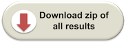
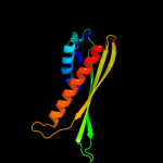
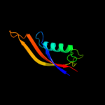
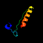
| 1 | c2jxpA_
|
|
 |
100.0 |
16 |
PDB header:lipoprotein
Chain: A: PDB Molecule:putative lipoprotein b;
PDBTitle: solution nmr structure of uncharacterized lipoprotein b2 from nitrosomonas europaea. northeast structural genomics3 target ner45a
|
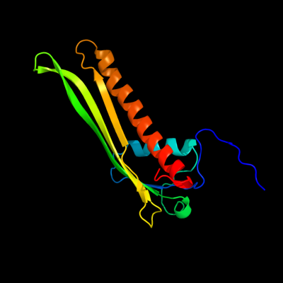
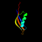
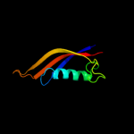
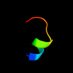
| 2 | c2r76A_
|
|
 |
100.0 |
34 |
PDB header:lipoprotein
Chain: A: PDB Molecule:rare lipoprotein b;
PDBTitle: crystal structure of the rare lipoprotein b (so_1173) from shewanella2 oneidensis, northeast structural genomics consortium target sor91a
|
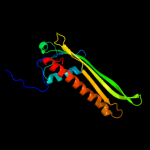
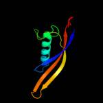
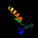
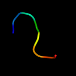
| 3 | c3bf2A_
|
|
 |
100.0 |
27 |
PDB header:lipoprotein
Chain: A: PDB Molecule:putative lipoprotein;
PDBTitle: crystal structure of the a1ksw9_neimf protein from2 neisseria meningitidis. northeast structural genomics3 consortium target mr36a
|
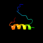
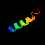
| 4 | c3oqtP_
|
|
 |
35.9 |
17 |
PDB header:flavoprotein
Chain: P: PDB Molecule:rv1498a protein;
PDBTitle: crystal structure of rv1498a protein from mycobacterium tuberculosis
|
| 5 | c2x5eA_
|
|
 |
35.3 |
60 |
PDB header:unknown function
Chain: A: PDB Molecule:upf0271 protein pa4511;
PDBTitle: crystal structure of the hypothetical protein pa4511 from2 pseudomonas aeruginosa
|
| 6 | d1xw8a_
|
|
 |
34.6 |
60 |
Fold:7-stranded beta/alpha barrel
Superfamily:Glycoside hydrolase/deacetylase
Family:LamB/YcsF-like |
| 7 | c3onrI_
|
|
 |
30.2 |
14 |
PDB header:metal binding protein
Chain: I: PDB Molecule:protein transport protein sece2;
PDBTitle: crystal structure of the calcium chelating immunodominant antigen,2 calcium dodecin (rv0379),from mycobacterium tuberculosis with a novel3 calcium-binding site
|
| 8 | c2vxaL_
|
|
 |
30.0 |
24 |
PDB header:flavoprotein
Chain: L: PDB Molecule:dodecin;
PDBTitle: h.halophila dodecin in complex with riboflavin
|
| 9 | d2ux9a1
|
|
 |
29.4 |
19 |
Fold:Dodecin subunit-like
Superfamily:Dodecin-like
Family:Dodecin-like |
| 10 | d1zlqa1
|
|
 |
18.4 |
12 |
Fold:Periplasmic binding protein-like II
Superfamily:Periplasmic binding protein-like II
Family:Phosphate binding protein-like |
| 11 | d2dfaa1
|
|
 |
18.2 |
29 |
Fold:7-stranded beta/alpha barrel
Superfamily:Glycoside hydrolase/deacetylase
Family:LamB/YcsF-like |
| 12 | c2obkE_
|
|
 |
17.7 |
50 |
PDB header:structural genomics, unknown function
Chain: E: PDB Molecule:selt/selw/selh selenoprotein domain;
PDBTitle: x-ray structure of the putative se binding protein from pseudomonas2 fluorescens. northeast structural genomics consortium target plr6.
|
| 13 | d1v6ta_
|
|
 |
17.6 |
13 |
Fold:7-stranded beta/alpha barrel
Superfamily:Glycoside hydrolase/deacetylase
Family:LamB/YcsF-like |
| 14 | c3mgjA_
|
|
 |
16.6 |
15 |
PDB header:structural genomics, unknown function
Chain: A: PDB Molecule:uncharacterized protein mj1480;
PDBTitle: crystal structure of the saccharop_dh_n domain of mj14802 protein from methanococcus jannaschii. northeast structural3 genomics consortium target mjr83a.
|
| 15 | c2ojlB_
|
|
 |
16.5 |
50 |
PDB header:structural genomics, unknown function
Chain: B: PDB Molecule:hypothetical protein;
PDBTitle: crystal structure of q7waf1_borpa from bordetella parapertussis.2 northeast structural genomics target bpr68.
|
| 16 | c2grvC_
|
|
 |
15.7 |
17 |
PDB header:biosynthetic protein
Chain: C: PDB Molecule:lpqw;
PDBTitle: crystal structure of lpqw
|
| 17 | d1a6ca3
|
|
 |
15.7 |
36 |
Fold:Nucleoplasmin-like/VP (viral coat and capsid proteins)
Superfamily:Positive stranded ssRNA viruses
Family:Comoviridae-like VP |
| 18 | c3dexA_
|
|
 |
15.5 |
38 |
PDB header:structural genomics, unknown function
Chain: A: PDB Molecule:sav_2001;
PDBTitle: crystal structure of sav_2001 protein from streptomyces2 avermitilis, northeast structural genomics consortium3 target svr107.
|
| 19 | c2kncA_
|
|
 |
15.2 |
35 |
PDB header:cell adhesion
Chain: A: PDB Molecule:integrin alpha-iib;
PDBTitle: platelet integrin alfaiib-beta3 transmembrane-cytoplasmic2 heterocomplex
|
| 20 | c2vsvB_
|
|
 |
14.6 |
30 |
PDB header:protein-binding
Chain: B: PDB Molecule:rhophilin-2;
PDBTitle: crystal structure of the pdz domain of human rhophilin-2
|
| 21 | d2fa8a1 |
|
not modelled |
14.1 |
50 |
Fold:Thioredoxin fold
Superfamily:Thioredoxin-like
Family:Selenoprotein W-related |
| 22 | d1b8qa_ |
|
not modelled |
14.0 |
17 |
Fold:PDZ domain-like
Superfamily:PDZ domain-like
Family:PDZ domain |
| 23 | c2p0gB_ |
|
not modelled |
13.9 |
50 |
PDB header:structural genomics, unknown function
Chain: B: PDB Molecule:selenoprotein w-related protein;
PDBTitle: crystal structure of selenoprotein w-related protein from2 vibrio cholerae. northeast structural genomics target vcr75
|
| 24 | c3od1A_ |
|
not modelled |
13.8 |
9 |
PDB header:transferase
Chain: A: PDB Molecule:atp phosphoribosyltransferase regulatory subunit;
PDBTitle: the crystal structure of an atp phosphoribosyltransferase regulatory2 subunit/histidyl-trna synthetase from bacillus halodurans c
|
| 25 | c2pxgA_ |
|
not modelled |
13.3 |
50 |
PDB header:membrane protein
Chain: A: PDB Molecule:outer membrane protein;
PDBTitle: nmr solution structure of omla
|
| 26 | d1x61a2 |
|
not modelled |
13.1 |
33 |
Fold:Glucocorticoid receptor-like (DNA-binding domain)
Superfamily:Glucocorticoid receptor-like (DNA-binding domain)
Family:LIM domain |
| 27 | d2fnea1 |
|
not modelled |
13.1 |
15 |
Fold:PDZ domain-like
Superfamily:PDZ domain-like
Family:PDZ domain |
| 28 | d1nn4a_ |
|
not modelled |
12.7 |
26 |
Fold:Ribose/Galactose isomerase RpiB/AlsB
Superfamily:Ribose/Galactose isomerase RpiB/AlsB
Family:Ribose/Galactose isomerase RpiB/AlsB |
| 29 | c3s5pA_ |
|
not modelled |
12.1 |
24 |
PDB header:isomerase
Chain: A: PDB Molecule:ribose 5-phosphate isomerase;
PDBTitle: crystal structure of ribose-5-phosphate isomerase b rpib from giardia2 lamblia
|
| 30 | c2a40F_ |
|
not modelled |
11.8 |
60 |
PDB header:structural protein
Chain: F: PDB Molecule:wiskott-aldrich syndrome protein family member 2;
PDBTitle: ternary complex of the wh2 domain of wave with actin-dnase i
|
| 31 | c2a40C_ |
|
not modelled |
11.8 |
60 |
PDB header:structural protein
Chain: C: PDB Molecule:wiskott-aldrich syndrome protein family member 2;
PDBTitle: ternary complex of the wh2 domain of wave with actin-dnase i
|
| 32 | c2v90E_ |
|
not modelled |
10.4 |
26 |
PDB header:protein-binding
Chain: E: PDB Molecule:pdz domain-containing protein 3;
PDBTitle: crystal structure of the 3rd pdz domain of intestine- and2 kidney-enriched pdz domain ikepp (pdzd3)
|
| 33 | d2ex2a1 |
|
not modelled |
9.8 |
11 |
Fold:beta-lactamase/transpeptidase-like
Superfamily:beta-lactamase/transpeptidase-like
Family:Dac-like |
| 34 | c1iclA_ |
|
not modelled |
9.6 |
42 |
PDB header:de novo protein
Chain: A: PDB Molecule:th1ox;
PDBTitle: solution structure of designed beta-sheet mini-protein th1ox
|
| 35 | c3ol0C_ |
|
not modelled |
9.0 |
50 |
PDB header:de novo protein
Chain: C: PDB Molecule:de novo designed monomer trefoil-fold sub-domain which
PDBTitle: crystal structure of monofoil-4p homo-trimer: de novo designed monomer2 trefoil-fold sub-domain which forms homo-trimer assembly
|
| 36 | d1qbea_ |
|
not modelled |
8.9 |
21 |
Fold:RNA bacteriophage capsid protein
Superfamily:RNA bacteriophage capsid protein
Family:RNA bacteriophage capsid protein |
| 37 | c3lvuB_ |
|
not modelled |
8.5 |
8 |
PDB header:transport protein
Chain: B: PDB Molecule:abc transporter, periplasmic substrate-binding protein;
PDBTitle: crystal structure of abc transporter, periplasmic substrate-binding2 protein spo2066 from silicibacter pomeroyi
|
| 38 | d2c7na1 |
|
not modelled |
8.3 |
75 |
Fold:Glucocorticoid receptor-like (DNA-binding domain)
Superfamily:Glucocorticoid receptor-like (DNA-binding domain)
Family:A20-like zinc finger |
| 39 | c2db5A_ |
|
not modelled |
8.3 |
13 |
PDB header:protein binding
Chain: A: PDB Molecule:inad-like protein;
PDBTitle: solution structure of the first pdz domain of inad-like2 protein
|
| 40 | c2c7mA_ |
|
not modelled |
7.9 |
75 |
PDB header:protein-binding
Chain: A: PDB Molecule:rab guanine nucleotide exchange factor 1;
PDBTitle: human rabex-5 residues 1-74 in complex with ubiquitin
|
| 41 | c3v2gA_ |
|
not modelled |
7.8 |
18 |
PDB header:oxidoreductase
Chain: A: PDB Molecule:3-oxoacyl-[acyl-carrier-protein] reductase;
PDBTitle: crystal structure of a dehydrogenase/reductase from sinorhizobium2 meliloti 1021
|
| 42 | d1hu3a_ |
|
not modelled |
7.7 |
13 |
Fold:alpha-alpha superhelix
Superfamily:ARM repeat
Family:MIF4G domain-like |
| 43 | c3ghfA_ |
|
not modelled |
7.7 |
18 |
PDB header:cell cycle
Chain: A: PDB Molecule:septum site-determining protein minc;
PDBTitle: crystal structure of the septum site-determining protein2 minc from salmonella typhimurium
|
| 44 | c2kzyA_ |
|
not modelled |
7.2 |
75 |
PDB header:metal binding protein
Chain: A: PDB Molecule:zfand5 protein (zinc finger protein 216 (predicted),
PDBTitle: solution nmr structure of the znf216 a20 zinc finger
|
| 45 | d1vpta_ |
|
not modelled |
7.1 |
38 |
Fold:S-adenosyl-L-methionine-dependent methyltransferases
Superfamily:S-adenosyl-L-methionine-dependent methyltransferases
Family:mRNA cap methylase |
| 46 | d2ccqa1 |
|
not modelled |
7.1 |
13 |
Fold:PUG domain-like
Superfamily:PUG domain-like
Family:PUG domain |
| 47 | d1g9oa_ |
|
not modelled |
7.1 |
21 |
Fold:PDZ domain-like
Superfamily:PDZ domain-like
Family:PDZ domain |
| 48 | c2fgyA_ |
|
not modelled |
6.9 |
38 |
PDB header:lyase
Chain: A: PDB Molecule:carboxysome shell polypeptide;
PDBTitle: beta carbonic anhydrase from the carboxysomal shell of2 halothiobacillus neapolitanus (csosca)
|
| 49 | d1uf2c1 |
|
not modelled |
6.8 |
67 |
Fold:A virus capsid protein alpha-helical domain
Superfamily:A virus capsid protein alpha-helical domain
Family:Phytoreovirus capsid |
| 50 | c1vp3A_ |
|
not modelled |
6.6 |
38 |
PDB header:methyltransferase
Chain: A: PDB Molecule:vp39;
PDBTitle: vaccinia virus protein vp39 in complex with s-adenosylhomocysteine
|
| 51 | c1v39A_ |
|
not modelled |
6.6 |
38 |
PDB header:methyltransferase
Chain: A: PDB Molecule:vp39;
PDBTitle: dc26 mutant of vaccinia virus protein vp39 in complex with s-2 adenosylhomocysteine and m7g(5')pppg
|
| 52 | c2k1aA_ |
|
not modelled |
6.6 |
32 |
PDB header:cell adhesion
Chain: A: PDB Molecule:integrin alpha-iib;
PDBTitle: bicelle-embedded integrin alpha(iib) transmembrane segment
|
| 53 | c3k7pA_ |
|
not modelled |
6.6 |
10 |
PDB header:isomerase
Chain: A: PDB Molecule:ribose 5-phosphate isomerase;
PDBTitle: structure of mutant of ribose 5-phosphate isomerase type b from2 trypanosoma cruzi.
|
| 54 | c3m1pA_ |
|
not modelled |
6.6 |
10 |
PDB header:isomerase
Chain: A: PDB Molecule:ribose 5-phosphate isomerase;
PDBTitle: structure of ribose 5-phosphate isomerase type b from trypanosoma2 cruzi, soaked with allose-6-phosphate
|
| 55 | c3l3bA_ |
|
not modelled |
6.6 |
67 |
PDB header:biosynthetic protein
Chain: A: PDB Molecule:es1 family protein;
PDBTitle: crystal structure of isoprenoid biosynthesis protein with2 amidotransferase-like domain from ehrlichia chaffeensis at 1.90a3 resolution
|
| 56 | c2iwnA_ |
|
not modelled |
6.3 |
17 |
PDB header:signaling protein
Chain: A: PDB Molecule:multiple pdz domain protein;
PDBTitle: 3rd pdz domain of multiple pdz domain protein mpdz (casp2 target)
|
| 57 | c3ftoA_ |
|
not modelled |
6.3 |
15 |
PDB header:peptide binding protein
Chain: A: PDB Molecule:oligopeptide-binding protein oppa;
PDBTitle: crystal structure of oppa in a open conformation
|
| 58 | c3ry3B_ |
|
not modelled |
6.3 |
21 |
PDB header:transport protein
Chain: B: PDB Molecule:putative solute-binding protein;
PDBTitle: putative solute-binding protein from yersinia pestis.
|
| 59 | d1w5da1 |
|
not modelled |
6.3 |
12 |
Fold:beta-lactamase/transpeptidase-like
Superfamily:beta-lactamase/transpeptidase-like
Family:Dac-like |
| 60 | d1ncfa3 |
|
not modelled |
6.1 |
43 |
Fold:TNF receptor-like
Superfamily:TNF receptor-like
Family:TNF receptor-like |
| 61 | c1ztyA_ |
|
not modelled |
6.1 |
12 |
PDB header:sugar binding protein, signaling protein
Chain: A: PDB Molecule:chitin oligosaccharide binding protein;
PDBTitle: crystal structure of the chitin oligasaccharide binding2 protein
|
| 62 | c2ogpA_ |
|
not modelled |
5.8 |
10 |
PDB header:signaling protein
Chain: A: PDB Molecule:partitioning-defective 3 homolog;
PDBTitle: solution structure of the second pdz domain of par-3
|
| 63 | c3j00Z_ |
|
not modelled |
5.8 |
18 |
PDB header:ribosome/ribosomal protein
Chain: Z: PDB Molecule:cell division protein ftsq;
PDBTitle: structure of the ribosome-secye complex in the membrane environment
|
| 64 | d1um1a_ |
|
not modelled |
5.7 |
18 |
Fold:PDZ domain-like
Superfamily:PDZ domain-like
Family:PDZ domain |
| 65 | d1vp8a_ |
|
not modelled |
5.7 |
30 |
Fold:Pyruvate kinase C-terminal domain-like
Superfamily:PK C-terminal domain-like
Family:MTH1675-like |
| 66 | d1jjcb5 |
|
not modelled |
5.6 |
25 |
Fold:Class II aaRS and biotin synthetases
Superfamily:Class II aaRS and biotin synthetases
Family:Class II aminoacyl-tRNA synthetase (aaRS)-like, catalytic domain |
| 67 | c1vdfB_ |
|
not modelled |
5.6 |
13 |
PDB header:extracellular matrix protein
Chain: B: PDB Molecule:cartilage oligomeric matrix protein;
PDBTitle: assembly domain of cartilage oligomeric matrix protein
|
| 68 | d2d5ua1 |
|
not modelled |
5.6 |
13 |
Fold:PUG domain-like
Superfamily:PUG domain-like
Family:PUG domain |
| 69 | c2npbA_ |
|
not modelled |
5.5 |
25 |
PDB header:oxidoreductase
Chain: A: PDB Molecule:selenoprotein w;
PDBTitle: nmr solution structure of mouse selw
|
| 70 | c2iwoA_ |
|
not modelled |
5.5 |
13 |
PDB header:signaling protein
Chain: A: PDB Molecule:multiple pdz domain protein;
PDBTitle: 12th pdz domain of multiple pdz domain protein mpdz (casp2 target)
|
| 71 | d1k32a1 |
|
not modelled |
5.5 |
25 |
Fold:PDZ domain-like
Superfamily:PDZ domain-like
Family:Tail specific protease PDZ domain |
| 72 | d2vvpa1 |
|
not modelled |
5.4 |
20 |
Fold:Ribose/Galactose isomerase RpiB/AlsB
Superfamily:Ribose/Galactose isomerase RpiB/AlsB
Family:Ribose/Galactose isomerase RpiB/AlsB |
| 73 | c3he8A_ |
|
not modelled |
5.1 |
23 |
PDB header:isomerase
Chain: A: PDB Molecule:ribose-5-phosphate isomerase;
PDBTitle: structural study of clostridium thermocellum ribose-5-phosphate2 isomerase b
|
| 74 | c2kr1A_ |
|
not modelled |
5.1 |
38 |
PDB header:ligase
Chain: A: PDB Molecule:ubiquitin protein ligase e3a;
PDBTitle: solution nmr structure of zinc binding n-terminal domain of ubiquitin-2 protein ligase e3a from homo sapiens. northeast structural genomics3 consortium (nesg) target hr3662
|
| 75 | d2d9ia1 |
|
not modelled |
5.1 |
21 |
Fold:IF3-like
Superfamily:SMR domain-like
Family:Smr domain |