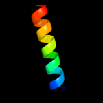
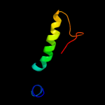
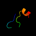
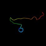
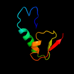
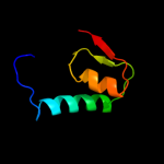
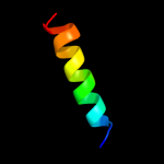
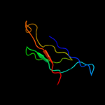
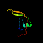
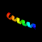
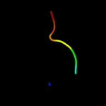
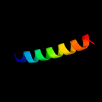
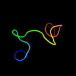
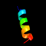
1 c3gw6F_
32.3
45
PDB header: chaperoneChain: F: PDB Molecule: endo-n-acetylneuraminidase;PDBTitle: intramolecular chaperone
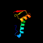
2 c3g79A_
29.9
31
PDB header: oxidoreductaseChain: A: PDB Molecule: ndp-n-acetyl-d-galactosaminuronic acid dehydrogenase;PDBTitle: crystal structure of ndp-n-acetyl-d-galactosaminuronic acid2 dehydrogenase from methanosarcina mazei go1
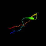
3 c1zy7A_
25.2
35
PDB header: hydrolaseChain: A: PDB Molecule: rna-specific adenosine deaminase b1, isoformPDBTitle: crystal structure of the catalytic domain of an adenosine2 deaminase that acts on rna (hadar2) bound to inositol3 hexakisphosphate (ihp)
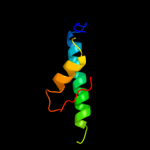
4 d2plga1
24.2
31
Fold: Secretion chaperone-likeSuperfamily: Type III secretory system chaperone-likeFamily: Tll0839-like5 c3plnA_
23.2
19
PDB header: oxidoreductaseChain: A: PDB Molecule: udp-glucose 6-dehydrogenase;PDBTitle: crystal structure of klebsiella pneumoniae udp-glucose 6-dehydrogenase2 complexed with udp-glucose
6 c1mv8A_
20.9
28
PDB header: oxidoreductaseChain: A: PDB Molecule: gdp-mannose 6-dehydrogenase;PDBTitle: 1.55 a crystal structure of a ternary complex of gdp-mannose2 dehydrogenase from psuedomonas aeruginosa
7 d3er7a1
20.1
41
Fold: Cystatin-likeSuperfamily: NTF2-likeFamily: Exig0174-like8 c1dliA_
19.1
19
PDB header: oxidoreductaseChain: A: PDB Molecule: udp-glucose dehydrogenase;PDBTitle: the first structure of udp-glucose dehydrogenase (udpgdh) reveals the2 catalytic residues necessary for the two-fold oxidation
9 c3prjB_
17.8
22
PDB header: oxidoreductaseChain: B: PDB Molecule: udp-glucose 6-dehydrogenase;PDBTitle: role of packing defects in the evolution of allostery and induced fit2 in human udp-glucose dehydrogenase.
10 c3ci9B_
15.2
29
PDB header: transcriptionChain: B: PDB Molecule: heat shock factor-binding protein 1;PDBTitle: crystal structure of the human hsbp1
11 c3ojlA_
15.1
28
PDB header: oxidoreductaseChain: A: PDB Molecule: cap5o;PDBTitle: native structure of the udp-n-acetyl-mannosamine dehydrogenase cap5o2 from staphylococcus aureus
12 c2y0dB_
15.0
33
PDB header: oxidoreductaseChain: B: PDB Molecule: udp-glucose dehydrogenase;PDBTitle: bcec mutation y10k
13 c3m06F_
14.0
43
PDB header: protein bindingChain: F: PDB Molecule: tnf receptor-associated factor 2;PDBTitle: crystal structure of traf2
14 d1ttaa_
13.0
18
Fold: Prealbumin-likeSuperfamily: Transthyretin (synonym: prealbumin)Family: Transthyretin (synonym: prealbumin)15 c2keqA_
13.0
23
PDB header: splicingChain: A: PDB Molecule: dna polymerase iii alpha subunit, nucleic acidPDBTitle: solution structure of dnae intein from nostoc punctiforme
16 c1aq5C_
12.5
45
PDB header: coiled-coilChain: C: PDB Molecule: cartilage matrix protein;PDBTitle: high-resolution solution nmr structure of the trimeric coiled-coil2 domain of chicken cartilage matrix protein, 20 structures
17 c2lkaA_
11.8
63
PDB header: toxinChain: A: PDB Molecule: toxin ts16;PDBTitle: new tricks of an old fold: structural versatility of scorpion toxins2 with common cysteine spacing
18 c2oqqB_
10.8
29
PDB header: transcriptionChain: B: PDB Molecule: transcription factor hy5;PDBTitle: crystal structure of hy5 leucine zipper homodimer from2 arabidopsis thaliana
19 d1rwha1
10.4
32
Fold: alpha/alpha toroidSuperfamily: Chondroitin AC/alginate lyaseFamily: Hyaluronate lyase-like catalytic, N-terminal domain20 c3rylB_
10.2
44
PDB header: protein bindingChain: B: PDB Molecule: protein vpa1370;PDBTitle: dimerization domain of vibrio parahemolyticus vopl
21 d1f53a_
not modelled
10.1
29
Fold: gamma-Crystallin-likeSuperfamily: gamma-Crystallin-likeFamily: Killer toxin-like protein SKLP22 c3c8xA_
not modelled
10.1
17
PDB header: transferaseChain: A: PDB Molecule: ephrin type-a receptor 2;PDBTitle: crystal structure of the ligand binding domain of human ephrin a22 (epha2) receptor protein kinase
23 c1cosA_
not modelled
10.0
50
PDB header: alpha-helical bundleChain: A: PDB Molecule: coiled serine;PDBTitle: crystal structure of a synthetic triple-stranded alpha-2 helical bundle
24 c1cosC_
not modelled
10.0
50
PDB header: alpha-helical bundleChain: C: PDB Molecule: coiled serine;PDBTitle: crystal structure of a synthetic triple-stranded alpha-2 helical bundle
25 c1cosB_
not modelled
10.0
50
PDB header: alpha-helical bundleChain: B: PDB Molecule: coiled serine;PDBTitle: crystal structure of a synthetic triple-stranded alpha-2 helical bundle
26 c3gg2B_
not modelled
9.9
45
PDB header: oxidoreductaseChain: B: PDB Molecule: sugar dehydrogenase, udp-glucose/gdp-mannosePDBTitle: crystal structure of udp-glucose 6-dehydrogenase from2 porphyromonas gingivalis bound to product udp-glucuronate
27 c3f59A_
not modelled
9.7
45
PDB header: structural proteinChain: A: PDB Molecule: ankyrin-1;PDBTitle: crystal structure of zu5-ank, the spectrin binding region of human2 erythroid ankyrin
28 c2h1xB_
not modelled
9.6
23
PDB header: hydrolaseChain: B: PDB Molecule: 5-hydroxyisourate hydrolase (formerly known asPDBTitle: crystal structure of 5-hydroxyisourate hydrolase (formerly2 known as trp, transthyretin related protein)
29 d2it9a1
not modelled
9.2
42
Fold: ssDNA-binding transcriptional regulator domainSuperfamily: ssDNA-binding transcriptional regulator domainFamily: PMN2A0962/syc2379c-like30 d2bvca1
not modelled
8.9
11
Fold: beta-Grasp (ubiquitin-like)Superfamily: Glutamine synthetase, N-terminal domainFamily: Glutamine synthetase, N-terminal domain31 c1coiA_
not modelled
8.8
50
PDB header: alpha-helical bundleChain: A: PDB Molecule: coil-vald;PDBTitle: designed trimeric coiled coil-vald
32 d1mv8a1
not modelled
8.6
55
Fold: 6-phosphogluconate dehydrogenase C-terminal domain-likeSuperfamily: 6-phosphogluconate dehydrogenase C-terminal domain-likeFamily: UDP-glucose/GDP-mannose dehydrogenase dimerisation domain33 d1f52a1
not modelled
8.3
11
Fold: beta-Grasp (ubiquitin-like)Superfamily: Glutamine synthetase, N-terminal domainFamily: Glutamine synthetase, N-terminal domain34 d1dlja1
not modelled
8.2
43
Fold: 6-phosphogluconate dehydrogenase C-terminal domain-likeSuperfamily: 6-phosphogluconate dehydrogenase C-terminal domain-likeFamily: UDP-glucose/GDP-mannose dehydrogenase dimerisation domain35 c2jgoC_
not modelled
7.8
50
PDB header: de novo proteinChain: C: PDB Molecule: coil ser l9c;PDBTitle: stucture of the arsenated de novo designed peptide coil ser2 l9c
36 c3ljmA_
not modelled
7.8
50
PDB header: de novo proteinChain: A: PDB Molecule: coil ser l9c;PDBTitle: structure of de novo designed apo peptide coil ser l9c
37 c2jgoA_
not modelled
7.8
50
PDB header: de novo proteinChain: A: PDB Molecule: coil ser l9c;PDBTitle: stucture of the arsenated de novo designed peptide coil ser2 l9c
38 c3ljmB_
not modelled
7.8
50
PDB header: de novo proteinChain: B: PDB Molecule: coil ser l9c;PDBTitle: structure of de novo designed apo peptide coil ser l9c
39 c3ljmC_
not modelled
7.8
50
PDB header: de novo proteinChain: C: PDB Molecule: coil ser l9c;PDBTitle: structure of de novo designed apo peptide coil ser l9c
40 c2jgoB_
not modelled
7.8
50
PDB header: de novo proteinChain: B: PDB Molecule: coil ser l9c;PDBTitle: stucture of the arsenated de novo designed peptide coil ser2 l9c
41 d1vq3a_
not modelled
7.5
17
Fold: PurS-likeSuperfamily: PurS-likeFamily: PurS subunit of FGAM synthetase42 c3hroA_
not modelled
7.5
35
PDB header: transport proteinChain: A: PDB Molecule: transient receptor potential (trp) channelPDBTitle: crystal structure of a c-terminal coiled coil domain of2 transient receptor potential (trp) channel subfamily p3 member 2 (trpp2, polycystic kidney disease 2)
43 c2pnvA_
not modelled
7.5
20
PDB header: membrane proteinChain: A: PDB Molecule: small conductance calcium-activated potassiumPDBTitle: crystal structure of the leucine zipper domain of small-2 conductance ca2+-activated k+ (skca) channel from rattus3 norvegicus
44 c3rj1S_
not modelled
7.4
31
PDB header: transcriptionChain: S: PDB Molecule: mediator of rna polymerase ii transcription subunit 18;PDBTitle: architecture of the mediator head module
45 d2nvna1
not modelled
7.2
50
Fold: ssDNA-binding transcriptional regulator domainSuperfamily: ssDNA-binding transcriptional regulator domainFamily: PMN2A0962/syc2379c-like46 d1hn0a2
not modelled
7.1
38
Fold: Galactose-binding domain-likeSuperfamily: Galactose-binding domain-likeFamily: Chondroitin ABC lyase I, N-terminal domain47 d1tfpa_
not modelled
7.0
25
Fold: Prealbumin-likeSuperfamily: Transthyretin (synonym: prealbumin)Family: Transthyretin (synonym: prealbumin)48 c3ssbJ_
not modelled
6.9
57
PDB header: hydrolase/hydrolase inhibitorChain: J: PDB Molecule: inducible metalloproteinase inhibitor protein;PDBTitle: structure of insect metalloproteinase inhibitor in complex with2 thermolysin
49 c1q2kA_
not modelled
6.7
46
PDB header: toxinChain: A: PDB Molecule: neurotoxin bmk37;PDBTitle: solution structure of bmbktx1 a new potassium channel2 blocker from the chinese scorpion buthus martensi karsch
50 d1k8ib2
not modelled
6.5
37
Fold: MHC antigen-recognition domainSuperfamily: MHC antigen-recognition domainFamily: MHC antigen-recognition domain51 c3e8yX_
not modelled
6.5
46
PDB header: toxinChain: X: PDB Molecule: potassium channel toxin alpha-ktx 19.1;PDBTitle: xray structure of scorpion toxin bmbktx1
52 c2fwvA_
not modelled
6.2
24
PDB header: structural genomics, unknown functionChain: A: PDB Molecule: hypothetical protein mtubf_01000852;PDBTitle: crystal structure of rv0813
53 c2jmzA_
not modelled
6.1
13
PDB header: unknown functionChain: A: PDB Molecule: hypothetical protein mj0781;PDBTitle: solution structure of a klba intein precursor from2 methanococcus jannaschii
54 d1eaic_
not modelled
6.0
50
Fold: Serine protease inhibitorsSuperfamily: Serine protease inhibitorsFamily: ATI-like55 d1mylb_
not modelled
5.9
50
Fold: Ribbon-helix-helixSuperfamily: Ribbon-helix-helixFamily: Arc/Mnt-like phage repressors56 c1i4oC_
not modelled
5.9
67
PDB header: apoptosis/hydrolaseChain: C: PDB Molecule: baculoviral iap repeat-containing protein 4;PDBTitle: crystal structure of the xiap/caspase-7 complex
57 d1f86a_
not modelled
5.9
18
Fold: Prealbumin-likeSuperfamily: Transthyretin (synonym: prealbumin)Family: Transthyretin (synonym: prealbumin)58 d1un7a1
not modelled
5.9
40
Fold: Composite domain of metallo-dependent hydrolasesSuperfamily: Composite domain of metallo-dependent hydrolasesFamily: N-acetylglucosamine-6-phosphate deacetylase, NagA59 d1t11a2
not modelled
5.9
17
Fold: Ribosome binding domain-likeSuperfamily: Trigger factor ribosome-binding domainFamily: Trigger factor ribosome-binding domain60 c3u5ga_
not modelled
5.9
43
PDB header: ribosomeChain: A: PDB Molecule: 40s ribosomal protein s0-a;PDBTitle: the structure of the eukaryotic ribosome at 3.0 a resolution
61 d1oo2a_
not modelled
5.9
21
Fold: Prealbumin-likeSuperfamily: Transthyretin (synonym: prealbumin)Family: Transthyretin (synonym: prealbumin)62 c1i7wB_
not modelled
5.8
40
PDB header: cell adhesionChain: B: PDB Molecule: epithelial-cadherin;PDBTitle: beta-catenin/phosphorylated e-cadherin complex
63 c2xzn5_
not modelled
5.8
43
PDB header: ribosomeChain: 5: PDB Molecule: ribosomal protein s26e containing protein;PDBTitle: crystal structure of the eukaryotic 40s ribosomal2 subunit in complex with initiation factor 1. this file3 contains the 40s subunit and initiation factor for4 molecule 2
64 c2zw2B_
not modelled
5.6
17
PDB header: ligaseChain: B: PDB Molecule: putative uncharacterized protein sts178;PDBTitle: crystal structure of formylglycinamide ribonucleotide amidotransferase2 iii from sulfolobus tokodaii (stpurs)
65 c2dadA_
not modelled
5.6
20
PDB header: oncoproteinChain: A: PDB Molecule: absent in melanoma 1 protein;PDBTitle: solution structure of the fifth crystall domain of the non-2 lens protein, absent in melanoma 1
66 c3batB_
not modelled
5.6
33
PDB header: contractile proteinChain: B: PDB Molecule: myosin heavy chain, striated muscle/generalPDBTitle: crystal structure of the n-terminal region of the scallop2 myosin rod, monoclinic (p21) form
67 d2uubc1
not modelled
5.4
36
Fold: Alpha-lytic protease prodomain-likeSuperfamily: Prokaryotic type KH domain (KH-domain type II)Family: Prokaryotic type KH domain (KH-domain type II)68 d1kgia_
not modelled
5.3
18
Fold: Prealbumin-likeSuperfamily: Transthyretin (synonym: prealbumin)Family: Transthyretin (synonym: prealbumin)69 d1okia1
not modelled
5.3
44
Fold: gamma-Crystallin-likeSuperfamily: gamma-Crystallin-likeFamily: Crystallins/Ca-binding development proteins70 d1ataa_
not modelled
5.2
42
Fold: Serine protease inhibitorsSuperfamily: Serine protease inhibitorsFamily: ATI-like71 d1rhoa_
not modelled
5.2
14
Fold: Immunoglobulin-like beta-sandwichSuperfamily: E set domainsFamily: RhoGDI-like72 d1u58a2
not modelled
5.1
38
Fold: MHC antigen-recognition domainSuperfamily: MHC antigen-recognition domainFamily: MHC antigen-recognition domain73 d1t6ca2
not modelled
5.1
21
Fold: Ribonuclease H-like motifSuperfamily: Actin-like ATPase domainFamily: Ppx/GppA phosphatase


