1 c1qd6C_
100.0
100
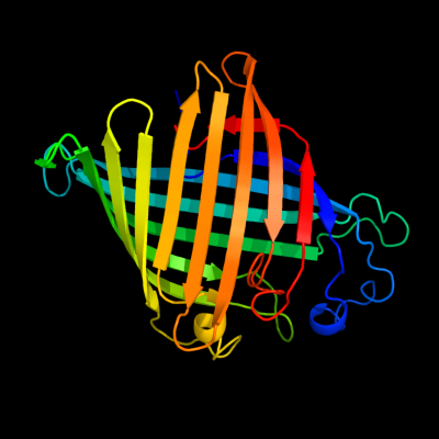
PDB header: membrane proteinChain: C: PDB Molecule: protein (outer membrane phospholipase (ompla));PDBTitle: outer membrane phospholipase a from escherichia coli
2 c1fw3A_
100.0
99
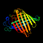
PDB header: hydrolase, membrane proteinChain: A: PDB Molecule: outer membrane phospholipase a;PDBTitle: outer membrane phospholipase a from escherichia coli
3 c2iwvD_
82.8
15
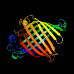
PDB header: ion channelChain: D: PDB Molecule: outer membrane protein g;PDBTitle: structure of the monomeric outer membrane porin ompg in the2 open and closed conformation
4 c2vveB_
50.8
26
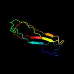
PDB header: viral proteinChain: B: PDB Molecule: spike protein p1;PDBTitle: crystal structure of the stem and receptor binding domain2 of the spike protein p1 from bacteriophage pm2
5 d2pora_
40.7
17
Fold: Transmembrane beta-barrelsSuperfamily: PorinsFamily: Porin6 c2w0cL_
29.0
22
PDB header: virusChain: L: PDB Molecule: protein 2;PDBTitle: x-ray structure of the entire lipid-containing2 bacteriophage pm2
7 c2wjqA_
19.0
16
PDB header: transport proteinChain: A: PDB Molecule: probable n-acetylneuraminic acid outer membrane channelPDBTitle: nanc porin structure in hexagonal crystal form.
8 d2fgqx1
17.3
18
Fold: Transmembrane beta-barrelsSuperfamily: PorinsFamily: Porin9 c2vvdA_
14.7
22
PDB header: viral proteinChain: A: PDB Molecule: spike protein p1;PDBTitle: crystal structure of the receptor binding domain of the2 spike protein p1 from bacteriophage pm2
10 d1xrsb2
14.2
60
Fold: Dodecin subunit-likeSuperfamily: D-lysine 5,6-aminomutase beta subunit KamE, N-terminal domainFamily: D-lysine 5,6-aminomutase beta subunit KamE, N-terminal domain11 c2kz3A_
13.6
21
PDB header: unknown functionChain: A: PDB Molecule: putative uncharacterized protein rad51l3;PDBTitle: backbone 1h, 13c, and 15n chemical shift assignments for human rad51d2 from 1 to 83
12 d1wpua1
8.8
25
Fold: Hut operon positive regulatory protein HutPSuperfamily: Hut operon positive regulatory protein HutPFamily: Hut operon positive regulatory protein HutP13 d1w0na_
8.8
33
Fold: Galactose-binding domain-likeSuperfamily: Galactose-binding domain-likeFamily: Family 36 carbohydrate binding module, CBM3614 d3buxb3
8.0
36
Fold: SH2-likeSuperfamily: SH2 domainFamily: SH2 domain15 d1aisa1
7.1
27
Fold: TBP-likeSuperfamily: TATA-box binding protein-likeFamily: TATA-box binding protein (TBP), C-terminal domain16 d1mp9a1
6.7
27
Fold: TBP-likeSuperfamily: TATA-box binding protein-likeFamily: TATA-box binding protein (TBP), C-terminal domain17 c2w56B_
6.5
30
PDB header: unknown functionChain: B: PDB Molecule: vc0508;PDBTitle: structure of the hypothetical protein vc0508 from vibrio cholerae2 vsp-ii pathogenicity island
18 d1pg5a1
6.1
33
Fold: ATC-likeSuperfamily: Aspartate/ornithine carbamoyltransferaseFamily: Aspartate/ornithine carbamoyltransferase19 d1uwka_
6.0
36
Fold: UrocanaseSuperfamily: UrocanaseFamily: Urocanase20 c3al0B_
6.0
36
PDB header: ligase/rnaChain: B: PDB Molecule: aspartyl/glutamyl-trna(asn/gln) amidotransferase subunit b;PDBTitle: crystal structure of the glutamine transamidosome from thermotoga2 maritima in the glutamylation state.
21 c3btpB_
not modelled
5.9
67
PDB header: dna binding protein, chaperoneChain: B: PDB Molecule: protein vire1;PDBTitle: crystal structure of agrobacterium tumefaciens vire2 in complex with2 its chaperone vire1: a novel fold and implications for dna binding
22 d2ogqa1
not modelled
5.6
31
Fold: Polo-box domainSuperfamily: Polo-box domainFamily: Polo-box duplicated region23 d2zfga1
not modelled
5.4
18
Fold: Transmembrane beta-barrelsSuperfamily: PorinsFamily: Porin24 c2fknC_
not modelled
5.4
50
PDB header: lyaseChain: C: PDB Molecule: urocanate hydratase;PDBTitle: crystal structure of urocanase from bacillus subtilis
25 c1pg5A_
not modelled
5.2
35
PDB header: transferaseChain: A: PDB Molecule: aspartate carbamoyltransferase;PDBTitle: crystal structure of the unligated (t-state) aspartate2 transcarbamoylase from the extremely thermophilic archaeon sulfolobus3 acidocaldarius