Phyre Home Retrieve Phyre Job Id
Emaill.a.kelley@imperial.ac.uk
DescriptionP0CF26
DateThu Jan 5 11:30:48 GMT 2012
Unique Job ID4cd80077a41492bb

Summary 

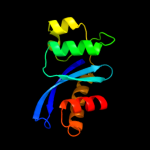
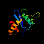
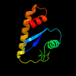
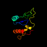
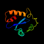
Top model
Image coloured by rainbow N → C terminus
Model (left) based on template d1hyva_
Top template information
Fold:Ribonuclease H-like motif
Superfamily:Ribonuclease H-like
Family:Retroviral integrase, catalytic domain
Confidence and coverage
Confidence: 97.0% Coverage: 65%
109 residues ( 65% of your sequence) have been modelled with 97.0% confidence by the single highest scoring template.
3D viewing
Interactive 3D view in Jmol

Sequence analysis 

Secondary structure and disorder prediction 

   1........10.........20.........30.........40.........50.........60
Sequence  MPGNRPHYGRWPQHDFPPFKKLRPQSVTSRIQPGSDVIVCAEMDEQWGYVGAKSRQRWLF
Secondary structure 


SS confidence 


Disorder  ???????


?????????????????


Disorder confidence 


   .........70.........80.........90.........100.........110.........120
Sequence  YAYDRLRKTVVAHVFGERTMATLGRLMSLLSPFDVVIWMTDGWPLYESRLKGKLHVISKR
Secondary structure 


SS confidence 


Disorder 


?


Disorder confidence 


   .........130.........140.........150.........160.......
Sequence  YTQRIERHNLNLRQHLARLGRKSLSFSKSVEQHDKVIGHYLNIKHYQ
Secondary structure 


SS confidence 


Disorder 


???
Disorder confidence 


Confidence Key
High(9)                    Low (0)
?Disordered
Alpha helix
Beta strand

Domain analysis 

Hover over an aligned region to see model and summary info

Please note, only up to the top 20 hits are modelled to reduce computer load

RankAligned region

PDB 1hyv chain A

3D model

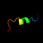
Region: 42 - 157
Aligned: 109
Modelled: 116
Confidence: 97.0%
Identity: 15%
Fold: Ribonuclease H-like motif
Superfamily: Ribonuclease H-like
Family: Retroviral integrase, catalytic domain

Phyre2

PDB 1cxq chain A

3D model

Region: 40 - 165
Aligned: 124
Modelled: 126
Confidence: 96.6%
Identity: 16%
Fold: Ribonuclease H-like motif
Superfamily: Ribonuclease H-like
Family: Retroviral integrase, catalytic domain

Phyre2

PDB 1exq chain A

3D model

Region: 55 - 157
Aligned: 103
Modelled: 103
Confidence: 96.2%
Identity: 12%
Fold: Ribonuclease H-like motif
Superfamily: Ribonuclease H-like
Family: Retroviral integrase, catalytic domain

Phyre2

PDB 1ex4 chain A

3D model

Region: 42 - 157
Aligned: 110
Modelled: 116
Confidence: 95.0%
Identity: 16%
PDB header:viral protein
Chain: A: PDB Molecule:integrase;
PDBTitle: hiv-1 integrase catalytic core and c-terminal domain

Phyre2

PDB 1k6y chain B

3D model

Region: 55 - 157
Aligned: 98
Modelled: 98
Confidence: 95.0%
Identity: 15%
PDB header:transferase
Chain: B: PDB Molecule:integrase;
PDBTitle: crystal structure of a two-domain fragment of hiv-1 integrase

Phyre2

PDB 3f9k chain V

3D model

Region: 55 - 157
Aligned: 103
Modelled: 103
Confidence: 94.3%
Identity: 14%
PDB header:viral protein, recombination
Chain: V: PDB Molecule:integrase;
PDBTitle: two domain fragment of hiv-2 integrase in complex with ledgf ibd

Phyre2

PDB 1asu chain A

3D model

Region: 40 - 166
Aligned: 120
Modelled: 127
Confidence: 93.8%
Identity: 16%
Fold: Ribonuclease H-like motif
Superfamily: Ribonuclease H-like
Family: Retroviral integrase, catalytic domain

Phyre2

PDB 1c6v chain A

3D model

Region: 55 - 157
Aligned: 103
Modelled: 103
Confidence: 93.6%
Identity: 16%
Fold: Ribonuclease H-like motif
Superfamily: Ribonuclease H-like
Family: Retroviral integrase, catalytic domain

Phyre2

PDB 3nf9 chain A

3D model

Region: 55 - 157
Aligned: 98
Modelled: 98
Confidence: 91.1%
Identity: 16%
PDB header:hydrolase/hydrolase inhibitor
Chain: A: PDB Molecule:integrase;
PDBTitle: structural basis for a new mechanism of inhibition of hiv integrase2 identified by fragment screening and structure based design

Phyre2

PDB 1c0m chain A

3D model

Region: 40 - 141
Aligned: 100
Modelled: 102
Confidence: 90.9%
Identity: 18%
PDB header:transferase
Chain: A: PDB Molecule:protein (integrase);
PDBTitle: crystal structure of rsv two-domain integrase

Phyre2

PDB 1c0m chain A domain 2

3D model

Region: 40 - 160
Aligned: 119
Modelled: 121
Confidence: 90.7%
Identity: 18%
Fold: Ribonuclease H-like motif
Superfamily: Ribonuclease H-like
Family: Retroviral integrase, catalytic domain

Phyre2

PDB 3dlr chain A

3D model

Region: 40 - 160
Aligned: 118
Modelled: 121
Confidence: 89.3%
Identity: 11%
PDB header:transferase
Chain: A: PDB Molecule:integrase;
PDBTitle: crystal structure of the catalytic core domain from pfv2 integrase

Phyre2

PDB 1bco chain A

3D model

Region: 58 - 167
Aligned: 108
Modelled: 110
Confidence: 80.3%
Identity: 10%
PDB header:transposase
Chain: A: PDB Molecule:bacteriophage mu transposase;
PDBTitle: bacteriophage mu transposase core domain

Phyre2

PDB 3kks chain B

3D model

Region: 55 - 167
Aligned: 111
Modelled: 113
Confidence: 69.6%
Identity: 13%
PDB header:dna binding protein
Chain: B: PDB Molecule:integrase;
PDBTitle: crystal structure of catalytic core domain of biv integrase in crystal2 form ii

Phyre2

PDB 1bco chain A domain 2

3D model

Region: 58 - 167
Aligned: 106
Modelled: 110
Confidence: 60.2%
Identity: 11%
Fold: Ribonuclease H-like motif
Superfamily: Ribonuclease H-like
Family: mu transposase, core domain

Phyre2

PDB 3hpg chain C

3D model

Region: 55 - 157
Aligned: 98
Modelled: 103
Confidence: 47.7%
Identity: 14%
PDB header:transferase
Chain: C: PDB Molecule:integrase;
PDBTitle: visna virus integrase (residues 1-219) in complex with ledgf2 ibd: examples of open integrase dimer-dimer interfaces

Phyre2

PDB 2k8f chain B

3D model

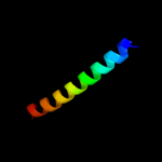
Region: 77 - 95
Aligned: 19
Modelled: 19
Confidence: 42.7%
Identity: 26%
PDB header:transferase/transcription
Chain: B: PDB Molecule:cellular tumor antigen p53;
PDBTitle: structural basis for the regulation of p53 function by p300

Phyre2

PDB 3he5 chain D

3D model

Region: 129 - 158
Aligned: 30
Modelled: 30
Confidence: 31.1%
Identity: 30%
PDB header:de novo protein
Chain: D: PDB Molecule:synzip2;
PDBTitle: heterospecific coiled-coil pair synzip2:synzip1

Phyre2

PDB 2l14 chain B

3D model

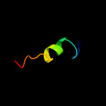
Region: 77 - 95
Aligned: 19
Modelled: 19
Confidence: 17.5%
Identity: 26%
PDB header:protein binding
Chain: B: PDB Molecule:cellular tumor antigen p53;
PDBTitle: structure of cbp nuclear coactivator binding domain in complex with2 p53 tad

Phyre2

PDB 3l2t chain B

3D model

Region: 52 - 157
Aligned: 102
Modelled: 106
Confidence: 13.3%
Identity: 11%
PDB header:recombination/dna
Chain: B: PDB Molecule:integrase;
PDBTitle: crystal structure of the prototype foamy virus (pfv) intasome in2 complex with magnesium and mk0518 (raltegravir)

Phyre2
1

d1hyva_
2

d1cxqa_
3

d1exqa_
4

c1ex4A_
5

c1k6yB_
6

c3f9kV_
7

d1asua_
8

d1c6va_
9

c3nf9A_
10

c1c0mA_
11

d1c0ma2
12

c3dlrA_
13

c1bcoA_
14

c3kksB_
15

d1bcoa2
16

c3hpgC_
17

c2k8fB_
18

c3he5D_
19

c2l14B_
20

c3l2tB_
21


22


23


24


25


26


27


28


29


30


31


32


33


34


35


36


37


38


39


40


41


42


43


44


45


46


47


Detailed template information 

#
Template Alignment Coverage3D Model Confidence
% i.d. Template Information
1d1hyva_


97.0 15 Fold:Ribonuclease H-like motif
Superfamily:Ribonuclease H-like
Family:Retroviral integrase, catalytic domain
2d1cxqa_


96.6 16 Fold:Ribonuclease H-like motif
Superfamily:Ribonuclease H-like
Family:Retroviral integrase, catalytic domain
3d1exqa_


96.2 12 Fold:Ribonuclease H-like motif
Superfamily:Ribonuclease H-like
Family:Retroviral integrase, catalytic domain
4c1ex4A_


95.0 16 PDB header:viral protein
Chain: A: PDB Molecule:integrase;
PDBTitle: hiv-1 integrase catalytic core and c-terminal domain
5c1k6yB_


95.0 15 PDB header:transferase
Chain: B: PDB Molecule:integrase;
PDBTitle: crystal structure of a two-domain fragment of hiv-1 integrase
6c3f9kV_


94.3 14 PDB header:viral protein, recombination
Chain: V: PDB Molecule:integrase;
PDBTitle: two domain fragment of hiv-2 integrase in complex with ledgf ibd
7d1asua_


93.8 16 Fold:Ribonuclease H-like motif
Superfamily:Ribonuclease H-like
Family:Retroviral integrase, catalytic domain
8d1c6va_


93.6 16 Fold:Ribonuclease H-like motif
Superfamily:Ribonuclease H-like
Family:Retroviral integrase, catalytic domain
9c3nf9A_


91.1 16 PDB header:hydrolase/hydrolase inhibitor
Chain: A: PDB Molecule:integrase;
PDBTitle: structural basis for a new mechanism of inhibition of hiv integrase2 identified by fragment screening and structure based design
10c1c0mA_


90.9 18 PDB header:transferase
Chain: A: PDB Molecule:protein (integrase);
PDBTitle: crystal structure of rsv two-domain integrase
11d1c0ma2


90.7 18 Fold:Ribonuclease H-like motif
Superfamily:Ribonuclease H-like
Family:Retroviral integrase, catalytic domain
12c3dlrA_


89.3 11 PDB header:transferase
Chain: A: PDB Molecule:integrase;
PDBTitle: crystal structure of the catalytic core domain from pfv2 integrase
13c1bcoA_


80.3 10 PDB header:transposase
Chain: A: PDB Molecule:bacteriophage mu transposase;
PDBTitle: bacteriophage mu transposase core domain
14c3kksB_


69.6 13 PDB header:dna binding protein
Chain: B: PDB Molecule:integrase;
PDBTitle: crystal structure of catalytic core domain of biv integrase in crystal2 form ii
15d1bcoa2


60.2 11 Fold:Ribonuclease H-like motif
Superfamily:Ribonuclease H-like
Family:mu transposase, core domain
16c3hpgC_


47.7 14 PDB header:transferase
Chain: C: PDB Molecule:integrase;
PDBTitle: visna virus integrase (residues 1-219) in complex with ledgf2 ibd: examples of open integrase dimer-dimer interfaces
17c2k8fB_


42.7 26 PDB header:transferase/transcription
Chain: B: PDB Molecule:cellular tumor antigen p53;
PDBTitle: structural basis for the regulation of p53 function by p300
18c3he5D_


31.1 30 PDB header:de novo protein
Chain: D: PDB Molecule:synzip2;
PDBTitle: heterospecific coiled-coil pair synzip2:synzip1
19c2l14B_


17.5 26 PDB header:protein binding
Chain: B: PDB Molecule:cellular tumor antigen p53;
PDBTitle: structure of cbp nuclear coactivator binding domain in complex with2 p53 tad
20c3l2tB_


13.3 11 PDB header:recombination/dna
Chain: B: PDB Molecule:integrase;
PDBTitle: crystal structure of the prototype foamy virus (pfv) intasome in2 complex with magnesium and mk0518 (raltegravir)
21c1q2iA_


not modelled 13.3 21 PDB header:antitumor protein
Chain: A: PDB Molecule:pnc27;
PDBTitle: nmr solution structure of a peptide from the mdm-2 binding2 domain of the p53 protein that is selectively cytotoxic to3 cancer cells
22c3dacB_


not modelled 9.6 33 PDB header:cell cycle
Chain: B: PDB Molecule:cellular tumor antigen p53;
PDBTitle: structure of the human mdmx protein bound to the p53 tumor2 suppressor transactivation domain
23c2glfB_


not modelled 9.5 11 PDB header:hydrolase
Chain: B: PDB Molecule:probable m18-family aminopeptidase 1;
PDBTitle: crystal structure of aminipeptidase (m18 family) from thermotoga2 maritima
24c3dacP_


not modelled 9.4 33 PDB header:cell cycle
Chain: P: PDB Molecule:cellular tumor antigen p53;
PDBTitle: structure of the human mdmx protein bound to the p53 tumor2 suppressor transactivation domain
25d1v6fa_


not modelled 9.4 9 Fold:Gelsolin-like
Superfamily:Actin depolymerizing proteins
Family:Cofilin-like
26c3a2aC_


not modelled 9.3 69 PDB header:transport protein
Chain: C: PDB Molecule:voltage-gated hydrogen channel 1;
PDBTitle: the structure of the carboxyl-terminal domain of the human voltage-2 gated proton channel hv1
27d1flga_


not modelled 7.8 23 Fold:8-bladed beta-propeller
Superfamily:Quinoprotein alcohol dehydrogenase-like
Family:Quinoprotein alcohol dehydrogenase-like
28d1xb4a1


not modelled 7.7 43 Fold:DNA/RNA-binding 3-helical bundle
Superfamily:"Winged helix" DNA-binding domain
Family:Vacuolar sorting protein domain
29d1e42a2


not modelled 7.4 5 Fold:Subdomain of clathrin and coatomer appendage domain
Superfamily:Subdomain of clathrin and coatomer appendage domain
Family:Clathrin adaptor appendage, alpha and beta chain-specific domain
30c1xb4C_


not modelled 7.1 43 PDB header:unknown function
Chain: C: PDB Molecule:hypothetical 23.6 kda protein in yuh1-ura8
PDBTitle: crystal structure of subunit vps25 of the endosomal2 trafficking complex escrt-ii
31c2obnA_


not modelled 7.1 21 PDB header:unknown function
Chain: A: PDB Molecule:hypothetical protein;
PDBTitle: crystal structure of a duf1611 family protein (ava_3511) from anabaena2 variabilis atcc 29413 at 2.30 a resolution
32d2bg1a1


not modelled 7.0 18 Fold:beta-lactamase/transpeptidase-like
Superfamily:beta-lactamase/transpeptidase-like
Family:beta-Lactamase/D-ala carboxypeptidase
33d1lyva_


not modelled 6.6 22 Fold:(Phosphotyrosine protein) phosphatases II
Superfamily:(Phosphotyrosine protein) phosphatases II
Family:Higher-molecular-weight phosphotyrosine protein phosphatases
34c3t38B_


not modelled 6.5 15 PDB header:oxidoreductase
Chain: B: PDB Molecule:arsenate reductase;
PDBTitle: corynebacterium glutamicum thioredoxin-dependent arsenate reductase2 cg_arsc1'
35d1ce7b2


not modelled 6.4 31 Fold:beta-Trefoil
Superfamily:Ricin B-like lectins
Family:Ricin B-like
36c2zmeD_


not modelled 6.2 43 PDB header:protein transport
Chain: D: PDB Molecule:vacuolar protein-sorting-associated protein 25;
PDBTitle: integrated structural and functional model of the human escrt-ii2 complex
37c1o7dE_


not modelled 6.1 31 PDB header:hydrolase
Chain: E: PDB Molecule:lysosomal alpha-mannosidase;
PDBTitle: the structure of the bovine lysosomal a-mannosidase2 suggests a novel mechanism for low ph activation
38d1y7ea2


not modelled 6.1 11 Fold:Phosphorylase/hydrolase-like
Superfamily:Zn-dependent exopeptidases
Family:Bacterial dinuclear zinc exopeptidases
39c3cuqD_


not modelled 6.0 43 PDB header:protein transport
Chain: D: PDB Molecule:vacuolar protein-sorting-associated protein 25;
PDBTitle: integrated structural and functional model of the human escrt-ii2 complex
40c2zkrl_


not modelled 5.9 16 PDB header:ribosomal protein/rna
Chain: L: PDB Molecule:rna expansion segment es20;
PDBTitle: structure of a mammalian ribosomal 60s subunit within an2 80s complex obtained by docking homology models of the rna3 and proteins into an 8.7 a cryo-em map
41d2c5wb1


not modelled 5.7 18 Fold:beta-lactamase/transpeptidase-like
Superfamily:beta-lactamase/transpeptidase-like
Family:beta-Lactamase/D-ala carboxypeptidase
42c3aa0C_


not modelled 5.6 63 PDB header:protein binding
Chain: C: PDB Molecule:21mer peptide from leucine-rich repeat-containing protein
PDBTitle: crystal structure of actin capping protein in complex with the cp-2 binding motif derived from carmil
43c2wmyH_


not modelled 5.4 19 PDB header:hydrolase
Chain: H: PDB Molecule:putative acid phosphatase wzb;
PDBTitle: crystal structure of the tyrosine phosphatase wzb from2 escherichia coli k30 in complex with sulphate.
44c3lk2T_


not modelled 5.2 63 PDB header:protein binding
Chain: T: PDB Molecule:leucine-rich repeat-containing protein 16a;
PDBTitle: crystal structure of capz bound to the uncapping motif from carmil
45d1vqqa3


not modelled 5.2 18 Fold:beta-lactamase/transpeptidase-like
Superfamily:beta-lactamase/transpeptidase-like
Family:beta-Lactamase/D-ala carboxypeptidase
46c3pp5A_


not modelled 5.1 19 PDB header:structural protein
Chain: A: PDB Molecule:brk1;
PDBTitle: high-resolution structure of the trimeric scar/wave complex precursor2 brk1
47d2olua2


not modelled 5.1 24 Fold:beta-lactamase/transpeptidase-like
Superfamily:beta-lactamase/transpeptidase-like
Family:beta-Lactamase/D-ala carboxypeptidase

Binding site prediction 

Due to computational demand, binding site predictions are not run for batch jobs

If you want to predict binding sites, please manually submit your model of choice to 3DLigandSite


Phyre is for academic use only

Please cite: Protein structure prediction on the web: a case study using the Phyre server
Kelley LA and Sternberg MJE. Nature Protocols 4, 363 - 371 (2009) [pdf] [Import into BibTeX]
 
If you use the binding site predictions from 3DLigandSite, please also cite:
3DLigandSite: predicting ligand-binding sites using similar structures.
Wass MN, Kelley LA and Sternberg MJ Nucleic Acids Research 38, W469-73 (2010) [PubMed]
 
© Structural Bioinformatics Group
Imperial College London
Lawrence Kelley, Benjamin Jefferys
Disclaimer
Terms and Conditions
Component software
Template detection: HHpred 1.51
Secondary structure prediction: Psi-pred 2.5
Disorder prediction: Disopred 2.4
Transmembrane prediction: Memsat_SVM
Multi-template modelling and ab initio: Poing 1.0