Phyre Home Retrieve Phyre Job Id
Emaill.a.kelley@imperial.ac.uk
DescriptionP77180
DateThu Jan 5 12:26:02 GMT 2012
Unique Job ID49bc6e93bf598471

Summary 

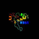
Top model
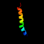
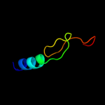
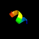
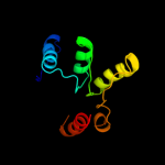
Image coloured by rainbow N → C terminus
Model (left) based on template d1jb0i_
Top template information
Fold:Single transmembrane helix
Superfamily:Subunit VIII of photosystem I reaction centre, PsaI
Family:Subunit VIII of photosystem I reaction centre, PsaI
Confidence and coverage
Confidence: 40.2% Coverage: 13%
29 residues ( 13% of your sequence) have been modelled with 40.2% confidence by the single highest scoring template.
You may wish to submit your sequence to Phyrealarm. This will automatically scan your sequence every week for new potential templates as they appear in the Phyre2 library.
Please note: You must be registered and logged in to use Phyrealarm.
3D viewing
Interactive 3D view in Jmol

Sequence analysis 

Secondary structure and disorder prediction 

   1........10.........20.........30.........40.........50.........60
Sequence  MREQIKQDIDLIEILFYLKKKIRVILFIMAICMAMVLLFLYINKDNIKVIYSLKINQTTP
Secondary structure 


SS confidence 


Disorder  ???


?


?

?
Disorder confidence 


   .........70.........80.........90.........100.........110.........120
Sequence  GILVSCDSNNNFACQTTMTEDVIQRITTFFHTSPDVKNREIRLEWSGDKRALPTAEEEIS
Secondary structure 


SS confidence 


Disorder 


?


Disorder confidence 


   .........130.........140.........150.........160.........170.........180
Sequence  RVQASIIKWYASEYHNGRQVLDEIQTPSAINSELYTKMIYLTRNWSLYPNGDGCVTISSP
Secondary structure 


SS confidence 


Disorder 


???
?


?
?
?


Disorder confidence 


   .........190.........200.........210.........220..
Sequence  EIKNKYPAAICLALGFFLSIVISVMFCLVKKMVDEYQQNSGQ
Secondary structure 


SS confidence 


Disorder 

?


?????
Disorder confidence 


Confidence Key
High(9)                    Low (0)
?Disordered
Alpha helix
Beta strand

Domain analysis 

Hover over an aligned region to see model and summary info

Please note, only up to the top 20 hits are modelled to reduce computer load

RankAligned region

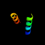
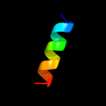
PDB 1jb0 chain I

3D model

Region: 17 - 45
Aligned: 29
Modelled: 29
Confidence: 40.2%
Identity: 28%
Fold: Single transmembrane helix
Superfamily: Subunit VIII of photosystem I reaction centre, PsaI
Family: Subunit VIII of photosystem I reaction centre, PsaI

Phyre2

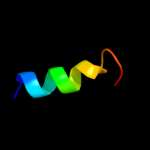
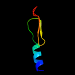
PDB 3lw5 chain I

3D model

Region: 29 - 44
Aligned: 16
Modelled: 16
Confidence: 37.0%
Identity: 38%
PDB header:photosynthesis
Chain: I: PDB Molecule:photosystem i reaction center subunit viii;
PDBTitle: improved model of plant photosystem i

Phyre2

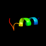
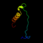
PDB 2wse chain I

3D model

Region: 29 - 44
Aligned: 16
Modelled: 16
Confidence: 37.0%
Identity: 38%
PDB header:photosynthesis
Chain: I: PDB Molecule:photosystem i reaction center subunit viii;
PDBTitle: improved model of plant photosystem i

Phyre2

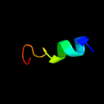
PDB 2o01 chain I

3D model

Region: 29 - 44
Aligned: 16
Modelled: 16
Confidence: 30.9%
Identity: 38%
PDB header:photosynthesis
Chain: I: PDB Molecule:photosystem i reaction center subunit viii;
PDBTitle: the structure of a plant photosystem i supercomplex at 3.42 angstrom resolution

Phyre2

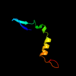
PDB 2nlv chain A domain 1

3D model

Region: 77 - 110
Aligned: 34
Modelled: 34
Confidence: 23.4%
Identity: 18%
Fold: XisI-like
Superfamily: XisI-like
Family: XisI-like

Phyre2

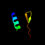
PDB 1n8i chain A

3D model

Region: 117 - 144
Aligned: 28
Modelled: 28
Confidence: 21.4%
Identity: 36%
Fold: TIM beta/alpha-barrel
Superfamily: Malate synthase G
Family: Malate synthase G

Phyre2

PDB 3d7q chain B

3D model

Region: 77 - 110
Aligned: 34
Modelled: 34
Confidence: 19.2%
Identity: 24%
PDB header:unknown function
Chain: B: PDB Molecule:xisi protein-like;
PDBTitle: crystal structure of a xisi-like protein (npun_ar114) from nostoc2 punctiforme pcc 73102 at 2.30 a resolution

Phyre2

PDB 2czv chain C domain 1

3D model

Region: 85 - 134
Aligned: 48
Modelled: 50
Confidence: 17.1%
Identity: 21%
Fold: Ferredoxin-like
Superfamily: Rnp2-like
Family: Rpp14/Pop5-like

Phyre2

PDB 2hxj chain A domain 1

3D model

Region: 75 - 181
Aligned: 87
Modelled: 105
Confidence: 11.5%
Identity: 16%
Fold: FinO-like
Superfamily: FinO-like
Family: FinO-like

Phyre2

PDB 1cf1 chain A domain 2

3D model

Region: 47 - 75
Aligned: 27
Modelled: 29
Confidence: 10.8%
Identity: 33%
Fold: Immunoglobulin-like beta-sandwich
Superfamily: E set domains
Family: Arrestin/Vps26-like

Phyre2

PDB 2axt chain H domain 1

3D model

Region: 28 - 42
Aligned: 15
Modelled: 15
Confidence: 10.4%
Identity: 47%
Fold: Single transmembrane helix
Superfamily: Photosystem II 10 kDa phosphoprotein PsbH
Family: PsbH-like

Phyre2

PDB 2pgg chain A domain 1

3D model

Region: 127 - 174
Aligned: 46
Modelled: 48
Confidence: 10.3%
Identity: 22%
Fold: DNA/RNA polymerases
Superfamily: DNA/RNA polymerases
Family: RNA-dependent RNA-polymerase

Phyre2

PDB 1wi9 chain A

3D model

Region: 79 - 107
Aligned: 29
Modelled: 29
Confidence: 10.2%
Identity: 38%
Fold: DNA/RNA-binding 3-helical bundle
Superfamily: "Winged helix" DNA-binding domain
Family: PCI domain (PINT motif)

Phyre2

PDB 2v52 chain M

3D model

Region: 17 - 24
Aligned: 8
Modelled: 8
Confidence: 10.0%
Identity: 75%
PDB header:structural protein/contractile protein
Chain: M: PDB Molecule:mkl/myocardin-like protein 1;
PDBTitle: structure of mal-rpel2 complexed to g-actin

Phyre2

PDB 3ady chain A

3D model

Region: 87 - 108
Aligned: 21
Modelled: 22
Confidence: 9.9%
Identity: 19%
PDB header:proton transport
Chain: A: PDB Molecule:dotd;
PDBTitle: crystal structure of dotd from legionella

Phyre2

PDB 2adz chain A domain 1

3D model

Region: 157 - 181
Aligned: 25
Modelled: 25
Confidence: 9.6%
Identity: 20%
Fold: PH domain-like barrel
Superfamily: PH domain-like
Family: Pleckstrin-homology domain (PH domain)

Phyre2

PDB 2ozb chain B domain 1

3D model

Region: 110 - 195
Aligned: 71
Modelled: 86
Confidence: 9.2%
Identity: 11%
Fold: Nop domain
Superfamily: Nop domain
Family: Nop domain

Phyre2

PDB 1li4 chain A domain 2

3D model

Region: 103 - 202
Aligned: 89
Modelled: 100
Confidence: 8.7%
Identity: 15%
Fold: Flavodoxin-like
Superfamily: Formate/glycerate dehydrogenase catalytic domain-like
Family: S-adenosylhomocystein hydrolase

Phyre2

PDB 2efv chain A domain 1

3D model

Region: 138 - 198
Aligned: 57
Modelled: 61
Confidence: 7.1%
Identity: 14%
Fold: Ribbon-helix-helix
Superfamily: Ribbon-helix-helix
Family: MJ0366-like

Phyre2

PDB 1ew4 chain A

3D model

Region: 108 - 148
Aligned: 40
Modelled: 41
Confidence: 6.4%
Identity: 18%
Fold: N domain of copper amine oxidase-like
Superfamily: Frataxin/Nqo15-like
Family: Frataxin-like

Phyre2
1

d1jb0i_
2

c3lw5I_
3

c2wseI_
4

c2o01I_
5

d2nlva1
6

d1n8ia_
7

c3d7qB_
8

d2czvc1
9

d2hxja1
10

d1cf1a2
11

d2axth1
12

d2pgga1
13

d1wi9a_
14

c2v52M_
15

c3adyA_
16

d2adza1
17

d2ozbb1
18

d1li4a2
19

d2efva1
20

d1ew4a_
21


22


23


24


25


26


27


28


29


30


Detailed template information 

#
Template Alignment Coverage3D Model Confidence
% i.d. Template Information
1d1jb0i_


40.2 28 Fold:Single transmembrane helix
Superfamily:Subunit VIII of photosystem I reaction centre, PsaI
Family:Subunit VIII of photosystem I reaction centre, PsaI
2c3lw5I_


37.0 38 PDB header:photosynthesis
Chain: I: PDB Molecule:photosystem i reaction center subunit viii;
PDBTitle: improved model of plant photosystem i
3c2wseI_


37.0 38 PDB header:photosynthesis
Chain: I: PDB Molecule:photosystem i reaction center subunit viii;
PDBTitle: improved model of plant photosystem i
4c2o01I_


30.9 38 PDB header:photosynthesis
Chain: I: PDB Molecule:photosystem i reaction center subunit viii;
PDBTitle: the structure of a plant photosystem i supercomplex at 3.42 angstrom resolution
5d2nlva1


23.4 18 Fold:XisI-like
Superfamily:XisI-like
Family:XisI-like
6d1n8ia_


21.4 36 Fold:TIM beta/alpha-barrel
Superfamily:Malate synthase G
Family:Malate synthase G
7c3d7qB_


19.2 24 PDB header:unknown function
Chain: B: PDB Molecule:xisi protein-like;
PDBTitle: crystal structure of a xisi-like protein (npun_ar114) from nostoc2 punctiforme pcc 73102 at 2.30 a resolution
8d2czvc1


17.1 21 Fold:Ferredoxin-like
Superfamily:Rnp2-like
Family:Rpp14/Pop5-like
9d2hxja1


11.5 16 Fold:FinO-like
Superfamily:FinO-like
Family:FinO-like
10d1cf1a2


10.8 33 Fold:Immunoglobulin-like beta-sandwich
Superfamily:E set domains
Family:Arrestin/Vps26-like
11d2axth1


10.4 47 Fold:Single transmembrane helix
Superfamily:Photosystem II 10 kDa phosphoprotein PsbH
Family:PsbH-like
12d2pgga1


10.3 22 Fold:DNA/RNA polymerases
Superfamily:DNA/RNA polymerases
Family:RNA-dependent RNA-polymerase
13d1wi9a_


10.2 38 Fold:DNA/RNA-binding 3-helical bundle
Superfamily:"Winged helix" DNA-binding domain
Family:PCI domain (PINT motif)
14c2v52M_


10.0 75 PDB header:structural protein/contractile protein
Chain: M: PDB Molecule:mkl/myocardin-like protein 1;
PDBTitle: structure of mal-rpel2 complexed to g-actin
15c3adyA_


9.9 19 PDB header:proton transport
Chain: A: PDB Molecule:dotd;
PDBTitle: crystal structure of dotd from legionella
16d2adza1


9.6 20 Fold:PH domain-like barrel
Superfamily:PH domain-like
Family:Pleckstrin-homology domain (PH domain)
17d2ozbb1


9.2 11 Fold:Nop domain
Superfamily:Nop domain
Family:Nop domain
18d1li4a2


8.7 15 Fold:Flavodoxin-like
Superfamily:Formate/glycerate dehydrogenase catalytic domain-like
Family:S-adenosylhomocystein hydrolase
19d2efva1


7.1 14 Fold:Ribbon-helix-helix
Superfamily:Ribbon-helix-helix
Family:MJ0366-like
20d1ew4a_


6.4 18 Fold:N domain of copper amine oxidase-like
Superfamily:Frataxin/Nqo15-like
Family:Frataxin-like
21c2p7vA_


not modelled 6.3 15 PDB header:transcription
Chain: A: PDB Molecule:regulator of sigma d;
PDBTitle: crystal structure of the escherichia coli regulator of sigma 70, rsd,2 in complex with sigma 70 domain 4
22c2xi1A_


not modelled 6.3 33 PDB header:viral protein
Chain: A: PDB Molecule:nef;
PDBTitle: crystal structure of the hiv-1 nef sequenced from a patient's sample
23d1uuja_


not modelled 6.0 30 Fold:Lissencephaly-1 protein (Lis-1, PAF-AH alpha) N-terminal domain
Superfamily:Lissencephaly-1 protein (Lis-1, PAF-AH alpha) N-terminal domain
Family:Lissencephaly-1 protein (Lis-1, PAF-AH alpha) N-terminal domain
24d2qw6a1


not modelled 6.0 50 Fold:post-AAA+ oligomerization domain-like
Superfamily:post-AAA+ oligomerization domain-like
Family:MgsA/YrvN C-terminal domain-like
25c2qw6A_


not modelled 6.0 50 PDB header:hydrolase
Chain: A: PDB Molecule:aaa atpase, central region;
PDBTitle: crystal structure of the c-terminal domain of an aaa atpase from2 enterococcus faecium do
26c2kw3C_


not modelled 5.9 23 PDB header:dna binding protein
Chain: C: PDB Molecule:regulatory factor x-associated protein;
PDBTitle: heterotrimeric interaction between rfx5 and rfxap
27c2r70A_


not modelled 5.8 22 PDB header:transferase
Chain: A: PDB Molecule:infectious bursal virus vp1 polymerase;
PDBTitle: crystal structure of infectious bursal disease virus vp12 polymerase, cocrystallized with an oligopeptide mimicking3 the vp3 c-terminus.
28c3ik5A_


not modelled 5.6 25 PDB header:viral protein/signaling protein
Chain: A: PDB Molecule:protein nef;
PDBTitle: sivmac239 nef in complex with tcr zeta itam 1 polypeptide2 (a63-r80)
29d1ayra2


not modelled 5.5 33 Fold:Immunoglobulin-like beta-sandwich
Superfamily:E set domains
Family:Arrestin/Vps26-like
30d1efnb_


not modelled 5.4 25 Fold:Regulatory factor Nef
Superfamily:Regulatory factor Nef
Family:Regulatory factor Nef

Binding site prediction 

Due to computational demand, binding site predictions are not run for batch jobs

If you want to predict binding sites, please manually submit your model of choice to 3DLigandSite


Phyre is for academic use only

Please cite: Protein structure prediction on the web: a case study using the Phyre server
Kelley LA and Sternberg MJE. Nature Protocols 4, 363 - 371 (2009) [pdf] [Import into BibTeX]
 
If you use the binding site predictions from 3DLigandSite, please also cite:
3DLigandSite: predicting ligand-binding sites using similar structures.
Wass MN, Kelley LA and Sternberg MJ Nucleic Acids Research 38, W469-73 (2010) [PubMed]
 
© Structural Bioinformatics Group
Imperial College London
Lawrence Kelley, Benjamin Jefferys
Disclaimer
Terms and Conditions
Component software
Template detection: HHpred 1.51
Secondary structure prediction: Psi-pred 2.5
Disorder prediction: Disopred 2.4
Transmembrane prediction: Memsat_SVM
Multi-template modelling and ab initio: Poing 1.0