1 c3k3gA_
85.9
12
PDB header: transport proteinChain: A: PDB Molecule: urea transporter;PDBTitle: crystal structure of the urea transporter from desulfovibrio vulgaris2 bound to 1,3-dimethylurea
2 c2ksfA_
54.6
16
PDB header: transferaseChain: A: PDB Molecule: sensor protein kdpd;PDBTitle: backbone structure of the membrane domain of e. coli2 histidine kinase receptor kdpd, center for structures of3 membrane proteins (csmp) target 4312c
3 d2r6gf1
36.2
11
Fold: MalF N-terminal region-likeSuperfamily: MalF N-terminal region-likeFamily: MalF N-terminal region-like4 d1ymga1
34.6
13
Fold: Aquaporin-likeSuperfamily: Aquaporin-likeFamily: Aquaporin-like5 c1ymgA_
34.6
13
PDB header: membrane proteinChain: A: PDB Molecule: lens fiber major intrinsic protein;PDBTitle: the channel architecture of aquaporin o at 2.2 angstrom resolution
6 c3llqB_
26.9
20
PDB header: membrane proteinChain: B: PDB Molecule: aquaporin z 2;PDBTitle: aquaporin structure from plant pathogen agrobacterium tumerfaciens
7 c2b5fD_
26.7
17
PDB header: transport protein,membrane proteinChain: D: PDB Molecule: aquaporin;PDBTitle: crystal structure of the spinach aquaporin sopip2;1 in an2 open conformation to 3.9 resolution
8 d1rc2a_
22.3
17
Fold: Aquaporin-likeSuperfamily: Aquaporin-likeFamily: Aquaporin-like9 c3k07A_
20.7
18
PDB header: transport proteinChain: A: PDB Molecule: cation efflux system protein cusa;PDBTitle: crystal structure of cusa
10 d1fx8a_
13.6
14
Fold: Aquaporin-likeSuperfamily: Aquaporin-likeFamily: Aquaporin-like11 c1ldaA_
13.6
14
PDB header: transport proteinChain: A: PDB Molecule: glycerol uptake facilitator protein;PDBTitle: crystal structure of the e. coli glycerol facilitator (glpf) without2 substrate glycerol
12 d1iwga7
13.2
17
Fold: Multidrug efflux transporter AcrB transmembrane domainSuperfamily: Multidrug efflux transporter AcrB transmembrane domainFamily: Multidrug efflux transporter AcrB transmembrane domain13 c2yvxD_
12.4
14
PDB header: transport proteinChain: D: PDB Molecule: mg2+ transporter mgte;PDBTitle: crystal structure of magnesium transporter mgte
14 d1kpla_
11.4
18
Fold: Clc chloride channelSuperfamily: Clc chloride channelFamily: Clc chloride channel15 c3d9sB_
10.8
11
PDB header: membrane proteinChain: B: PDB Molecule: aquaporin-5;PDBTitle: human aquaporin 5 (aqp5) - high resolution x-ray structure
16 c2kr6A_
10.2
17
PDB header: hydrolaseChain: A: PDB Molecule: presenilin-1;PDBTitle: solution structure of presenilin-1 ctf subunit
17 d2nr9a1
9.2
7
Fold: Rhomboid-likeSuperfamily: Rhomboid-likeFamily: Rhomboid-like18 c2w2eA_
8.9
16
PDB header: membrane proteinChain: A: PDB Molecule: aquaporin;PDBTitle: 1.15 angstrom crystal structure of p.pastoris aquaporin,2 aqy1, in a closed conformation at ph 3.5
19 c2d57A_
8.8
7
PDB header: transport proteinChain: A: PDB Molecule: aquaporin-4;PDBTitle: double layered 2d crystal structure of aquaporin-4 (aqp4m23) at 3.2 a2 resolution by electron crystallography
20 c2ht2B_
8.1
19
PDB header: membrane proteinChain: B: PDB Molecule: h(+)/cl(-) exchange transporter clca;PDBTitle: structure of the escherichia coli clc chloride channel2 y445h mutant and fab complex
21 c2oarA_
not modelled
7.8
20
PDB header: membrane proteinChain: A: PDB Molecule: large-conductance mechanosensitive channel;PDBTitle: mechanosensitive channel of large conductance (mscl)
22 d2oara1
not modelled
7.6
20
Fold: Gated mechanosensitive channelSuperfamily: Gated mechanosensitive channelFamily: Gated mechanosensitive channel23 c1oy8A_
not modelled
7.0
11
PDB header: membrane proteinChain: A: PDB Molecule: acriflavine resistance protein b;PDBTitle: structural basis of multiple drug binding capacity of the acrb2 multidrug efflux pump
24 d1h6ia_
not modelled
6.8
14
Fold: Aquaporin-likeSuperfamily: Aquaporin-likeFamily: Aquaporin-like25 c1ciiA_
not modelled
6.7
14
PDB header: transmembrane proteinChain: A: PDB Molecule: colicin ia;PDBTitle: colicin ia
26 c3dh4A_
not modelled
6.6
11
PDB header: transport proteinChain: A: PDB Molecule: sodium/glucose cotransporter;PDBTitle: crystal structure of sodium/sugar symporter with bound galactose from2 vibrio parahaemolyticus
27 c3mk7F_
not modelled
6.5
16
PDB header: oxidoreductaseChain: F: PDB Molecule: cytochrome c oxidase, cbb3-type, subunit p;PDBTitle: the structure of cbb3 cytochrome oxidase
28 c3nd0A_
not modelled
6.5
19
PDB header: transport proteinChain: A: PDB Molecule: sll0855 protein;PDBTitle: x-ray crystal structure of a slow cyanobacterial cl-/h+ antiporter
29 c3hzqA_
not modelled
6.5
17
PDB header: membrane proteinChain: A: PDB Molecule: large-conductance mechanosensitive channel;PDBTitle: structure of a tetrameric mscl in an expanded intermediate2 state
30 c2jo1A_
not modelled
6.4
19
PDB header: hydrolase regulatorChain: A: PDB Molecule: phospholemman;PDBTitle: structure of the na,k-atpase regulatory protein fxyd1 in2 micelles
31 d2yvxa3
not modelled
5.9
14
Fold: MgtE membrane domain-likeSuperfamily: MgtE membrane domain-likeFamily: MgtE membrane domain-like32 c3iyzA_
not modelled
5.9
11
PDB header: transport proteinChain: A: PDB Molecule: aquaporin-4;PDBTitle: structure of aquaporin-4 s180d mutant at 10.0 a resolution from2 electron micrograph
33 d1s7ba_
not modelled
5.7
12
Fold: Multidrug resistance efflux transporter EmrESuperfamily: Multidrug resistance efflux transporter EmrEFamily: Multidrug resistance efflux transporter EmrE34 c2zxeG_
not modelled
5.6
16
PDB header: hydrolase/transport proteinChain: G: PDB Molecule: phospholemman-like protein;PDBTitle: crystal structure of the sodium - potassium pump in the e2.2k+.pi2 state
35 d2o7ta2
not modelled
5.2
18
Fold: Tetracyclin repressor-like, C-terminal domainSuperfamily: Tetracyclin repressor-like, C-terminal domainFamily: Tetracyclin repressor-like, C-terminal domain36 d1j4na_
not modelled
5.2
13
Fold: Aquaporin-likeSuperfamily: Aquaporin-likeFamily: Aquaporin-like37 d1k1fa_
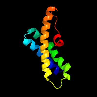
not modelled
5.1
20
Fold: Bcr-Abl oncoprotein oligomerization domainSuperfamily: Bcr-Abl oncoprotein oligomerization domainFamily: Bcr-Abl oncoprotein oligomerization domain