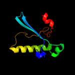
1 d1l5ia_
74.1
16
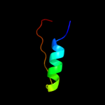
Fold: Origin of replication-binding domain, RBD-likeSuperfamily: Origin of replication-binding domain, RBD-likeFamily: DNA-binding domain of REP protein2 c3dkxA_
69.9
31
PDB header: replicationChain: A: PDB Molecule: replication protein repb;PDBTitle: crystal structure of the replication initiator protein2 encoded on plasmid pmv158 (repb), trigonal form, to 2.7 ang3 resolution
3 c2x3gA_
61.1
24
PDB header: viral proteinChain: A: PDB Molecule: sirv1 hypothetical protein orf119;PDBTitle: crystal structure of the hypothetical protein orf119 from2 sulfolobus islandicus rod-shaped virus 1
4 c2ns6A_
25.7
18
PDB header: hydrolaseChain: A: PDB Molecule: mobilization protein a;PDBTitle: crystal structure of the minimal relaxase domain of moba2 from plasmid r1162
5 d2nzca1
17.4
17
Fold: Ferredoxin-likeSuperfamily: ACT-likeFamily: TM1266-like6 c2hw0A_
11.5
9
PDB header: hydrolase, replicationChain: A: PDB Molecule: replicase;PDBTitle: nmr solution structure of the nuclease domain from the2 replicator initiator protein from porcine circovirus pcv2
7 c2hwtA_
10.2
9
PDB header: replication, hydrolaseChain: A: PDB Molecule: putative replicase-associated protein;PDBTitle: nmr solution structure of the master-rep protein nuclease2 domain (2-95) from the faba bean necrotic yellows virus
8 d1m55a_
10.2
11
Fold: Origin of replication-binding domain, RBD-likeSuperfamily: Origin of replication-binding domain, RBD-likeFamily: Replication protein Rep, nuclease domain9 c3maxB_
9.9
26
PDB header: hydrolaseChain: B: PDB Molecule: histone deacetylase 2;PDBTitle: crystal structure of human hdac2 complexed with an n-(2-aminophenyl)2 benzamide
10 c2h1xB_
9.9
36
PDB header: hydrolaseChain: B: PDB Molecule: 5-hydroxyisourate hydrolase (formerly known asPDBTitle: crystal structure of 5-hydroxyisourate hydrolase (formerly2 known as trp, transthyretin related protein)
11 d1f86a_
9.8
12
Fold: Prealbumin-likeSuperfamily: Transthyretin (synonym: prealbumin)Family: Transthyretin (synonym: prealbumin)12 c3ew8A_
9.6
19
PDB header: hydrolaseChain: A: PDB Molecule: histone deacetylase 8;PDBTitle: crystal structure analysis of human hdac8 d101l variant
13 c3ugsB_
9.1
19
PDB header: transferaseChain: B: PDB Molecule: undecaprenyl pyrophosphate synthase;PDBTitle: crystal structure of a probable undecaprenyl diphosphate synthase2 (upps) from campylobacter jejuni
14 d1t64a_
9.0
19
Fold: Arginase/deacetylaseSuperfamily: Arginase/deacetylaseFamily: Histone deacetylase, HDAC15 d1ttaa_
9.0
12
Fold: Prealbumin-likeSuperfamily: Transthyretin (synonym: prealbumin)Family: Transthyretin (synonym: prealbumin)16 c2elzA_
8.9
40
PDB header: transcriptionChain: A: PDB Molecule: zinc finger protein 224;PDBTitle: solution structure of the 17th zf-c2h2 domain from human2 zinc finger protein 224
17 d2b7ta1
8.5
11
Fold: dsRBD-likeSuperfamily: dsRNA-binding domain-likeFamily: Double-stranded RNA-binding domain (dsRBD)18 d1tfpa_
8.1
18
Fold: Prealbumin-likeSuperfamily: Transthyretin (synonym: prealbumin)Family: Transthyretin (synonym: prealbumin)19 c2h0eA_
7.9
31
PDB header: hydrolaseChain: A: PDB Molecule: transthyretin-like protein pucm;PDBTitle: crystal structure of pucm in the absence of substrate
20 c2gpzC_
7.8
29
PDB header: hydrolaseChain: C: PDB Molecule: transthyretin-like protein;PDBTitle: transthyretin-like protein from salmonella dublin
21 d2b7va1
not modelled
7.4
9
Fold: dsRBD-likeSuperfamily: dsRNA-binding domain-likeFamily: Double-stranded RNA-binding domain (dsRBD)22 c2jepB_
not modelled
6.7
16
PDB header: hydrolaseChain: B: PDB Molecule: xyloglucanase;PDBTitle: native family 5 xyloglucanase from paenibacillus pabuli
23 d1oo2a_
not modelled
6.6
25
Fold: Prealbumin-likeSuperfamily: Transthyretin (synonym: prealbumin)Family: Transthyretin (synonym: prealbumin)24 c2jttD_
not modelled
6.1
21
PDB header: calcium binding protein/antitumor proteiChain: D: PDB Molecule: calcyclin-binding protein;PDBTitle: solution structure of calcium loaded s100a6 bound to c-2 terminal siah-1 interacting protein
25 c2yttA_
not modelled
6.0
30
PDB header: transcriptionChain: A: PDB Molecule: zinc finger protein 473;PDBTitle: solution structure of the c2h2 type zinc finger (region 204-2 236) of human zinc finger protein 473
26 c2entA_
not modelled
5.9
40
PDB header: transcriptionChain: A: PDB Molecule: krueppel-like factor 15;PDBTitle: solution structure of the second c2h2-type zinc finger2 domain from human krueppel-like factor 15
27 d1luza_
not modelled
5.4
25
Fold: OB-foldSuperfamily: Nucleic acid-binding proteinsFamily: Cold shock DNA-binding domain-like28 d1kgia_
not modelled
5.3
19
Fold: Prealbumin-likeSuperfamily: Transthyretin (synonym: prealbumin)Family: Transthyretin (synonym: prealbumin)29 d1ohea1
not modelled
5.1
20
Fold: (Phosphotyrosine protein) phosphatases IISuperfamily: (Phosphotyrosine protein) phosphatases IIFamily: Dual specificity phosphatase-like