1 d2okqa1
100.0
100
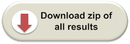
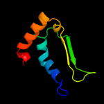
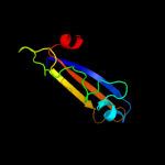
Fold: Ferredoxin-likeSuperfamily: Dimeric alpha+beta barrelFamily: YbaA-like2 c2okqB_
100.0
100
PDB header: structural genomics, unknown functionChain: B: PDB Molecule: hypothetical protein ybaa;PDBTitle: crystal structure of unknown conserved ybaa protein from2 shigella flexneri
3 d1vqsa_
87.4
19
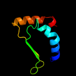
Fold: Ferredoxin-likeSuperfamily: Dimeric alpha+beta barrelFamily: NIPSNAP4 d1vqya1
86.1
19
Fold: Ferredoxin-likeSuperfamily: Dimeric alpha+beta barrelFamily: NIPSNAP5 d2fiua1
80.5
23
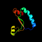
Fold: Ferredoxin-likeSuperfamily: Dimeric alpha+beta barrelFamily: Atu0297-like6 c3lo3E_
75.2
18
PDB header: structure genomics, unknown functionChain: E: PDB Molecule: uncharacterized conserved protein;PDBTitle: the crystal structure of a conserved functionally unknown2 protein from colwellia psychrerythraea 34h.
7 c2wghA_
53.4
14
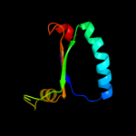
PDB header: oxidoreductaseChain: A: PDB Molecule: ribonucleoside-diphosphate reductase largePDBTitle: human ribonucleotide reductase r1 subunit (rrm1) in complex2 with datp and mg.
8 c3nfqA_
52.2
25
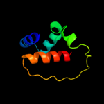
PDB header: transcriptionChain: A: PDB Molecule: transcription factor iws1;PDBTitle: crystal structure of the conserved central domain of yeast spn1/iws1
9 c2cvuA_
51.4
19
PDB header: oxidoreductaseChain: A: PDB Molecule: ribonucleoside-diphosphate reductase large chainPDBTitle: structures of yeast ribonucleotide reductase i
10 c3hnfA_
45.2
14
PDB header: oxidoreductaseChain: A: PDB Molecule: ribonucleoside-diphosphate reductase large subunit;PDBTitle: crystal structure of human ribonucleotide reductase 1 bound to the2 effectors ttp and datp
11 c2xpnA_
44.7
18
PDB header: transcriptionChain: A: PDB Molecule: iws1;PDBTitle: crystal structure of a spt6-iws1(spn1) complex from2 encephalitozoon cuniculi, form i
12 c3dcaC_
33.2
19
PDB header: structural genomics, unknown functionChain: C: PDB Molecule: rpa0582;PDBTitle: crystal structure of the rpa0582- protein of unknown2 function from rhodopseudomonas palustris- a structural3 genomics target
13 c3bm7A_
31.0
21
PDB header: oxidoreductaseChain: A: PDB Molecule: protein of unknown function with ferredoxin-like fold;PDBTitle: crystal structure of a putative antibiotic biosynthesis monooxygenase2 (cc_2132) from caulobacter crescentus cb15 at 1.35 a resolution
14 c2rilA_
29.9
22
PDB header: oxidoreductaseChain: A: PDB Molecule: antibiotic biosynthesis monooxygenase;PDBTitle: crystal structure of a putative monooxygenase (yp_001095275.1) from2 shewanella loihica pv-4 at 1.26 a resolution
15 c1pemA_
22.9
19
PDB header: oxidoreductaseChain: A: PDB Molecule: ribonucleoside-diphosphate reductase 2 alphaPDBTitle: ribonucleotide reductase protein r1e from salmonella2 typhimurium
16 d1iuja_
21.9
16
Fold: Ferredoxin-likeSuperfamily: Dimeric alpha+beta barrelFamily: PG130-like17 d1edqa1
21.1
27
Fold: Immunoglobulin-like beta-sandwichSuperfamily: E set domainsFamily: E-set domains of sugar-utilizing enzymes18 d1peqa2
20.6
21
Fold: PFL-like glycyl radical enzymesSuperfamily: PFL-like glycyl radical enzymesFamily: R1 subunit of ribonucleotide reductase, C-terminal domain19 d1xbwa_
19.4
17
Fold: Ferredoxin-likeSuperfamily: Dimeric alpha+beta barrelFamily: PG130-like20 c3chgB_
17.5
9
PDB header: ligand binding proteinChain: B: PDB Molecule: glycine betaine-binding protein;PDBTitle: the compatible solute-binding protein opuac from bacillus2 subtilis in complex with dmsa
21 c2qlxA_
not modelled
17.3
21
PDB header: isomeraseChain: A: PDB Molecule: l-rhamnose mutarotase;PDBTitle: crystal structure of rhamnose mutarotase rhau of rhizobium2 leguminosarum in complex with l-rhamnose
22 c2qlwA_
not modelled
17.2
21
PDB header: isomeraseChain: A: PDB Molecule: rhau;PDBTitle: crystal structure of rhamnose mutarotase rhau of rhizobium2 leguminosarum
23 d2k8ea1
not modelled
16.6
27
Fold: YegP-likeSuperfamily: YegP-likeFamily: YegP-like24 d1rlra2
not modelled
16.2
9
Fold: PFL-like glycyl radical enzymesSuperfamily: PFL-like glycyl radical enzymesFamily: R1 subunit of ribonucleotide reductase, C-terminal domain25 d2k49a2
not modelled
15.9
22
Fold: YegP-likeSuperfamily: YegP-likeFamily: YegP-like26 d1iwpa_
not modelled
14.3
39
Fold: TIM beta/alpha-barrelSuperfamily: Cobalamin (vitamin B12)-dependent enzymesFamily: Diol dehydratase, alpha subunit27 d1x4ha1
not modelled
12.5
9
Fold: Ferredoxin-likeSuperfamily: RNA-binding domain, RBDFamily: Canonical RBD28 d1fnxh2
not modelled
12.5
14
Fold: Ferredoxin-likeSuperfamily: RNA-binding domain, RBDFamily: Canonical RBD29 d2ix0a1
not modelled
12.4
22
Fold: OB-foldSuperfamily: Nucleic acid-binding proteinsFamily: Cold shock DNA-binding domain-like30 d1imta2
not modelled
11.7
56
Fold: Knottins (small inhibitors, toxins, lectins)Superfamily: Colipase-likeFamily: Colipase-like31 d2qtva2
not modelled
11.1
50
Fold: Common fold of diphtheria toxin/transcription factors/cytochrome fSuperfamily: beta-sandwich domain of Sec23/24Family: beta-sandwich domain of Sec23/2432 c3r1rB_
not modelled
10.6
8
PDB header: complex (oxidoreductase/peptide)Chain: B: PDB Molecule: ribonucleotide reductase r1 protein;PDBTitle: ribonucleotide reductase r1 protein with amppnp occupying2 the activity site from escherichia coli
33 d3bida1
not modelled
9.8
28
Fold: YegP-likeSuperfamily: YegP-likeFamily: YegP-like34 d2omoa1
not modelled
9.4
19
Fold: Ferredoxin-likeSuperfamily: Dimeric alpha+beta barrelFamily: PA3566-like35 d1eexa_
not modelled
9.3
35
Fold: TIM beta/alpha-barrelSuperfamily: Cobalamin (vitamin B12)-dependent enzymesFamily: Diol dehydratase, alpha subunit36 c1xjeA_
not modelled
9.3
25
PDB header: oxidoreductaseChain: A: PDB Molecule: ribonucleotide reductase, b12-dependent;PDBTitle: structural mechanism of allosteric substrate specificity in a2 ribonucleotide reductase: dttp-gdp complex
37 c3gh7A_
not modelled
8.8
13
PDB header: hydrolaseChain: A: PDB Molecule: beta-hexosaminidase;PDBTitle: crystal structure of beta-hexosaminidase from paenibacillus2 sp. ts12 in complex with galnac
38 d2k7ia1
not modelled
8.1
26
Fold: YegP-likeSuperfamily: YegP-likeFamily: YegP-like39 c2k7iB_
not modelled
8.1
26
PDB header: structural genomics, unknown functionChain: B: PDB Molecule: upf0339 protein atu0232;PDBTitle: solution nmr structure of protein atu0232 from agrobacterium2 tumefaciens. northeast structural genomics consortium (nesg) target3 att3. ontario center for structural proteomics target atc0223.
40 d2zdpa1
not modelled
7.4
8
Fold: Ferredoxin-likeSuperfamily: Dimeric alpha+beta barrelFamily: PG130-like41 c3p6yD_
not modelled
7.4
13
PDB header: rna binding protein/rnaChain: D: PDB Molecule: cleavage and polyadenylation specificity factor subunit 6;PDBTitle: cf im25-cf im68-uguaa complex
42 c2i38A_
not modelled
7.1
26
PDB header: rna binding protein/chimeraChain: A: PDB Molecule: fusion protein consists of immunoglobin g-PDBTitle: solution structure of the rrm of srp20
43 c2kdbA_
not modelled
6.6
19
PDB header: protein bindingChain: A: PDB Molecule: homocysteine-responsive endoplasmic reticulum-PDBTitle: solution structure of human ubiquitin-like domain of2 herpud2_9_85, northeast structural genomics consortium3 (nesg) target ht53a
44 c2k49A_
not modelled
6.6
21
PDB header: structural genomics, unknown functionChain: A: PDB Molecule: upf0339 protein so_3888;PDBTitle: solution nmr structure of upf0339 protein so3888 from shewanella2 oneidensis. northeast structural genomics consortium target sor190
45 c2f3jA_
not modelled
6.5
17
PDB header: transport proteinChain: A: PDB Molecule: rna and export factor binding protein 2;PDBTitle: the solution structure of the ref2-i mrna export factor2 (residues 1-155).
46 c1ylhA_
not modelled
5.6
14
PDB header: lyaseChain: A: PDB Molecule: phosphoenolpyruvate carboxykinase;PDBTitle: crystal structure of phosphoenolpyruvate carboxykinase from2 actinobaccilus succinogenes in complex with manganese and3 pyruvate
47 d1v3va1
not modelled
5.5
25
Fold: GroES-likeSuperfamily: GroES-likeFamily: Alcohol dehydrogenase-like, N-terminal domain48 d1tz0a_
not modelled
5.3
24
Fold: Ferredoxin-likeSuperfamily: Dimeric alpha+beta barrelFamily: PG130-like49 c2omoC_
not modelled
5.3
19
PDB header: oxidoreductaseChain: C: PDB Molecule: duf176;PDBTitle: putative antibiotic biosynthesis monooxygenase from nitrosomonas2 europaea