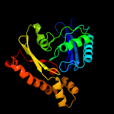
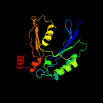
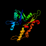
1 c3h1qB_
99.9
19
PDB header: structural proteinChain: B: PDB Molecule: ethanolamine utilization protein eutj;PDBTitle: crystal structure of ethanolamine utilization protein eutj from2 carboxydothermus hydrogenoformans
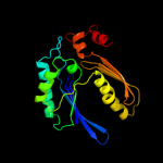
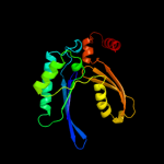
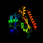
2 c2floA_
99.9
14
PDB header: hydrolaseChain: A: PDB Molecule: exopolyphosphatase;PDBTitle: crystal structure of exopolyphosphatase (ppx) from e. coli o157:h7
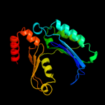
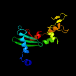
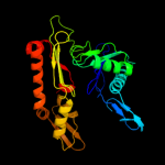
3 c3hi0B_
99.9
16
PDB header: hydrolaseChain: B: PDB Molecule: putative exopolyphosphatase;PDBTitle: crystal structure of putative exopolyphosphatase (17739545) from2 agrobacterium tumefaciens str. c58 (dupont) at 2.30 a resolution
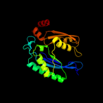
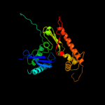
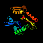
4 c1t6dB_
99.9
18
PDB header: hydrolaseChain: B: PDB Molecule: exopolyphosphatase;PDBTitle: miras phasing of the aquifex aeolicus ppx/gppa phosphatase: crystal2 structure of the type ii variant
5 c3cerD_
99.9
19
PDB header: structural genomics, unknown functionChain: D: PDB Molecule: possible exopolyphosphatase-like protein;PDBTitle: crystal structure of the exopolyphosphatase-like protein2 q8g5j2. northeast structural genomics consortium target3 blr13
6 c3mdqA_
99.9
15
PDB header: hydrolaseChain: A: PDB Molecule: exopolyphosphatase;PDBTitle: crystal structure of an exopolyphosphatase (chu_0316) from cytophaga2 hutchinsonii atcc 33406 at 1.50 a resolution
7 c2d0oA_
99.9
14
PDB header: chaperoneChain: A: PDB Molecule: diol dehydratase-reactivating factor largePDBTitle: strcuture of diol dehydratase-reactivating factor complexed2 with adp and mg2+
8 c1e4gT_
99.9
14
PDB header: bacterial cell divisionChain: T: PDB Molecule: cell division protein ftsa;PDBTitle: ftsa (atp-bound form) from thermotoga maritima
9 c1jcgA_
99.8
17
PDB header: structural proteinChain: A: PDB Molecule: rod shape-determining protein mreb;PDBTitle: mreb from thermotoga maritima, amppnp
10 c2v7yA_
99.8
19
PDB header: chaperoneChain: A: PDB Molecule: chaperone protein dnak;PDBTitle: crystal structure of the molecular chaperone dnak from2 geobacillus kaustophilus hta426 in post-atp hydrolysis3 state
11 c3iucC_
99.8
18
PDB header: chaperoneChain: C: PDB Molecule: heat shock 70kda protein 5 (glucose-regulatedPDBTitle: crystal structure of the human 70kda heat shock protein 52 (bip/grp78) atpase domain in complex with adp
12 c1dkgD_
99.8
18
PDB header: complex (hsp24/hsp70)Chain: D: PDB Molecule: molecular chaperone dnak;PDBTitle: crystal structure of the nucleotide exchange factor grpe2 bound to the atpase domain of the molecular chaperone dnak
13 c3d2fC_
99.8
16
PDB header: chaperoneChain: C: PDB Molecule: heat shock protein homolog sse1;PDBTitle: crystal structure of a complex of sse1p and hsp70
14 c2ychA_
99.8
20
PDB header: cell cycleChain: A: PDB Molecule: competence protein pilm;PDBTitle: pilm-piln type iv pilus biogenesis complex
15 c2v7zA_
99.7
20
PDB header: chaperoneChain: A: PDB Molecule: heat shock cognate 71 kda protein;PDBTitle: crystal structure of the 70-kda heat shock cognate protein2 from rattus norvegicus in post-atp hydrolysis state
16 c2khoA_
99.7
17
PDB header: chaperoneChain: A: PDB Molecule: heat shock protein 70;PDBTitle: nmr-rdc / xray structure of e. coli hsp70 (dnak) chaperone2 (1-605) complexed with adp and substrate
17 c3dwlB_
99.7
12
PDB header: structural proteinChain: B: PDB Molecule: actin-related protein 3;PDBTitle: crystal structure of fission yeast arp2/3 complex lacking the arp22 subunit
18 c1hpmA_
99.7
19
PDB header: hydrolase (acting on acid anhydrides)Chain: A: PDB Molecule: 44k atpase fragment (n-terminal) of 7o kd heat-PDBTitle: how potassium affects the activity of the molecular2 chaperone hsc70. ii. potassium binds specifically in the3 atpase active site
19 d1jcea2
99.7
20
Fold: Ribonuclease H-like motifSuperfamily: Actin-like ATPase domainFamily: Actin/HSP7020 c1o1f4_
99.7
11
PDB header: contractile proteinChain: 4: PDB Molecule: skeletal muscle actin;PDBTitle: molecular models of averaged rigor crossbridges from2 tomograms of insect flight muscle
21 d1e4ft2
not modelled
99.6
11
Fold: Ribonuclease H-like motifSuperfamily: Actin-like ATPase domainFamily: Actin/HSP7022 c2p9lA_
not modelled
99.6
13
PDB header: structural proteinChain: A: PDB Molecule: actin-like protein 3;PDBTitle: crystal structure of bovine arp2/3 complex
23 c2fsnB_
not modelled
99.6
15
PDB header: structural proteinChain: B: PDB Molecule: hypothetical protein ta0583;PDBTitle: crystal structure of ta0583, an archaeal actin homolog, complex with2 adp
24 c1mwmA_
not modelled
99.6
17
PDB header: structural proteinChain: A: PDB Molecule: parm;PDBTitle: parm from plasmid r1 adp form
25 c3qb0C_
not modelled
99.5
16
PDB header: structural proteinChain: C: PDB Molecule: actin-related protein 4;PDBTitle: crystal structure of actin-related protein arp4 from s. cerevisiae2 complexed with atp
26 d1u6za2
not modelled
99.5
13
Fold: Ribonuclease H-like motifSuperfamily: Actin-like ATPase domainFamily: Ppx/GppA phosphatase27 d1t6ca1
not modelled
99.4
17
Fold: Ribonuclease H-like motifSuperfamily: Actin-like ATPase domainFamily: Ppx/GppA phosphatase28 c3aapA_
not modelled
99.4
12
PDB header: hydrolaseChain: A: PDB Molecule: ectonucleoside triphosphate diphosphohydrolase i;PDBTitle: crystal structure of lp1ntpdase from legionella pneumophila
29 c3zx2A_
not modelled
99.3
13
PDB header: hydrolaseChain: A: PDB Molecule: ectonucleoside triphosphate diphosphohydrolase 1;PDBTitle: ntpdase1 in complex with decavanadate
30 c3js6A_
not modelled
99.2
11
PDB header: unknown functionChain: A: PDB Molecule: uncharacterized parm protein;PDBTitle: crystal structure of apo psk41 parm protein
31 d2e8aa2
not modelled
99.1
20
Fold: Ribonuclease H-like motifSuperfamily: Actin-like ATPase domainFamily: Actin/HSP7032 d1dkgd2
not modelled
99.0
19
Fold: Ribonuclease H-like motifSuperfamily: Actin-like ATPase domainFamily: Actin/HSP7033 d1bupa2
not modelled
98.9
20
Fold: Ribonuclease H-like motifSuperfamily: Actin-like ATPase domainFamily: Actin/HSP7034 c3cj9A_
not modelled
98.8
11
PDB header: hydrolaseChain: A: PDB Molecule: ectonucleoside triphosphate diphosphohydrolase 2;PDBTitle: structure of rattus norvegicus ntpdase2 in complex with2 calcium, amp and phosphate
35 d2zgya2
not modelled
98.8
14
Fold: Ribonuclease H-like motifSuperfamily: Actin-like ATPase domainFamily: Actin/HSP7036 d2fsja1
not modelled
98.8
13
Fold: Ribonuclease H-like motifSuperfamily: Actin-like ATPase domainFamily: Ta0583-like37 d1huxa_
not modelled
98.8
16
Fold: Ribonuclease H-like motifSuperfamily: Actin-like ATPase domainFamily: BadF/BadG/BcrA/BcrD-like38 d1jcea1
not modelled
98.5
15
Fold: Ribonuclease H-like motifSuperfamily: Actin-like ATPase domainFamily: Actin/HSP7039 d1u6za3
not modelled
98.3
19
Fold: Ribonuclease H-like motifSuperfamily: Actin-like ATPase domainFamily: Ppx/GppA phosphatase40 d1t6ca2
not modelled
98.3
17
Fold: Ribonuclease H-like motifSuperfamily: Actin-like ATPase domainFamily: Ppx/GppA phosphatase41 d2fxua2
not modelled
98.2
15
Fold: Ribonuclease H-like motifSuperfamily: Actin-like ATPase domainFamily: Actin/HSP7042 d1k8kb1
not modelled
98.2
17
Fold: Ribonuclease H-like motifSuperfamily: Actin-like ATPase domainFamily: Actin/HSP7043 d1nm1a2
not modelled
98.1
15
Fold: Ribonuclease H-like motifSuperfamily: Actin-like ATPase domainFamily: Actin/HSP7044 c3cetA_
not modelled
98.1
13
PDB header: structural genomics, unknown functionChain: A: PDB Molecule: conserved archaeal protein;PDBTitle: crystal structure of the pantheonate kinase-like protein2 q6m145 at the resolution 1.8 a. northeast structural3 genomics consortium target mrr63
45 d1k8ka2
not modelled
98.0
14
Fold: Ribonuclease H-like motifSuperfamily: Actin-like ATPase domainFamily: Actin/HSP7046 c1nbwA_
not modelled
98.0
18
PDB header: hydrolaseChain: A: PDB Molecule: glycerol dehydratase reactivase alpha subunit;PDBTitle: glycerol dehydratase reactivase
47 d2hf3a2
not modelled
97.9
15
Fold: Ribonuclease H-like motifSuperfamily: Actin-like ATPase domainFamily: Actin/HSP7048 c2p9kB_
not modelled
97.9
20
PDB header: structural proteinChain: B: PDB Molecule: actin-like protein 2;PDBTitle: crystal structure of bovine arp2/3 complex co-crystallized2 with atp and crosslinked with glutaraldehyde
49 c3agrB_
not modelled
97.8
17
PDB header: hydrolaseChain: B: PDB Molecule: nucleoside triphosphate hydrolase;PDBTitle: crystal structure of nucleoside triphosphate hydrolases from neospora2 caninum
50 d1yaga2
not modelled
97.8
15
Fold: Ribonuclease H-like motifSuperfamily: Actin-like ATPase domainFamily: Actin/HSP7051 c4a5bA_
not modelled
97.7
17
PDB header: hydrolaseChain: A: PDB Molecule: nucleoside-triphosphatase 2;PDBTitle: crystal structure of the c258s/c268s variant of toxoplasma gondii2 nucleoside triphosphate diphosphohydrolase 1 (ntpdase1)
52 d1e4ft1
not modelled
97.7
16
Fold: Ribonuclease H-like motifSuperfamily: Actin-like ATPase domainFamily: Actin/HSP7053 d1bupa1
not modelled
97.5
16
Fold: Ribonuclease H-like motifSuperfamily: Actin-like ATPase domainFamily: Actin/HSP7054 d2e8aa1
not modelled
97.4
17
Fold: Ribonuclease H-like motifSuperfamily: Actin-like ATPase domainFamily: Actin/HSP7055 d2d0oa3
not modelled
97.2
12
Fold: Ribonuclease H-like motifSuperfamily: Actin-like ATPase domainFamily: ATPase domain of dehydratase reactivase alpha subunit56 d1nbwa3
not modelled
97.1
14
Fold: Ribonuclease H-like motifSuperfamily: Actin-like ATPase domainFamily: ATPase domain of dehydratase reactivase alpha subunit57 d2ewsa1
not modelled
97.0
17
Fold: Ribonuclease H-like motifSuperfamily: Actin-like ATPase domainFamily: Fumble-like58 c1z6rC_
not modelled
96.6
15
PDB header: transcriptionChain: C: PDB Molecule: mlc protein;PDBTitle: crystal structure of mlc from escherichia coli
59 d1zc6a1
not modelled
96.5
11
Fold: Ribonuclease H-like motifSuperfamily: Actin-like ATPase domainFamily: BadF/BadG/BcrA/BcrD-like60 d1dkgd1
not modelled
96.4
18
Fold: Ribonuclease H-like motifSuperfamily: Actin-like ATPase domainFamily: Actin/HSP7061 d1c0fa1
not modelled
96.4
8
Fold: Ribonuclease H-like motifSuperfamily: Actin-like ATPase domainFamily: Actin/HSP7062 d2fxua1
not modelled
96.3
9
Fold: Ribonuclease H-like motifSuperfamily: Actin-like ATPase domainFamily: Actin/HSP7063 d2ch5a2
not modelled
95.7
11
Fold: Ribonuclease H-like motifSuperfamily: Actin-like ATPase domainFamily: BadF/BadG/BcrA/BcrD-like64 c3vgkB_
not modelled
95.5
16
PDB header: transferaseChain: B: PDB Molecule: glucokinase;PDBTitle: crystal structure of a rok family glucokinase from streptomyces2 griseus
65 d2p3ra1
not modelled
95.3
19
Fold: Ribonuclease H-like motifSuperfamily: Actin-like ATPase domainFamily: Glycerol kinase66 d2d0oa2
not modelled
95.1
20
Fold: Ribonuclease H-like motifSuperfamily: Actin-like ATPase domainFamily: ATPase domain of dehydratase reactivase alpha subunit67 c1woqB_
not modelled
94.9
20
PDB header: transferaseChain: B: PDB Molecule: inorganic polyphosphate/atp-glucomannokinase;PDBTitle: crystal structure of inorganic polyphosphate/atp-glucomannokinase from2 arthrobacter sp. strain km at 1.8 a resolution
68 c2ch5D_
not modelled
94.8
12
PDB header: transferaseChain: D: PDB Molecule: nagk protein;PDBTitle: crystal structure of human n-acetylglucosamine kinase in2 complex with n-acetylglucosamine
69 c1z05A_
not modelled
94.6
16
PDB header: transcriptionChain: A: PDB Molecule: transcriptional regulator, rok family;PDBTitle: crystal structure of the rok family transcriptional regulator, homolog2 of e.coli mlc protein.
70 c2cgkB_
not modelled
94.5
16
PDB header: transferaseChain: B: PDB Molecule: l-rhamnulose kinase;PDBTitle: crystal structure of l-rhamnulose kinase from escherichia2 coli in an open uncomplexed conformation.
71 c3flcX_
not modelled
94.4
18
PDB header: transferaseChain: X: PDB Molecule: glycerol kinase;PDBTitle: crystal structure of the his-tagged h232r mutant of glycerol kinase2 from enterococcus casseliflavus with glycerol
72 c3htvA_
not modelled
94.4
17
PDB header: transferaseChain: A: PDB Molecule: d-allose kinase;PDBTitle: crystal structure of d-allose kinase (np_418508.1) from escherichia2 coli k12 at 1.95 a resolution
73 d1nbwa2
not modelled
94.2
20
Fold: Ribonuclease H-like motifSuperfamily: Actin-like ATPase domainFamily: ATPase domain of dehydratase reactivase alpha subunit74 c3g25B_
not modelled
94.1
16
PDB header: transferaseChain: B: PDB Molecule: glycerol kinase;PDBTitle: 1.9 angstrom crystal structure of glycerol kinase (glpk) from2 staphylococcus aureus in complex with glycerol.
75 c2ap1A_
not modelled
94.0
16
PDB header: transferaseChain: A: PDB Molecule: putative regulator protein;PDBTitle: crystal structure of the putative regulatory protein
76 c2aa4B_
not modelled
94.0
17
PDB header: transferaseChain: B: PDB Molecule: putative n-acetylmannosamine kinase;PDBTitle: crystal structure of escherichia coli putative n-2 acetylmannosamine kinase, new york structural genomics3 consortium
77 d1yaga1
not modelled
93.7
9
Fold: Ribonuclease H-like motifSuperfamily: Actin-like ATPase domainFamily: Actin/HSP7078 c2zf5O_
not modelled
93.7
17
PDB header: transferaseChain: O: PDB Molecule: glycerol kinase;PDBTitle: crystal structure of highly thermostable glycerol kinase from a2 hyperthermophilic archaeon
79 c3ezwD_
not modelled
93.7
18
PDB header: transferaseChain: D: PDB Molecule: glycerol kinase;PDBTitle: crystal structure of a hyperactive escherichia coli glycerol kinase2 mutant gly230 --> asp obtained using microfluidic crystallization3 devices
80 c3i8bA_
not modelled
93.6
19
PDB header: transferaseChain: A: PDB Molecule: xylulose kinase;PDBTitle: the crystal structure of xylulose kinase from2 bifidobacterium adolescentis
81 c2d4wA_
not modelled
93.4
14
PDB header: transferaseChain: A: PDB Molecule: glycerol kinase;PDBTitle: crystal structure of glycerol kinase from cellulomonas sp.2 nt3060
82 c1glbG_
not modelled
93.4
19
PDB header: phosphotransferaseChain: G: PDB Molecule: glycerol kinase;PDBTitle: structure of the regulatory complex of escherichia coli iiiglc with2 glycerol kinase
83 c3hz6A_
not modelled
93.4
9
PDB header: transferaseChain: A: PDB Molecule: xylulokinase;PDBTitle: crystal structure of xylulokinase from chromobacterium violaceum
84 c2dpnB_
not modelled
93.2
22
PDB header: transferaseChain: B: PDB Molecule: glycerol kinase;PDBTitle: crystal structure of the glycerol kinase from thermus2 thermophilus hb8
85 c3r8eA_
not modelled
93.2
16
PDB header: transferaseChain: A: PDB Molecule: hypothetical sugar kinase;PDBTitle: crystal structure of a hypothetical sugar kinase (chu_1875) from2 cytophaga hutchinsonii atcc 33406 at 1.65 a resolution
86 c2w40C_
not modelled
92.7
20
PDB header: transferaseChain: C: PDB Molecule: glycerol kinase, putative;PDBTitle: crystal structure of plasmodium falciparum glycerol kinase2 with bound glycerol
87 c3ifrB_
not modelled
92.6
17
PDB header: transferaseChain: B: PDB Molecule: carbohydrate kinase, fggy;PDBTitle: the crystal structure of xylulose kinase from rhodospirillum rubrum
88 c1zc6A_
not modelled
92.4
13
PDB header: structural genomics, unknown functionChain: A: PDB Molecule: probable n-acetylglucosamine kinase;PDBTitle: crystal structure of putative n-acetylglucosamine kinase from2 chromobacterium violaceum. northeast structural genomics target3 cvr23.
89 c3gg4B_
not modelled
92.0
18
PDB header: transferaseChain: B: PDB Molecule: glycerol kinase;PDBTitle: the crystal structure of glycerol kinase from yersinia2 pseudotuberculosis
90 c2e2pA_
not modelled
91.8
14
PDB header: transferaseChain: A: PDB Molecule: hexokinase;PDBTitle: crystal structure of sulfolobus tokodaii hexokinase in2 complex with adp
91 c2qm1D_
not modelled
91.7
17
PDB header: transferaseChain: D: PDB Molecule: glucokinase;PDBTitle: crystal structure of glucokinase from enterococcus faecalis
92 c3h6eB_
not modelled
91.6
18
PDB header: transferaseChain: B: PDB Molecule: carbohydrate kinase, fggy;PDBTitle: the crystal structure of a carbohydrate kinase from novosphingobium2 aromaticivorans
93 c3jvpA_
not modelled
91.2
10
PDB header: transferaseChain: A: PDB Molecule: ribulokinase;PDBTitle: crystal structure of ribulokinase from bacillus halodurans
94 c3gbtA_
not modelled
91.2
30
PDB header: transferaseChain: A: PDB Molecule: gluconate kinase;PDBTitle: crystal structure of gluconate kinase from lactobacillus acidophilus
95 d2hf3a1
not modelled
91.1
10
Fold: Ribonuclease H-like motifSuperfamily: Actin-like ATPase domainFamily: Actin/HSP7096 c1xupO_
not modelled
89.4
19
PDB header: transferaseChain: O: PDB Molecule: glycerol kinase;PDBTitle: enterococcus casseliflavus glycerol kinase complexed with glycerol
97 c2hoeA_
not modelled
89.2
12
PDB header: transferaseChain: A: PDB Molecule: n-acetylglucosamine kinase;PDBTitle: crystal structure of n-acetylglucosamine kinase (tm1224) from2 thermotoga maritima at 2.46 a resolution
98 d1r59o1
not modelled
88.4
23
Fold: Ribonuclease H-like motifSuperfamily: Actin-like ATPase domainFamily: Glycerol kinase99 d1z6ra2
not modelled
88.2
18
Fold: Ribonuclease H-like motifSuperfamily: Actin-like ATPase domainFamily: ROK100 c1zbsA_
not modelled
87.8
12
PDB header: structural genomics, unknown functionChain: A: PDB Molecule: hypothetical protein pg1100;PDBTitle: crystal structure of the putative n-acetylglucosamine kinase (pg1100)2 from porphyromonas gingivalis, northeast structural genomics target3 pgr18
101 d1k8ka1
not modelled
87.2
11
Fold: Ribonuclease H-like motifSuperfamily: Actin-like ATPase domainFamily: Actin/HSP70102 c3eo3B_
not modelled
85.9
12
PDB header: isomerase, transferaseChain: B: PDB Molecule: bifunctional udp-n-acetylglucosamine 2-epimerase/n-PDBTitle: crystal structure of the n-acetylmannosamine kinase domain of human2 gne protein
103 c2iirJ_
not modelled
85.0
23
PDB header: transferaseChain: J: PDB Molecule: acetate kinase;PDBTitle: acetate kinase from a hypothermophile thermotoga maritima
104 c2nlxA_
not modelled
84.5
39
PDB header: transferaseChain: A: PDB Molecule: xylulose kinase;PDBTitle: crystal structure of the apo e. coli xylulose kinase
105 d1woqa1
not modelled
84.4
19
Fold: Ribonuclease H-like motifSuperfamily: Actin-like ATPase domainFamily: ROK106 d1g99a2
not modelled
83.0
17
Fold: Ribonuclease H-like motifSuperfamily: Actin-like ATPase domainFamily: Acetokinase-like107 c3khyA_
not modelled
83.0
18
PDB header: transferaseChain: A: PDB Molecule: propionate kinase;PDBTitle: crystal structure of a propionate kinase from francisella2 tularensis subsp. tularensis schu s4
108 c3p4iA_
not modelled
82.1
18
PDB header: transferaseChain: A: PDB Molecule: acetate kinase;PDBTitle: crystal structure of acetate kinase from mycobacterium avium
109 d2hoea3
not modelled
80.1
12
Fold: Ribonuclease H-like motifSuperfamily: Actin-like ATPase domainFamily: ROK110 c3enoB_
not modelled
79.9
17
PDB header: hydrolase/unknown functionChain: B: PDB Molecule: putative o-sialoglycoprotein endopeptidase;PDBTitle: crystal structure of pyrococcus furiosus pcc1 in complex2 with thermoplasma acidophilum kae1
111 d1saza1
not modelled
79.0
13
Fold: Ribonuclease H-like motifSuperfamily: Actin-like ATPase domainFamily: Acetokinase-like112 d2e1za2
not modelled
78.2
18
Fold: Ribonuclease H-like motifSuperfamily: Actin-like ATPase domainFamily: Acetokinase-like113 c2i7pA_
not modelled
78.0
25
PDB header: transferaseChain: A: PDB Molecule: pantothenate kinase 3;PDBTitle: crystal structure of human pank3 in complex with accoa
114 d1z05a3
not modelled
77.3
15
Fold: Ribonuclease H-like motifSuperfamily: Actin-like ATPase domainFamily: ROK115 c3djcA_
not modelled
75.4
11
PDB header: transferaseChain: A: PDB Molecule: type iii pantothenate kinase;PDBTitle: crystal structure of pantothenate kinase from legionella pneumophila
116 d2aa4a1
not modelled
75.0
16
Fold: Ribonuclease H-like motifSuperfamily: Actin-like ATPase domainFamily: ROK117 c1tuuA_
not modelled
75.0
15
PDB header: transferaseChain: A: PDB Molecule: acetate kinase;PDBTitle: acetate kinase crystallized with atpgs
118 c2ivoC_
not modelled
74.9
15
PDB header: hydrolaseChain: C: PDB Molecule: up1;PDBTitle: structure of up1 protein
119 c3mixA_
72.9
16
PDB header: protein transportChain: A: PDB Molecule: flagellar biosynthesis protein flha;PDBTitle: crystal structure of the cytosolic domain of b. subtilis flha
120 c2h3gX_
not modelled
71.9
11
PDB header: biosynthetic proteinChain: X: PDB Molecule: biosynthetic protein;PDBTitle: structure of the type iii pantothenate kinase (coax) from bacillus2 anthracis