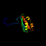
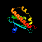
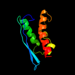
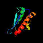
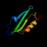
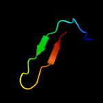
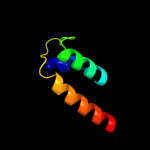
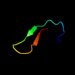
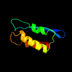
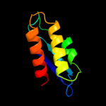
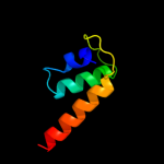
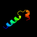
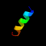
1 d1r9pa_
100.0
86
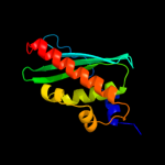
Fold: SufE/NifUSuperfamily: SufE/NifUFamily: NifU/IscU domain2 c2z7eB_
100.0
53
PDB header: biosynthetic proteinChain: B: PDB Molecule: nifu-like protein;PDBTitle: crystal structure of aquifex aeolicus iscu with bound [2fe-2 2s] cluster
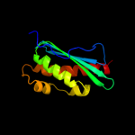
3 d1wfza_
100.0
74
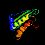
Fold: SufE/NifUSuperfamily: SufE/NifUFamily: NifU/IscU domain4 d1xjsa_
100.0
31
Fold: SufE/NifUSuperfamily: SufE/NifUFamily: NifU/IscU domain5 d1su0b_
100.0
38
Fold: SufE/NifUSuperfamily: SufE/NifUFamily: NifU/IscU domain6 c2qq4A_
100.0
32
PDB header: metal binding proteinChain: A: PDB Molecule: iron-sulfur cluster biosynthesis protein iscu;PDBTitle: crystal structure of iron-sulfur cluster biosynthesis2 protein iscu (ttha1736) from thermus thermophilus hb8
7 d1mzga_
96.2
13
Fold: SufE/NifUSuperfamily: SufE/NifUFamily: SufE-like8 d1ni7a_
94.9
21
Fold: SufE/NifUSuperfamily: SufE/NifUFamily: SufE-like9 c1wloA_
91.3
16
PDB header: structural genomics, unknown functionChain: A: PDB Molecule: sufe protein;PDBTitle: solution structure of the hypothetical protein from thermus2 thermophilus hb8
10 c2e5aA_
75.5
11
PDB header: ligaseChain: A: PDB Molecule: lipoyltransferase 1;PDBTitle: crystal structure of bovine lipoyltransferase in complex2 with lipoyl-amp
11 d1u0la1
67.4
22
Fold: OB-foldSuperfamily: Nucleic acid-binding proteinsFamily: Cold shock DNA-binding domain-like12 c2zkrr_
56.0
15
PDB header: ribosomal protein/rnaChain: R: PDB Molecule: rna expansion segment es39 part i;PDBTitle: structure of a mammalian ribosomal 60s subunit within an2 80s complex obtained by docking homology models of the rna3 and proteins into an 8.7 a cryo-em map
13 d1t9ha1
54.1
26
Fold: OB-foldSuperfamily: Nucleic acid-binding proteinsFamily: Cold shock DNA-binding domain-like14 d1v97a4
52.3
8
Fold: CO dehydrogenase flavoprotein C-domain-likeSuperfamily: CO dehydrogenase flavoprotein C-terminal domain-likeFamily: CO dehydrogenase flavoprotein C-terminal domain-like15 d1ffvc1
50.5
13
Fold: CO dehydrogenase flavoprotein C-domain-likeSuperfamily: CO dehydrogenase flavoprotein C-terminal domain-likeFamily: CO dehydrogenase flavoprotein C-terminal domain-like16 d1fs1b1
49.6
24
Fold: Skp1 dimerisation domain-likeSuperfamily: Skp1 dimerisation domain-likeFamily: Skp1 dimerisation domain-like17 d1fs2b1
42.2
24
Fold: Skp1 dimerisation domain-likeSuperfamily: Skp1 dimerisation domain-likeFamily: Skp1 dimerisation domain-like18 c3jywN_
38.3
18
PDB header: ribosomeChain: N: PDB Molecule: 60s ribosomal protein l17(a);PDBTitle: structure of the 60s proteins for eukaryotic ribosome based on cryo-em2 map of thermomyces lanuginosus ribosome at 8.9a resolution
19 d1nexa1
37.8
24
Fold: Skp1 dimerisation domain-likeSuperfamily: Skp1 dimerisation domain-likeFamily: Skp1 dimerisation domain-like20 c2kelB_
35.7
44
PDB header: transcription repressorChain: B: PDB Molecule: uncharacterized protein 56b;PDBTitle: structure of the transcription regulator svtr from the2 hyperthermophilic archaeal virus sirv1
21 d1i4ja_
not modelled
34.8
23
Fold: Ribosomal protein L22Superfamily: Ribosomal protein L22Family: Ribosomal protein L2222 d1n62c1
not modelled
33.6
15
Fold: CO dehydrogenase flavoprotein C-domain-likeSuperfamily: CO dehydrogenase flavoprotein C-terminal domain-likeFamily: CO dehydrogenase flavoprotein C-terminal domain-like23 c3cxoA_
not modelled
33.3
13
PDB header: lyaseChain: A: PDB Molecule: putative galactonate dehydratase;PDBTitle: crystal structure of l-rhamnonate dehydratase from2 salmonella typhimurium complexed with mg and 3-deoxy-l-3 rhamnonate
24 c4a17Q_
not modelled
33.2
16
PDB header: ribosomeChain: Q: PDB Molecule: rpl17;PDBTitle: t.thermophila 60s ribosomal subunit in complex with2 initiation factor 6. this file contains 5s rrna,3 5.8s rrna and proteins of molecule 2.
25 c3iz5V_
not modelled
33.0
18
PDB header: ribosomeChain: V: PDB Molecule: 60s ribosomal protein l17 (l22p);PDBTitle: localization of the large subunit ribosomal proteins into a 5.5 a2 cryo-em map of triticum aestivum translating 80s ribosome
26 c2ftcM_
not modelled
31.8
16
PDB header: ribosomeChain: M: PDB Molecule: mitochondrial ribosomal protein l22 isoform a;PDBTitle: structural model for the large subunit of the mammalian mitochondrial2 ribosome
27 d2ovra1
not modelled
31.7
24
Fold: Skp1 dimerisation domain-likeSuperfamily: Skp1 dimerisation domain-likeFamily: Skp1 dimerisation domain-like28 c1x2gB_
not modelled
28.7
18
PDB header: ligaseChain: B: PDB Molecule: lipoate-protein ligase a;PDBTitle: crystal structure of lipate-protein ligase a from2 escherichia coli
29 d1vqza1
not modelled
28.0
17
Fold: SufE/NifUSuperfamily: SufE/NifUFamily: SP1160 C-terminal domain-like30 d1t3qc1
not modelled
27.9
17
Fold: CO dehydrogenase flavoprotein C-domain-likeSuperfamily: CO dehydrogenase flavoprotein C-terminal domain-likeFamily: CO dehydrogenase flavoprotein C-terminal domain-like31 d1x2ga1
not modelled
27.6
26
Fold: SufE/NifUSuperfamily: SufE/NifUFamily: SP1160 C-terminal domain-like32 d2zjrp1
not modelled
25.9
23
Fold: Ribosomal protein L22Superfamily: Ribosomal protein L22Family: Ribosomal protein L2233 d2p92a1
not modelled
24.0
25
Fold: eIF1-likeSuperfamily: TM1457-likeFamily: TM1457-like34 c3bboU_
not modelled
23.6
23
PDB header: ribosomeChain: U: PDB Molecule: ribosomal protein l22;PDBTitle: homology model for the spinach chloroplast 50s subunit2 fitted to 9.4a cryo-em map of the 70s chlororibosome
35 d1jroa3
not modelled
21.5
13
Fold: CO dehydrogenase flavoprotein C-domain-likeSuperfamily: CO dehydrogenase flavoprotein C-terminal domain-likeFamily: CO dehydrogenase flavoprotein C-terminal domain-like36 c2p1nD_
not modelled
18.2
20
PDB header: signaling proteinChain: D: PDB Molecule: skp1-like protein 1a;PDBTitle: mechanism of auxin perception by the tir1 ubiqutin ligase
37 d2axoa1
not modelled
18.1
19
Fold: Thioredoxin foldSuperfamily: Thioredoxin-likeFamily: Atu2684-like38 c3hrdC_
not modelled
18.1
16
PDB header: oxidoreductaseChain: C: PDB Molecule: nicotinate dehydrogenase fad-subunit;PDBTitle: crystal structure of nicotinate dehydrogenase
39 d1vqor1
not modelled
15.8
24
Fold: Ribosomal protein L22Superfamily: Ribosomal protein L22Family: Ribosomal protein L2240 d1io2a_
not modelled
14.9
14
Fold: Ribonuclease H-like motifSuperfamily: Ribonuclease H-likeFamily: Ribonuclease H41 c1nexC_
not modelled
14.7
24
PDB header: ligase, cell cycleChain: C: PDB Molecule: centromere dna-binding protein complex cbf3PDBTitle: crystal structure of scskp1-sccdc4-cpd peptide complex
42 c2yv5A_
not modelled
14.3
17
PDB header: hydrolaseChain: A: PDB Molecule: yjeq protein;PDBTitle: crystal structure of yjeq from aquifex aeolicus
43 c1t9hA_
not modelled
14.0
24
PDB header: hydrolaseChain: A: PDB Molecule: probable gtpase engc;PDBTitle: the crystal structure of yloq, a circularly permuted gtpase.
44 c2axoA_
not modelled
13.8
19
PDB header: unknown functionChain: A: PDB Molecule: hypothetical protein atu2684;PDBTitle: x-ray crystal structure of protein agr_c_4864 from agrobacterium2 tumefaciens. northeast structural genomics consortium target atr35.
45 c3h5fB_
not modelled
13.0
27
PDB header: de novo proteinChain: B: PDB Molecule: coil ser l16l-pen;PDBTitle: switching the chirality of the metal environment alters the2 coordination mode in designed peptides.
46 c3h5fA_
not modelled
13.0
27
PDB header: de novo proteinChain: A: PDB Molecule: coil ser l16l-pen;PDBTitle: switching the chirality of the metal environment alters the2 coordination mode in designed peptides.
47 c3h5gA_
not modelled
13.0
27
PDB header: de novo proteinChain: A: PDB Molecule: coil ser l16d-pen;PDBTitle: switching the chirality of the metal environment alters the2 coordination mode in designed peptides.
48 c3h5gC_
not modelled
13.0
27
PDB header: de novo proteinChain: C: PDB Molecule: coil ser l16d-pen;PDBTitle: switching the chirality of the metal environment alters the2 coordination mode in designed peptides.
49 c3h5fC_
not modelled
13.0
27
PDB header: de novo proteinChain: C: PDB Molecule: coil ser l16l-pen;PDBTitle: switching the chirality of the metal environment alters the2 coordination mode in designed peptides.
50 c3h5gB_
not modelled
13.0
27
PDB header: de novo proteinChain: B: PDB Molecule: coil ser l16d-pen;PDBTitle: switching the chirality of the metal environment alters the2 coordination mode in designed peptides.
51 c2l55A_
not modelled
12.7
15
PDB header: metal binding proteinChain: A: PDB Molecule: silb,silver efflux protein, mfp component of the threePDBTitle: solution structure of the c-terminal domain of silb from cupriavidus2 metallidurans
52 d1rvka2
not modelled
12.5
9
Fold: Enolase N-terminal domain-likeSuperfamily: Enolase N-terminal domain-likeFamily: Enolase N-terminal domain-like53 c1zeqX_
not modelled
12.2
15
PDB header: metal binding proteinChain: X: PDB Molecule: cation efflux system protein cusf;PDBTitle: 1.5 a structure of apo-cusf residues 6-88 from escherichia2 coli
54 c1vqzA_
not modelled
12.0
17
PDB header: ligaseChain: A: PDB Molecule: lipoate-protein ligase, putative;PDBTitle: crystal structure of a putative lipoate-protein ligase a (sp_1160)2 from streptococcus pneumoniae tigr4 at 1.99 a resolution
55 c2ovqA_
not modelled
11.1
24
PDB header: transcription/cell cycleChain: A: PDB Molecule: s-phase kinase-associated protein 1a;PDBTitle: structure of the skp1-fbw7-cyclinedegc complex
56 c2p0iA_
not modelled
10.8
10
PDB header: lyaseChain: A: PDB Molecule: l-rhamnonate dehydratase;PDBTitle: crystal structure of l-rhamnonate dehydratase from gibberella zeae
57 c2jnvA_
not modelled
10.0
17
PDB header: metal transportChain: A: PDB Molecule: nifu-like protein 1, chloroplast;PDBTitle: solution structure of c-terminal domain of nifu-like2 protein from oryza sativa
58 c1u0lB_
not modelled
10.0
22
PDB header: hydrolaseChain: B: PDB Molecule: probable gtpase engc;PDBTitle: crystal structure of yjeq from thermotoga maritima
59 d1iloa_
not modelled
9.6
21
Fold: Thioredoxin foldSuperfamily: Thioredoxin-likeFamily: Thioltransferase60 d2if6a1
not modelled
8.8
18
Fold: Cysteine proteinasesSuperfamily: Cysteine proteinasesFamily: YiiX-like61 c3nyeA_
not modelled
8.8
20
PDB header: oxidoreductaseChain: A: PDB Molecule: d-arginine dehydrogenase;PDBTitle: crystal structure of pseudomonas aeruginosa d-arginine dehydrogenase2 in complex with imino-arginine
62 c3iz5w_
not modelled
8.0
35
PDB header: ribosomeChain: W: PDB Molecule: 60s ribosomal protein l22 (l22e);PDBTitle: localization of the large subunit ribosomal proteins into a 5.5 a2 cryo-em map of triticum aestivum translating 80s ribosome
63 c2fgxA_
not modelled
7.9
11
PDB header: structural genomics, unknown functionChain: A: PDB Molecule: putative thioredoxin;PDBTitle: solution nmr structure of protein ne2328 from nitrosomonas2 europaea. northeast structural genomics consortium target3 net3.
64 d1r8ja1
not modelled
7.6
29
Fold: KaiA/RbsU domainSuperfamily: KaiA/RbsU domainFamily: Circadian clock protein KaiA, C-terminal domain65 c2k8sA_
not modelled
7.4
21
PDB header: oxidoreductaseChain: A: PDB Molecule: thioredoxin;PDBTitle: solution nmr structure of dimeric thioredoxin-like protein2 ne0084 from nitrosomonas europea: northeast structural3 genomics target net6
66 d1ofcx1
not modelled
7.4
24
Fold: DNA/RNA-binding 3-helical bundleSuperfamily: Homeodomain-likeFamily: Myb/SANT domain67 d1lj8a3
not modelled
7.3
23
Fold: 6-phosphogluconate dehydrogenase C-terminal domain-likeSuperfamily: 6-phosphogluconate dehydrogenase C-terminal domain-likeFamily: Mannitol 2-dehydrogenase68 c2oz3F_
not modelled
7.2
13
PDB header: lyaseChain: F: PDB Molecule: mandelate racemase/muconate lactonizing enzyme;PDBTitle: crystal structure of l-rhamnonate dehydratase from azotobacter2 vinelandii
69 c3ic4A_
not modelled
7.2
5
PDB header: oxidoreductaseChain: A: PDB Molecule: glutaredoxin (grx-1);PDBTitle: the crystal structure of the glutaredoxin(grx-1) from archaeoglobus2 fulgidus
70 c3f4yF_
not modelled
7.0
41
PDB header: viral proteinChain: F: PDB Molecule: mutant peptide derived from hiv gp41 chr domain;PDBTitle: hiv gp41 six-helix bundle containing a mutant chr alpha-2 peptide sequence
71 d1egoa_
not modelled
7.0
21
Fold: Thioredoxin foldSuperfamily: Thioredoxin-likeFamily: Thioltransferase72 d1yeya2
not modelled
6.9
15
Fold: Enolase N-terminal domain-likeSuperfamily: Enolase N-terminal domain-likeFamily: Enolase N-terminal domain-like73 d1v2za_
not modelled
6.8
14
Fold: KaiA/RbsU domainSuperfamily: KaiA/RbsU domainFamily: Circadian clock protein KaiA, C-terminal domain74 d1rm6b1
not modelled
6.8
14
Fold: CO dehydrogenase flavoprotein C-domain-likeSuperfamily: CO dehydrogenase flavoprotein C-terminal domain-likeFamily: CO dehydrogenase flavoprotein C-terminal domain-like75 d1sv1a_
not modelled
6.7
14
Fold: KaiA/RbsU domainSuperfamily: KaiA/RbsU domainFamily: Circadian clock protein KaiA, C-terminal domain76 d1r5qa_
not modelled
6.5
7
Fold: KaiA/RbsU domainSuperfamily: KaiA/RbsU domainFamily: Circadian clock protein KaiA, C-terminal domain77 c2x6pA_
not modelled
6.4
29
PDB header: de novo proteinChain: A: PDB Molecule: coil ser l19c;PDBTitle: crystal structure of coil ser l19c
78 c2x6pC_
not modelled
6.4
29
PDB header: de novo proteinChain: C: PDB Molecule: coil ser l19c;PDBTitle: crystal structure of coil ser l19c
79 c2x6pB_
not modelled
6.4
29
PDB header: de novo proteinChain: B: PDB Molecule: coil ser l19c;PDBTitle: crystal structure of coil ser l19c
80 d2cona1
not modelled
6.4
30
Fold: Rubredoxin-likeSuperfamily: NOB1 zinc finger-likeFamily: NOB1 zinc finger-like81 d1nm3a1
not modelled
6.2
21
Fold: Thioredoxin foldSuperfamily: Thioredoxin-likeFamily: Thioltransferase82 c1cosB_
not modelled
6.1
29
PDB header: alpha-helical bundleChain: B: PDB Molecule: coiled serine;PDBTitle: crystal structure of a synthetic triple-stranded alpha-2 helical bundle
83 c1cosA_
not modelled
6.1
29
PDB header: alpha-helical bundleChain: A: PDB Molecule: coiled serine;PDBTitle: crystal structure of a synthetic triple-stranded alpha-2 helical bundle
84 c1cosC_
not modelled
6.1
29
PDB header: alpha-helical bundleChain: C: PDB Molecule: coiled serine;PDBTitle: crystal structure of a synthetic triple-stranded alpha-2 helical bundle
85 c1ffuF_
not modelled
6.1
14
PDB header: hydrolaseChain: F: PDB Molecule: cutm, flavoprotein of carbon monoxidePDBTitle: carbon monoxide dehydrogenase from hydrogenophaga2 pseudoflava which lacks the mo-pyranopterin moiety of the3 molybdenum cofactor
86 d2jnya1
not modelled
6.0
10
Fold: Trm112p-likeSuperfamily: Trm112p-likeFamily: Trm112p-like87 d1yq9h1
not modelled
6.0
28
Fold: HydB/Nqo4-likeSuperfamily: HydB/Nqo4-likeFamily: Nickel-iron hydrogenase, large subunit88 c2wpnB_
not modelled
5.9
28
PDB header: oxidoreductaseChain: B: PDB Molecule: periplasmic [nifese] hydrogenase, large subunit,PDBTitle: structure of the oxidised, as-isolated nifese hydrogenase2 from d. vulgaris hildenborough
89 c3pdgA_
not modelled
5.8
12
PDB header: unknown functionChain: A: PDB Molecule: fibronectin(iii)-like module;PDBTitle: structures of clostridium thermocellum cbha fibronectin(iii)-like2 modules
90 d1ryph_
not modelled
5.6
11
Fold: Ntn hydrolase-likeSuperfamily: N-terminal nucleophile aminohydrolases (Ntn hydrolases)Family: Proteasome subunits91 d2gl5a2
not modelled
5.5
13
Fold: Enolase N-terminal domain-likeSuperfamily: Enolase N-terminal domain-likeFamily: Enolase N-terminal domain-like92 c2kpiA_
not modelled
5.4
14
PDB header: structural genomics, unknown functionChain: A: PDB Molecule: uncharacterized protein sco3027;PDBTitle: solution nmr structure of streptomyces coelicolor sco30272 modeled with zn+2 bound, northeast structural genomics3 consortium target rr58
93 d1u07a_
not modelled
5.4
17
Fold: TolA/TonB C-terminal domainSuperfamily: TolA/TonB C-terminal domainFamily: TonB94 d1iyxa2
not modelled
5.2
20
Fold: Enolase N-terminal domain-likeSuperfamily: Enolase N-terminal domain-likeFamily: Enolase N-terminal domain-like95 c3b9jJ_
not modelled
5.2
7
PDB header: oxidoreductaseChain: J: PDB Molecule: xanthine oxidase;PDBTitle: structure of xanthine oxidase with 2-hydroxy-6-methylpurine
96 c3etrM_
not modelled
5.2
7
PDB header: oxidoreductaseChain: M: PDB Molecule: xanthine dehydrogenase/oxidase;PDBTitle: crystal structure of xanthine oxidase in complex with2 lumazine
97 d1rypi_
not modelled
5.2
19
Fold: Ntn hydrolase-likeSuperfamily: N-terminal nucleophile aminohydrolases (Ntn hydrolases)Family: Proteasome subunits98 c3hrdF_
not modelled
5.2
12
PDB header: oxidoreductaseChain: F: PDB Molecule: nicotinate dehydrogenase medium molybdopterinPDBTitle: crystal structure of nicotinate dehydrogenase
99 d1e3db_
not modelled
5.1
31
Fold: HydB/Nqo4-likeSuperfamily: HydB/Nqo4-likeFamily: Nickel-iron hydrogenase, large subunit


