| 1 | d1sg5a1
|
|
 |
100.0 |
100 |
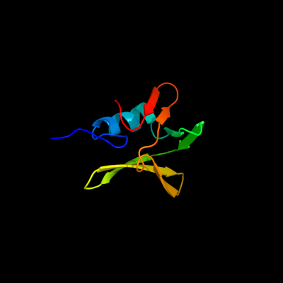
Fold:Rof/RNase P subunit-like
Superfamily:Rof/RNase P subunit-like
Family:Rof-like |
| 2 | c2e12B_
|
|
 |
54.5 |
22 |
PDB header:translation
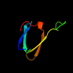
Chain: B: PDB Molecule:hypothetical protein xcc3642;
PDBTitle: the crystal structure of xc5848 from xanthomonas campestris2 adopting a novel variant of sm-like motif
|
| 3 | d1m5q1_
|
|
 |
51.9 |
16 |
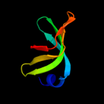
Fold:Sm-like fold
Superfamily:Sm-like ribonucleoproteins
Family:Sm motif of small nuclear ribonucleoproteins, SNRNP |
| 4 | c3cw15_
|
|
 |
41.4 |
25 |
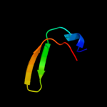
PDB header:splicing
Chain: 5: PDB Molecule:small nuclear ribonucleoprotein g;
PDB Fragment:residues 1-215;
PDBTitle: crystal structure of human spliceosomal u1 snrnp
|
| 5 | c1kq1W_
|
|
 |
40.6 |
13 |
PDB header:translation
Chain: W: PDB Molecule:host factor for q beta;
PDBTitle: 1.55 a crystal structure of the pleiotropic translational2 regulator, hfq
|
| 6 | c2cghB_
|
|
 |
40.1 |
19 |
PDB header:ligase
Chain: B: PDB Molecule:biotin ligase;
PDBTitle: crystal structure of biotin ligase from mycobacterium2 tuberculosis
|
| 7 | c3swnC_
|
|
 |
37.7 |
15 |
PDB header:rna binding protein
Chain: C: PDB Molecule:u6 snrna-associated sm-like protein lsm7;
PDBTitle: structure of the lsm657 complex: an assembly intermediate of the lsm12 7 and lsm2 8 rings
|
| 8 | c1b34A_
|
|
 |
30.7 |
27 |
PDB header:rna binding protein
Chain: A: PDB Molecule:protein (small nuclear ribonucleoprotein sm d1);
PDBTitle: crystal structure of the d1d2 sub-complex from the human snrnp core2 domain
|
| 9 | d1b34a_
|
|
 |
30.7 |
27 |
Fold:Sm-like fold
Superfamily:Sm-like ribonucleoproteins
Family:Sm motif of small nuclear ribonucleoproteins, SNRNP |
| 10 | c3swnT_
|
|
 |
27.6 |
19 |
PDB header:rna binding protein
Chain: T: PDB Molecule:u6 snrna-associated sm-like protein lsm6;
PDBTitle: structure of the lsm657 complex: an assembly intermediate of the lsm12 7 and lsm2 8 rings
|
| 11 | c3d6wA_
|
|
 |
26.6 |
8 |
PDB header:dna binding protein
Chain: A: PDB Molecule:methyl-accepting/dna response regulator;
PDBTitle: lyttr dna-binding domain of putative methyl-accepting/dna response2 regulator from bacillus cereus.
|
| 12 | c3cw1D_
|
|
 |
25.1 |
24 |
PDB header:splicing
Chain: D: PDB Molecule:small nuclear ribonucleoprotein sm d3;
PDBTitle: crystal structure of human spliceosomal u1 snrnp
|
| 13 | d1d3bb_
|
|
 |
22.9 |
21 |
Fold:Sm-like fold
Superfamily:Sm-like ribonucleoproteins
Family:Sm motif of small nuclear ribonucleoproteins, SNRNP |
| 14 | d1b34b_
|
|
 |
20.4 |
11 |
Fold:Sm-like fold
Superfamily:Sm-like ribonucleoproteins
Family:Sm motif of small nuclear ribonucleoproteins, SNRNP |
| 15 | c1b34B_
|
|
 |
20.4 |
11 |
PDB header:rna binding protein
Chain: B: PDB Molecule:protein (small nuclear ribonucleoprotein sm d2);
PDBTitle: crystal structure of the d1d2 sub-complex from the human snrnp core2 domain
|
| 16 | c3qiiA_
|
|
 |
19.3 |
17 |
PDB header:transcription regulator
Chain: A: PDB Molecule:phd finger protein 20;
PDBTitle: crystal structure of tudor domain 2 of human phd finger protein 20
|
| 17 | c2xk0A_
|
|
 |
18.1 |
13 |
PDB header:transcription
Chain: A: PDB Molecule:polycomb protein pcl;
PDBTitle: solution structure of the tudor domain from drosophila2 polycomblike (pcl)
|
| 18 | c3cw1Z_
|
|
 |
17.6 |
15 |
PDB header:splicing
Chain: Z: PDB Molecule:small nuclear ribonucleoprotein f;
PDBTitle: crystal structure of human spliceosomal u1 snrnp
|
| 19 | d1hk9a_
|
|
 |
17.2 |
19 |
Fold:Sm-like fold
Superfamily:Sm-like ribonucleoproteins
Family:Pleiotropic translational regulator Hfq |
| 20 | c2fwkB_
|
|
 |
16.9 |
5 |
PDB header:dna binding protein
Chain: B: PDB Molecule:u6 snrna-associated sm-like protein lsm5;
PDBTitle: crystal structure of cryptosporidium parvum u6 snrna-associated sm-2 like protein lsm5
|
| 21 | d1a9xb1 |
|
not modelled |
16.5 |
24 |
Fold:The "swivelling" beta/beta/alpha domain
Superfamily:Carbamoyl phosphate synthetase, small subunit N-terminal domain
Family:Carbamoyl phosphate synthetase, small subunit N-terminal domain |
| 22 | c3qhsD_ |
|
not modelled |
15.7 |
19 |
PDB header:rna binding protein
Chain: D: PDB Molecule:protein hfq;
PDBTitle: crystal structure of full-length hfq from escherichia coli
|
| 23 | d1i4k1_ |
|
not modelled |
15.2 |
15 |
Fold:Sm-like fold
Superfamily:Sm-like ribonucleoproteins
Family:Sm motif of small nuclear ribonucleoproteins, SNRNP |
| 24 | c3bw1A_ |
|
not modelled |
14.5 |
11 |
PDB header:rna binding protein
Chain: A: PDB Molecule:u6 snrna-associated sm-like protein lsm3;
PDBTitle: crystal structure of homomeric yeast lsm3 exhibiting novel octameric2 ring organisation
|
| 25 | d1th7a1 |
|
not modelled |
14.1 |
15 |
Fold:Sm-like fold
Superfamily:Sm-like ribonucleoproteins
Family:Sm motif of small nuclear ribonucleoproteins, SNRNP |
| 26 | c3cw1A_ |
|
not modelled |
13.5 |
16 |
PDB header:splicing
Chain: A: PDB Molecule:small nuclear ribonucleoprotein-associated proteins b and
PDBTitle: crystal structure of human spliceosomal u1 snrnp
|
| 27 | c3swnA_ |
|
not modelled |
13.3 |
10 |
PDB header:rna binding protein
Chain: A: PDB Molecule:u6 snrna-associated sm-like protein lsm5;
PDBTitle: structure of the lsm657 complex: an assembly intermediate of the lsm12 7 and lsm2 8 rings
|
| 28 | c3hsbB_ |
|
not modelled |
13.3 |
16 |
PDB header:rna binding protein/rna
Chain: B: PDB Molecule:protein hfq;
PDBTitle: crystal structure of ymah (hfq) from bacillus subtilis in complex with2 an rna aptamer
|
| 29 | c2ewnA_ |
|
not modelled |
12.6 |
11 |
PDB header:ligase, transcription
Chain: A: PDB Molecule:bira bifunctional protein;
PDBTitle: ecoli biotin repressor with co-repressor analog
|
| 30 | d1mgqa_ |
|
not modelled |
12.0 |
11 |
Fold:Sm-like fold
Superfamily:Sm-like ribonucleoproteins
Family:Sm motif of small nuclear ribonucleoproteins, SNRNP |
| 31 | c2ej9A_ |
|
not modelled |
11.9 |
9 |
PDB header:ligase
Chain: A: PDB Molecule:putative biotin ligase;
PDBTitle: crystal structure of biotin protein ligase from2 methanococcus jannaschii
|
| 32 | d1i8fa_ |
|
not modelled |
11.8 |
10 |
Fold:Sm-like fold
Superfamily:Sm-like ribonucleoproteins
Family:Sm motif of small nuclear ribonucleoproteins, SNRNP |
| 33 | d1jbma_ |
|
not modelled |
11.2 |
12 |
Fold:Sm-like fold
Superfamily:Sm-like ribonucleoproteins
Family:Sm motif of small nuclear ribonucleoproteins, SNRNP |
| 34 | d1d3ba_ |
|
not modelled |
11.2 |
23 |
Fold:Sm-like fold
Superfamily:Sm-like ribonucleoproteins
Family:Sm motif of small nuclear ribonucleoproteins, SNRNP |
| 35 | c3p8dB_ |
|
not modelled |
11.0 |
17 |
PDB header:protein binding
Chain: B: PDB Molecule:medulloblastoma antigen mu-mb-50.72;
PDBTitle: crystal structure of the second tudor domain of human phf20 (homodimer2 form)
|
| 36 | c3pgwQ_ |
|
not modelled |
10.3 |
13 |
PDB header:splicing/dna/rna
Chain: Q: PDB Molecule:sm b;
PDBTitle: crystal structure of human u1 snrnp
|
| 37 | d1xg8a_ |
|
not modelled |
10.2 |
23 |
Fold:Thioredoxin fold
Superfamily:Thioredoxin-like
Family:YuzD-like |
| 38 | c1ssfA_ |
|
not modelled |
9.5 |
11 |
PDB header:cell cycle
Chain: A: PDB Molecule:transformation related protein 53 binding
PDBTitle: solution structure of the mouse 53bp1 fragment (residues2 1463-1617)
|
| 39 | d1etha1 |
|
not modelled |
9.1 |
17 |
Fold:Lipase/lipooxygenase domain (PLAT/LH2 domain)
Superfamily:Lipase/lipooxygenase domain (PLAT/LH2 domain)
Family:Colipase-binding domain |
| 40 | d2zgwa1 |
|
not modelled |
9.1 |
27 |
Fold:SH3-like barrel
Superfamily:C-terminal domain of transcriptional repressors
Family:Biotin repressor (BirA) |
| 41 | c1wwtA_ |
|
not modelled |
8.9 |
29 |
PDB header:ligase
Chain: A: PDB Molecule:threonyl-trna synthetase, cytoplasmic;
PDBTitle: solution structure of the tgs domain from human threonyl-2 trna synthetase
|
| 42 | c1keeH_ |
|
not modelled |
8.8 |
22 |
PDB header:ligase
Chain: H: PDB Molecule:carbamoyl-phosphate synthetase small chain;
PDBTitle: inactivation of the amidotransferase activity of carbamoyl phosphate2 synthetase by the antibiotic acivicin
|
| 43 | d2fwka1 |
|
not modelled |
8.8 |
4 |
Fold:Sm-like fold
Superfamily:Sm-like ribonucleoproteins
Family:Sm motif of small nuclear ribonucleoproteins, SNRNP |
| 44 | c1y96C_ |
|
not modelled |
8.8 |
7 |
PDB header:rna binding protein
Chain: C: PDB Molecule:gem-associated protein 6;
PDBTitle: crystal structure of the gemin6/gemin7 heterodimer from the2 human smn complex
|
| 45 | d1d3bl_ |
|
not modelled |
8.7 |
16 |
Fold:Sm-like fold
Superfamily:Sm-like ribonucleoproteins
Family:Sm motif of small nuclear ribonucleoproteins, SNRNP |
| 46 | d1h641_ |
|
not modelled |
8.5 |
12 |
Fold:Sm-like fold
Superfamily:Sm-like ribonucleoproteins
Family:Sm motif of small nuclear ribonucleoproteins, SNRNP |
| 47 | c3mlqE_ |
|
not modelled |
8.0 |
21 |
PDB header:transferase/transcription
Chain: E: PDB Molecule:transcription-repair coupling factor;
PDBTitle: crystal structure of the thermus thermophilus transcription-repair2 coupling factor rna polymerase interacting domain with the thermus3 aquaticus rna polymerase beta1 domain
|
| 48 | d1u1sa1 |
|
not modelled |
7.8 |
19 |
Fold:Sm-like fold
Superfamily:Sm-like ribonucleoproteins
Family:Pleiotropic translational regulator Hfq |
| 49 | c2pvsB_ |
|
not modelled |
7.7 |
11 |
PDB header:hydrolase
Chain: B: PDB Molecule:pancreatic lipase-related protein 2;
PDBTitle: structure of human pancreatic lipase related protein 22 mutant n336q
|
| 50 | d1n9ra_ |
|
not modelled |
7.3 |
11 |
Fold:Sm-like fold
Superfamily:Sm-like ribonucleoproteins
Family:Sm motif of small nuclear ribonucleoproteins, SNRNP |
| 51 | c2advB_ |
|
not modelled |
6.7 |
17 |
PDB header:hydrolase
Chain: B: PDB Molecule:glutaryl 7- aminocephalosporanic acid acylase;
PDBTitle: crystal structures of glutaryl 7-aminocephalosporanic acid acylase:2 mutational study of activation mechanism
|
| 52 | c3mlqH_ |
|
not modelled |
5.9 |
21 |
PDB header:transferase/transcription
Chain: H: PDB Molecule:transcription-repair coupling factor;
PDBTitle: crystal structure of the thermus thermophilus transcription-repair2 coupling factor rna polymerase interacting domain with the thermus3 aquaticus rna polymerase beta1 domain
|
| 53 | c1n9sH_ |
|
not modelled |
5.8 |
9 |
PDB header:translation
Chain: H: PDB Molecule:small nuclear ribonucleoprotein f;
PDBTitle: crystal structure of yeast smf in spacegroup p43212
|
| 54 | d1kn0a_ |
|
not modelled |
5.8 |
29 |
Fold:dsRBD-like
Superfamily:dsRNA-binding domain-like
Family:The homologous-pairing domain of Rad52 recombinase |
| 55 | d1kq1a_ |
|
not modelled |
5.5 |
13 |
Fold:Sm-like fold
Superfamily:Sm-like ribonucleoproteins
Family:Pleiotropic translational regulator Hfq |
| 56 | c3pgwB_ |
|
not modelled |
5.5 |
13 |
PDB header:splicing/dna/rna
Chain: B: PDB Molecule:sm b;
PDBTitle: crystal structure of human u1 snrnp
|
| 57 | d1n9sc_ |
|
not modelled |
5.2 |
11 |
Fold:Sm-like fold
Superfamily:Sm-like ribonucleoproteins
Family:Sm motif of small nuclear ribonucleoproteins, SNRNP |
| 58 | d2hqxa1 |
|
not modelled |
5.0 |
13 |
Fold:SH3-like barrel
Superfamily:Tudor/PWWP/MBT
Family:Tudor domain |