 |
|
 |
| Summary |
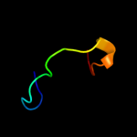
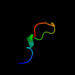
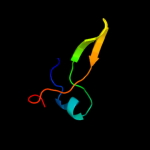
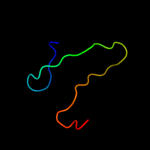
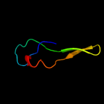
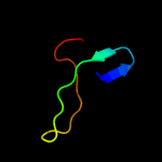
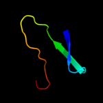
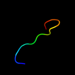
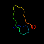
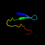
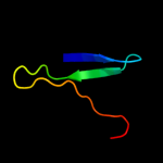
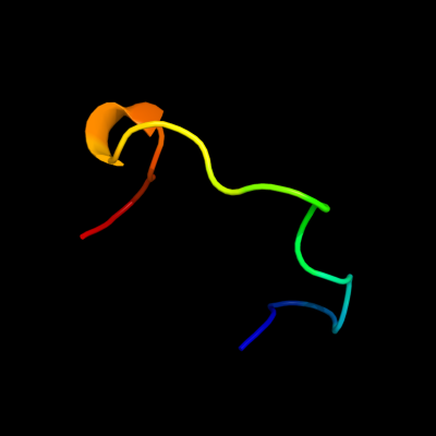
| Top model | ||||||||||||||||||||
|
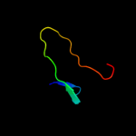
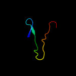
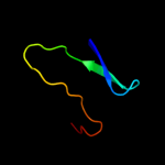
| |||||||||||||||||||

| Sequence analysis |
| Secondary structure and disorder prediction |
| 1 | . | . | . | . | . | . | . | . | 10 | . | . | . | . | . | . | . | . | . | 20 | . | . | . | . | . | . | . | . | . | 30 | . | . | . | . | . | . | . | . | . | 40 | . | . | . | . | . | . | . | . | . | 50 | . | . | . | . | . | . | . | . | . | 60 | |||||||||||||||||||||
| Sequence | M | K | F | G | M | G | S | A | Q | A | C | P | C | Q | V | P | R | A | A | S | T | T | W | V | P | C | Q | I | C | E | T | R | G | A | A | A | A | P | P | L | T | L | E | G | P | V | Q | S | H | H | G | T | P | A | L | T | Q | G | P | Q | ||||||||||||||||||||
| Secondary structure |  | | | | | |  |  | | | | | | | | |||||||||||||||||||||||||||||||||||||||||||||||||||||||||||||||||
| SS confidence | ||||||||||||||||||||||||||||||||||||||||||||||||||||||||||||||||||||||||||||||||
| Disorder | ? | ? | ? | ? | ? | ? | ? | ? | ? | ? | ? | ? | ? | ? | ? | ? | ? | ? | ? | ? | ? | ? | ? | ? | ? | ? | ? | ? | ? | ? | ? | |||||||||||||||||||||||||||||||||||||||||||||||||
| Disorder confidence | ||||||||||||||||||||||||||||||||||||||||||||||||||||||||||||||||||||||||||||||||
| . | . | . | . | . | . | . | . | . | 70 | . | . | . | . | . | . | . | . | . | 80 | . | . | . | . | . | . | . | . | . | 90 | . | . | . | . | . | . | . | . | . | 100 | . | ||||||||||||||||||||||||||||||||||||||||
| Sequence | S | P | R | D | G | A | Q | L | G | A | C | T | R | P | V | D | V | R | D | S | G | G | R | P | L | P | P | P | D | T | L | A | S | A | G | D | F | L | C | T | M | |||||||||||||||||||||||||||||||||||||||
| Secondary structure | | | | | | | | | | | | | | | | | | |||||||||||||||||||||||||||||||||||||||||||||||||||||||||||||||
| SS confidence | ||||||||||||||||||||||||||||||||||||||||||||||||||||||||||||||||||||||||||||||||
| Disorder | ? | ? | ? | ? | ? | ? | ? | ? | ? | ? | ? | ? | ? | ? | ? | ? | ? | ? | ? | ? | ? | ? | ? | ? | ||||||||||||||||||||||||||||||||||||||||||||||||||||||||
| Disorder confidence | ||||||||||||||||||||||||||||||||||||||||||||||||||||||||||||||||||||||||||||||||
| Confidence Key | |||||||||||
| High(9) | Low (0) | ||||||||||
| ? | Disordered |
| Alpha helix |
| Beta strand |
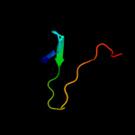
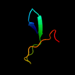
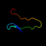
| Domain analysis |
Hover over an aligned region to see model and summary info
Please note, only up to the top 20 hits are modelled to reduce computer load
| |||||||||||||||||||||||||||||||||||||||||||||||||||||||||||||||||||||||||||||||||||||||||||||||||||||||||||||||||||||||||||||||||||||||||||||||||||||||||||||||||||||||||||||||
|