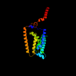
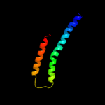
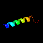
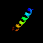
Region: 395 - 435 Aligned: 41 Modelled: 41 Confidence: 8.2% Identity: 10% Fold: Transmembrane helix hairpin Superfamily: Magnesium transport protein CorA, transmembrane region Family: Magnesium transport protein CorA, transmembrane region
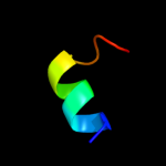
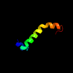
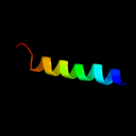
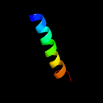
Region: 412 - 435 Aligned: 24 Modelled: 24 Confidence: 5.3% Identity: 21% PDB header:electron transport Chain: T: PDB Molecule:photosystem ii reaction center protein t; PDBTitle: crystal structure of monomeric form of cyanobacterial photosystem ii
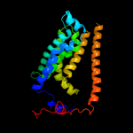
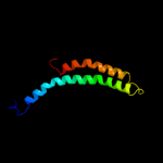
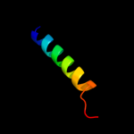
Region: 412 - 435 Aligned: 24 Modelled: 24 Confidence: 5.3% Identity: 21% PDB header:electron transport Chain: T: PDB Molecule:photosystem ii reaction center t protein; PDBTitle: crystal structure of photosystem ii from thermosynechococcus elongatus
Phyre2
21
22
23
24
25
26
27
28
Detailed template information
Binding site prediction
Due to computational demand, binding site predictions are not run for batch jobs
If you want to predict binding sites, please manually submit your model of choice to 3DLigandSite
Phyre is for academic use only
Please cite: Protein structure prediction on
the web: a case study using the Phyre server
Kelley LA and Sternberg MJE. Nature Protocols
4, 363 - 371 (2009) [pdf] [Import into BibTeX]
If you use the binding site
predictions from 3DLigandSite, please also cite:
3DLigandSite: predicting ligand-binding sites using similar structures.
Wass MN, Kelley LA and Sternberg
MJ Nucleic Acids Research 38, W469-73 (2010) [PubMed]