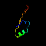
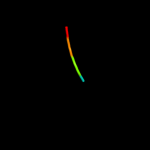
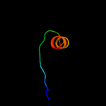
Fold:HAD-like Superfamily:HAD-like Family:Hypothetical protein VC0232
Confidence and coverage
Confidence:
45.7%
Coverage:
52%
33 residues ( 52% of your sequence) have been modelled with 45.7% confidence by the single highest scoring template.
You may wish to submit your sequence to Phyrealarm. This will automatically scan your sequence every week for new potential templates as they appear in the Phyre2 library.
Please note: You must be registered and logged in to use Phyrealarm.
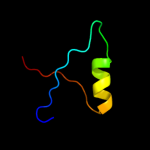
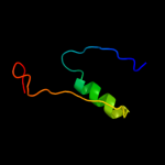
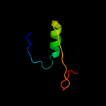
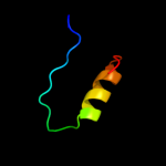
Region: 14 - 48 Aligned: 35 Modelled: 35 Confidence: 16.7% Identity: 20% PDB header:hydrolase Chain: A: PDB Molecule:cation-transporting atpase pacs; PDBTitle: solution structure of the n-terminal domain of the coppper(i) atpase2 pacs in its apo form
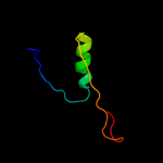
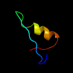
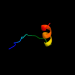
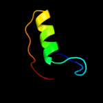
Region: 14 - 43 Aligned: 30 Modelled: 30 Confidence: 14.1% Identity: 27% PDB header:hydrolase, membrane protein Chain: X: PDB Molecule:zinc-transporting atpase; PDBTitle: solution structure of the n-terminal domain of the zinc(ii) atpase2 ziaa in its apo form
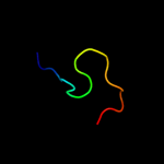
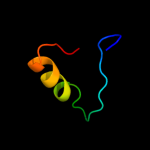
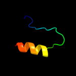
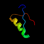
Region: 14 - 37 Aligned: 23 Modelled: 24 Confidence: 9.5% Identity: 17% PDB header:rna binding protein Chain: A: PDB Molecule:signal recognition particle 19 kda protein; PDBTitle: structures of srp54 and srp19, the two proteins assembling2 the ribonucleic core of the signal recognition particle3 from the archaeon pyrococcus furiosus.
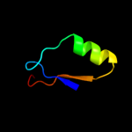
Region: 14 - 47 Aligned: 32 Modelled: 34 Confidence: 8.5% Identity: 19% PDB header:metal binding protein Chain: A: PDB Molecule:heavy metal binding protein; PDBTitle: nmr structure of heavy metal binding protein tm0320 from thermotoga2 maritima
Phyre2
21
22
23
24
25
26
27
28
29
30
31
32
33
34
35
36
37
38
39
40
41
42
Detailed template information
Binding site prediction
Due to computational demand, binding site predictions are not run for batch jobs
If you want to predict binding sites, please manually submit your model of choice to 3DLigandSite
Phyre is for academic use only
Please cite: Protein structure prediction on
the web: a case study using the Phyre server
Kelley LA and Sternberg MJE. Nature Protocols
4, 363 - 371 (2009) [pdf] [Import into BibTeX]
If you use the binding site
predictions from 3DLigandSite, please also cite:
3DLigandSite: predicting ligand-binding sites using similar structures.
Wass MN, Kelley LA and Sternberg
MJ Nucleic Acids Research 38, W469-73 (2010) [PubMed]