| 1 | c3ed6B_
|
|
 |
100.0 |
37 |
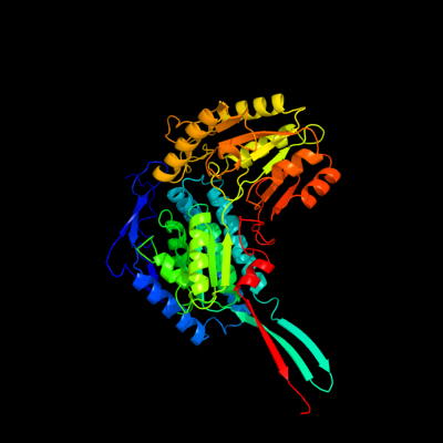
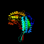
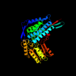
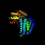
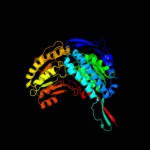
PDB header:oxidoreductase
Chain: B: PDB Molecule:betaine aldehyde dehydrogenase;
PDBTitle: 1.7 angstrom resolution crystal structure of betaine aldehyde2 dehydrogenase (betb) from staphylococcus aureus
|
| 2 | d1bxsa_
|
|
 |
100.0 |
40 |
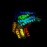
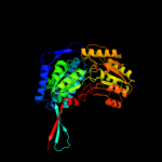
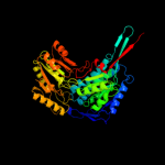
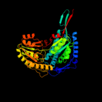
Fold:ALDH-like
Superfamily:ALDH-like
Family:ALDH-like |
| 3 | d1o9ja_
|
|
 |
100.0 |
38 |
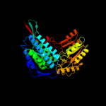
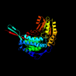
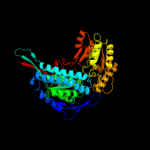
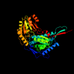
Fold:ALDH-like
Superfamily:ALDH-like
Family:ALDH-like |
| 4 | c2o2qA_
|
|
 |
100.0 |
37 |
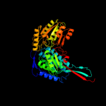
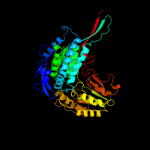
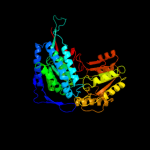
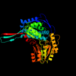
PDB header:oxidoreductase
Chain: A: PDB Molecule:formyltetrahydrofolate dehydrogenase;
PDBTitle: crystal structure of the c-terminal domain of rat2 10'formyltetrahydrofolate dehydrogenase in complex with nadp
|
| 5 | d1a4sa_
|
|
 |
100.0 |
33 |
Fold:ALDH-like
Superfamily:ALDH-like
Family:ALDH-like |
| 6 | d1o04a_
|
|
 |
100.0 |
42 |
Fold:ALDH-like
Superfamily:ALDH-like
Family:ALDH-like |
| 7 | d1ag8a_
|
|
 |
100.0 |
42 |
Fold:ALDH-like
Superfamily:ALDH-like
Family:ALDH-like |
| 8 | c2ve5H_
|
|
 |
100.0 |
40 |
PDB header:oxidoreductase
Chain: H: PDB Molecule:betaine aldehyde dehydrogenase;
PDBTitle: crystallographic structure of betaine aldehyde2 dehydrogenase from pseudomonas aeruginosa
|
| 9 | c3iwkB_
|
|
 |
100.0 |
38 |
PDB header:oxidoreductase
Chain: B: PDB Molecule:aminoaldehyde dehydrogenase;
PDBTitle: crystal structure of aminoaldehyde dehydrogenase 1 from2 pisum sativum (psamadh1)
|
| 10 | c2d4eB_
|
|
 |
100.0 |
43 |
PDB header:oxidoreductase
Chain: B: PDB Molecule:5-carboxymethyl-2-hydroxymuconate semialdehyde
PDBTitle: crystal structure of the hpcc from thermus thermophilus hb8
|
| 11 | c2jg7G_
|
|
 |
100.0 |
25 |
PDB header:oxidoreductase
Chain: G: PDB Molecule:antiquitin;
PDBTitle: crystal structure of seabream antiquitin and elucidation of2 its substrate specificity
|
| 12 | c3b4wA_
|
|
 |
100.0 |
37 |
PDB header:oxidoreductase
Chain: A: PDB Molecule:aldehyde dehydrogenase;
PDBTitle: crystal structure of mycobacterium tuberculosis aldehyde dehydrogenase2 complexed with nad+
|
| 13 | c3r31A_
|
|
 |
100.0 |
38 |
PDB header:oxidoreductase
Chain: A: PDB Molecule:betaine aldehyde dehydrogenase;
PDBTitle: crystal structure of betaine aldehyde dehydrogenase from agrobacterium2 tumefaciens
|
| 14 | c3ek1C_
|
|
 |
100.0 |
33 |
PDB header:oxidoreductase
Chain: C: PDB Molecule:aldehyde dehydrogenase;
PDBTitle: crystal structure of aldehyde dehydrogenase from brucella2 melitensis biovar abortus 2308
|
| 15 | c3rh9A_
|
|
 |
100.0 |
31 |
PDB header:oxidoreductase
Chain: A: PDB Molecule:succinate-semialdehyde dehydrogenase (nad(p)(+));
PDBTitle: the crystal structure of oxidoreductase from marinobacter aquaeolei
|
| 16 | d1wnda_
|
|
 |
100.0 |
37 |
Fold:ALDH-like
Superfamily:ALDH-like
Family:ALDH-like |
| 17 | d1uzba_
|
|
 |
100.0 |
30 |
Fold:ALDH-like
Superfamily:ALDH-like
Family:ALDH-like |
| 18 | c3qanB_
|
|
 |
100.0 |
30 |
PDB header:oxidoreductase
Chain: B: PDB Molecule:1-pyrroline-5-carboxylate dehydrogenase 1;
PDBTitle: crystal structure of 1-pyrroline-5-carboxylate dehydrogenase from2 bacillus halodurans
|
| 19 | c3ifgH_
|
|
 |
100.0 |
35 |
PDB header:oxidoreductase
Chain: H: PDB Molecule:succinate-semialdehyde dehydrogenase (nadp+);
PDBTitle: crystal structure of succinate-semialdehyde dehydrogenase from2 burkholderia pseudomallei, part 1 of 2
|
| 20 | c3jz4C_
|
|
 |
100.0 |
36 |
PDB header:oxidoreductase
Chain: C: PDB Molecule:succinate-semialdehyde dehydrogenase [nadp+];
PDBTitle: crystal structure of e. coli nadp dependent enzyme
|
| 21 | c3k2wD_ |
|
not modelled |
100.0 |
32 |
PDB header:oxidoreductase
Chain: D: PDB Molecule:betaine-aldehyde dehydrogenase;
PDBTitle: crystal structure of betaine-aldehyde dehydrogenase from2 pseudoalteromonas atlantica t6c
|
| 22 | c3i44A_ |
|
not modelled |
100.0 |
35 |
PDB header:oxidoreductase
Chain: A: PDB Molecule:aldehyde dehydrogenase;
PDBTitle: crystal structure of aldehyde dehydrogenase from bartonella2 henselae at 2.0a resolution
|
| 23 | c2hg2A_ |
|
not modelled |
100.0 |
30 |
PDB header:oxidoreductase
Chain: A: PDB Molecule:aldehyde dehydrogenase a;
PDBTitle: structure of lactaldehyde dehydrogenase
|
| 24 | d1euha_ |
|
not modelled |
100.0 |
33 |
Fold:ALDH-like
Superfamily:ALDH-like
Family:ALDH-like |
| 25 | c2w8qA_ |
|
not modelled |
100.0 |
33 |
PDB header:oxidoreductase
Chain: A: PDB Molecule:succinate-semialdehyde dehydrogenase,
PDBTitle: the crystal structure of human ssadh in complex with ssa.
|
| 26 | c3prlD_ |
|
not modelled |
100.0 |
32 |
PDB header:oxidoreductase
Chain: D: PDB Molecule:nadp-dependent glyceraldehyde-3-phosphate dehydrogenase;
PDBTitle: crystal structure of nadp-dependent glyceraldehyde-3-phosphate2 dehydrogenase from bacillus halodurans c-125
|
| 27 | c1t90B_ |
|
not modelled |
100.0 |
31 |
PDB header:oxidoreductase
Chain: B: PDB Molecule:probable methylmalonate-semialdehyde
PDBTitle: crystal structure of methylmalonate semialdehyde2 dehydrogenase from bacillus subtilis
|
| 28 | d1ky8a_ |
|
not modelled |
100.0 |
29 |
Fold:ALDH-like
Superfamily:ALDH-like
Family:ALDH-like |
| 29 | d1bi9a_ |
|
not modelled |
100.0 |
40 |
Fold:ALDH-like
Superfamily:ALDH-like
Family:ALDH-like |
| 30 | c3hazA_ |
|
not modelled |
100.0 |
31 |
PDB header:oxidoreductase
Chain: A: PDB Molecule:proline dehydrogenase;
PDBTitle: crystal structure of bifunctional proline utilization a2 (puta) protein
|
| 31 | c3ju8B_ |
|
not modelled |
100.0 |
32 |
PDB header:oxidoreductase
Chain: B: PDB Molecule:succinylglutamic semialdehyde dehydrogenase;
PDBTitle: crystal structure of succinylglutamic semialdehyde dehydrogenase from2 pseudomonas aeruginosa.
|
| 32 | c3rosA_ |
|
not modelled |
100.0 |
30 |
PDB header:oxidoreductase
Chain: A: PDB Molecule:nad-dependent aldehyde dehydrogenase;
PDBTitle: crystal structure of nad-dependent aldehyde dehydrogenase from2 lactobacillus acidophilus
|
| 33 | c3efvC_ |
|
not modelled |
100.0 |
35 |
PDB header:oxidoreductase
Chain: C: PDB Molecule:putative succinate-semialdehyde dehydrogenase;
PDBTitle: crystal structure of a putative succinate-semialdehyde dehydrogenase2 from salmonella typhimurium lt2 with bound nad
|
| 34 | c2vroB_ |
|
not modelled |
100.0 |
25 |
PDB header:oxidoreductase
Chain: B: PDB Molecule:aldehyde dehydrogenase;
PDBTitle: crystal structure of aldehyde dehydrogenase from2 burkholderia xenovorans lb400
|
| 35 | c3r64A_ |
|
not modelled |
100.0 |
30 |
PDB header:oxidoreductase
Chain: A: PDB Molecule:nad dependent benzaldehyde dehydrogenase;
PDBTitle: crystal structure of a nad-dependent benzaldehyde dehydrogenase from2 corynebacterium glutamicum
|
| 36 | c3pqaA_ |
|
not modelled |
100.0 |
35 |
PDB header:oxidoreductase
Chain: A: PDB Molecule:lactaldehyde dehydrogenase;
PDBTitle: crystal structure of glyceraldehyde-3-phosphate dehydrogenase gapn2 from methanocaldococcus jannaschii dsm 2661
|
| 37 | d1ad3a_ |
|
not modelled |
100.0 |
29 |
Fold:ALDH-like
Superfamily:ALDH-like
Family:ALDH-like |
| 38 | c3v4cB_ |
|
not modelled |
100.0 |
27 |
PDB header:oxidoreductase
Chain: B: PDB Molecule:aldehyde dehydrogenase (nadp+);
PDBTitle: crystal structure of a semialdehyde dehydrogenase from sinorhizobium2 meliloti 1021
|
| 39 | d1ez0a_ |
|
not modelled |
100.0 |
22 |
Fold:ALDH-like
Superfamily:ALDH-like
Family:ALDH-like |
| 40 | c3lnsD_ |
|
not modelled |
100.0 |
28 |
PDB header:oxidoreductase
Chain: D: PDB Molecule:benzaldehyde dehydrogenase;
PDBTitle: benzaldehyde dehydrogenase, a class 3 aldehyde dehydrogenase, with2 bound nadp+ and benzoate adduct
|
| 41 | c3k9dD_ |
|
not modelled |
100.0 |
17 |
PDB header:oxidoreductase
Chain: D: PDB Molecule:aldehyde dehydrogenase;
PDBTitle: crystal structure of probable aldehyde dehydrogenase from listeria2 monocytogenes egd-e
|
| 42 | d1o20a_ |
|
not modelled |
100.0 |
18 |
Fold:ALDH-like
Superfamily:ALDH-like
Family:ALDH-like |
| 43 | c3my7A_ |
|
not modelled |
100.0 |
18 |
PDB header:oxidoreductase
Chain: A: PDB Molecule:alcohol dehydrogenase/acetaldehyde dehydrogenase;
PDBTitle: the crystal structure of the acdh domain of an alcohol dehydrogenase2 from vibrio parahaemolyticus to 2.25a
|
| 44 | c2h5gA_ |
|
not modelled |
100.0 |
21 |
PDB header:oxidoreductase
Chain: A: PDB Molecule:delta 1-pyrroline-5-carboxylate synthetase;
PDBTitle: crystal structure of human pyrroline-5-carboxylate synthetase
|
| 45 | d1vlua_ |
|
not modelled |
100.0 |
19 |
Fold:ALDH-like
Superfamily:ALDH-like
Family:ALDH-like |
| 46 | c1vluB_ |
|
not modelled |
100.0 |
19 |
PDB header:oxidoreductase
Chain: B: PDB Molecule:gamma-glutamyl phosphate reductase;
PDBTitle: crystal structure of gamma-glutamyl phosphate reductase (yor323c) from2 saccharomyces cerevisiae at 2.40 a resolution
|
| 47 | d1k75a_ |
|
not modelled |
98.5 |
19 |
Fold:ALDH-like
Superfamily:ALDH-like
Family:L-histidinol dehydrogenase HisD |
| 48 | d1y5ea1 |
|
not modelled |
51.5 |
29 |
Fold:Molybdenum cofactor biosynthesis proteins
Superfamily:Molybdenum cofactor biosynthesis proteins
Family:MogA-like |
| 49 | d2g2ca1 |
|
not modelled |
35.2 |
16 |
Fold:Molybdenum cofactor biosynthesis proteins
Superfamily:Molybdenum cofactor biosynthesis proteins
Family:MogA-like |
| 50 | c2yvqA_ |
|
not modelled |
34.5 |
15 |
PDB header:ligase
Chain: A: PDB Molecule:carbamoyl-phosphate synthase;
PDBTitle: crystal structure of mgs domain of carbamoyl-phosphate2 synthetase from homo sapiens
|
| 51 | c3i0pA_ |
|
not modelled |
33.3 |
14 |
PDB header:oxidoreductase
Chain: A: PDB Molecule:malate dehydrogenase;
PDBTitle: crystal structure of malate dehydrogenase from entamoeba histolytica
|
| 52 | d1s7ia_ |
|
not modelled |
33.0 |
17 |
Fold:Ferredoxin-like
Superfamily:Dimeric alpha+beta barrel
Family:DGPF domain (Pfam 04946) |
| 53 | c1v9nA_ |
|
not modelled |
29.5 |
19 |
PDB header:oxidoreductase
Chain: A: PDB Molecule:malate dehydrogenase;
PDBTitle: structure of malate dehydrogenase from pyrococcus horikoshii ot3
|
| 54 | c2l69A_ |
|
not modelled |
29.2 |
19 |
PDB header:de novo protein
Chain: A: PDB Molecule:rossmann 2x3 fold protein;
PDBTitle: solution nmr structure of de novo designed protein, p-loop ntpase2 fold, northeast structural genomics consortium target or28
|
| 55 | d1rfma_ |
|
not modelled |
29.0 |
21 |
Fold:L-sulfolactate dehydrogenase-like
Superfamily:L-sulfolactate dehydrogenase-like
Family:L-sulfolactate dehydrogenase-like |
| 56 | d1wu2a3 |
|
not modelled |
27.7 |
21 |
Fold:Molybdenum cofactor biosynthesis proteins
Superfamily:Molybdenum cofactor biosynthesis proteins
Family:MoeA central domain-like |
| 57 | c3jtpB_ |
|
not modelled |
27.3 |
21 |
PDB header:protein binding
Chain: B: PDB Molecule:adapter protein meca 1;
PDBTitle: crystal structure of the c-terminal domain of meca
|
| 58 | c1vbiA_ |
|
not modelled |
27.0 |
26 |
PDB header:oxidoreductase
Chain: A: PDB Molecule:type 2 malate/lactate dehydrogenase;
PDBTitle: crystal structure of type 2 malate/lactate dehydrogenase from thermus2 thermophilus hb8
|
| 59 | d2bona1 |
|
not modelled |
26.9 |
17 |
Fold:NAD kinase/diacylglycerol kinase-like
Superfamily:NAD kinase/diacylglycerol kinase-like
Family:Diacylglycerol kinase-like |
| 60 | d1mkza_ |
|
not modelled |
26.8 |
25 |
Fold:Molybdenum cofactor biosynthesis proteins
Superfamily:Molybdenum cofactor biosynthesis proteins
Family:MogA-like |
| 61 | d1u0ta_ |
|
not modelled |
26.1 |
11 |
Fold:NAD kinase/diacylglycerol kinase-like
Superfamily:NAD kinase/diacylglycerol kinase-like
Family:NAD kinase-like |
| 62 | d1xxaa_ |
|
not modelled |
25.6 |
24 |
Fold:DCoH-like
Superfamily:C-terminal domain of arginine repressor
Family:C-terminal domain of arginine repressor |
| 63 | c3v4gA_ |
|
not modelled |
24.9 |
14 |
PDB header:dna binding protein
Chain: A: PDB Molecule:arginine repressor;
PDBTitle: 1.60 angstrom resolution crystal structure of an arginine repressor2 from vibrio vulnificus cmcp6
|
| 64 | d1xrha_ |
|
not modelled |
24.3 |
18 |
Fold:L-sulfolactate dehydrogenase-like
Superfamily:L-sulfolactate dehydrogenase-like
Family:L-sulfolactate dehydrogenase-like |
| 65 | d1uz5a3 |
|
not modelled |
23.8 |
15 |
Fold:Molybdenum cofactor biosynthesis proteins
Superfamily:Molybdenum cofactor biosynthesis proteins
Family:MoeA central domain-like |
| 66 | c2pjkA_ |
|
not modelled |
22.8 |
23 |
PDB header:structural genomics, unknown function
Chain: A: PDB Molecule:178aa long hypothetical molybdenum cofactor
PDBTitle: structure of hypothetical molybdenum cofactor biosynthesis2 protein b from sulfolobus tokodaii
|
| 67 | d1nxua_ |
|
not modelled |
22.7 |
21 |
Fold:L-sulfolactate dehydrogenase-like
Superfamily:L-sulfolactate dehydrogenase-like
Family:L-sulfolactate dehydrogenase-like |
| 68 | c2is8A_ |
|
not modelled |
22.1 |
12 |
PDB header:structural protein
Chain: A: PDB Molecule:molybdopterin biosynthesis enzyme, moab;
PDBTitle: crystal structure of the molybdopterin biosynthesis enzyme moab2 (ttha0341) from thermus theromophilus hb8
|
| 69 | c3uoeB_ |
|
not modelled |
21.7 |
11 |
PDB header:oxidoreductase
Chain: B: PDB Molecule:dehydrogenase;
PDBTitle: the crystal structure of dehydrogenase from sinorhizobium meliloti
|
| 70 | d1u2ca2 |
|
not modelled |
20.2 |
20 |
Fold:Dystroglycan, domain 2
Superfamily:Dystroglycan, domain 2
Family:Dystroglycan, domain 2 |
| 71 | d1wo8a1 |
|
not modelled |
19.6 |
15 |
Fold:Methylglyoxal synthase-like
Superfamily:Methylglyoxal synthase-like
Family:Methylglyoxal synthase, MgsA |
| 72 | c2ec4A_ |
|
not modelled |
19.1 |
16 |
PDB header:structural genomics, unknown function
Chain: A: PDB Molecule:fas-associated factor 1;
PDBTitle: solution structure of the uas domain from human fas-2 associated factor 1
|
| 73 | d2ftsa3 |
|
not modelled |
19.1 |
23 |
Fold:Molybdenum cofactor biosynthesis proteins
Superfamily:Molybdenum cofactor biosynthesis proteins
Family:MoeA central domain-like |
| 74 | c1wtjB_ |
|
not modelled |
19.1 |
14 |
PDB header:oxidoreductase
Chain: B: PDB Molecule:ureidoglycolate dehydrogenase;
PDBTitle: crystal structure of delta1-piperideine-2-carboxylate2 reductase from pseudomonas syringae pvar.tomato
|
| 75 | d1a9xa2 |
|
not modelled |
19.1 |
18 |
Fold:Methylglyoxal synthase-like
Superfamily:Methylglyoxal synthase-like
Family:Carbamoyl phosphate synthetase, large subunit allosteric, C-terminal domain |
| 76 | c2qguA_ |
|
not modelled |
18.4 |
18 |
PDB header:structural genomics, unknown function
Chain: A: PDB Molecule:probable signal peptide protein;
PDBTitle: three-dimensional structure of the phospholipid-binding protein from2 ralstonia solanacearum q8xv73_ralsq in complex with a phospholipid at3 the resolution 1.53 a. northeast structural genomics consortium4 target rsr89
|
| 77 | c1b4aA_ |
|
not modelled |
17.7 |
11 |
PDB header:repressor
Chain: A: PDB Molecule:arginine repressor;
PDBTitle: structure of the arginine repressor from bacillus stearothermophilus
|
| 78 | c1z2iA_ |
|
not modelled |
17.5 |
18 |
PDB header:oxidoreductase
Chain: A: PDB Molecule:malate dehydrogenase;
PDBTitle: crystal structure of agrobacterium tumefaciens malate2 dehydrogenase, new york structural genomics consortium
|
| 79 | d1srqa_ |
|
not modelled |
17.4 |
14 |
Fold:Rap/Ran-GAP
Superfamily:Rap/Ran-GAP
Family:Rap/Ran-GAP |
| 80 | c2v9vA_ |
|
not modelled |
16.9 |
10 |
PDB header:transcription
Chain: A: PDB Molecule:selenocysteine-specific elongation factor;
PDBTitle: crystal structure of moorella thermoacetica selb(377-511)
|
| 81 | c2yukA_ |
|
not modelled |
16.1 |
24 |
PDB header:transferase
Chain: A: PDB Molecule:myeloid/lymphoid or mixed-lineage leukemia
PDBTitle: solution structure of the hmg box of human myeloid/lymphoid2 or mixed-lineage leukemia protein 3 homolog
|
| 82 | c1gr0A_ |
|
not modelled |
16.1 |
16 |
PDB header:isomerase
Chain: A: PDB Molecule:inositol-3-phosphate synthase;
PDBTitle: myo-inositol 1-phosphate synthase from mycobacterium2 tuberculosis in complex with nad and zinc.
|
| 83 | c2crjA_ |
|
not modelled |
15.7 |
18 |
PDB header:gene regulation
Chain: A: PDB Molecule:swi/snf-related matrix-associated actin-
PDBTitle: solution structure of the hmg domain of mouse hmg domain2 protein hmgx2
|
| 84 | c3q3hA_ |
|
not modelled |
15.6 |
18 |
PDB header:transferase
Chain: A: PDB Molecule:hmw1c-like glycosyltransferase;
PDBTitle: crystal structure of the actinobacillus pleuropneumoniae hmw1c2 glycosyltransferase in complex with udp-glc
|
| 85 | c3ereD_ |
|
not modelled |
15.6 |
16 |
PDB header:dna binding protein/dna
Chain: D: PDB Molecule:arginine repressor;
PDBTitle: crystal structure of the arginine repressor protein from mycobacterium2 tuberculosis in complex with the dna operator
|
| 86 | d2ioja1 |
|
not modelled |
15.5 |
11 |
Fold:MurF and HprK N-domain-like
Superfamily:HprK N-terminal domain-like
Family:DRTGG domain |
| 87 | c3e7hA_ |
|
not modelled |
15.2 |
17 |
PDB header:transferase
Chain: A: PDB Molecule:dna-directed rna polymerase subunit beta;
PDBTitle: the crystal structure of the beta subunit of the dna-2 directed rna polymerase from vibrio cholerae o1 biovar3 eltor
|
| 88 | c3rfqC_ |
|
not modelled |
14.7 |
23 |
PDB header:biosynthetic protein
Chain: C: PDB Molecule:pterin-4-alpha-carbinolamine dehydratase moab2;
PDBTitle: crystal structure of pterin-4-alpha-carbinolamine dehydratase moab22 from mycobacterium marinum
|
| 89 | c2bonB_ |
|
not modelled |
13.9 |
15 |
PDB header:transferase
Chain: B: PDB Molecule:lipid kinase;
PDBTitle: structure of an escherichia coli lipid kinase (yegs)
|
| 90 | c3cinA_ |
|
not modelled |
13.7 |
23 |
PDB header:isomerase
Chain: A: PDB Molecule:myo-inositol-1-phosphate synthase-related protein;
PDBTitle: crystal structure of a myo-inositol-1-phosphate synthase-related2 protein (tm_1419) from thermotoga maritima msb8 at 1.70 a resolution
|
| 91 | c2nqqA_ |
|
not modelled |
13.5 |
13 |
PDB header:biosynthetic protein
Chain: A: PDB Molecule:molybdopterin biosynthesis protein moea;
PDBTitle: moea r137q
|
| 92 | d2f7wa1 |
|
not modelled |
13.4 |
17 |
Fold:Molybdenum cofactor biosynthesis proteins
Superfamily:Molybdenum cofactor biosynthesis proteins
Family:MogA-like |
| 93 | c2g8yB_ |
|
not modelled |
12.8 |
16 |
PDB header:oxidoreductase
Chain: B: PDB Molecule:malate/l-lactate dehydrogenases;
PDBTitle: the structure of a putative malate/lactate dehydrogenase from e. coli.
|
| 94 | d2p1ra1 |
|
not modelled |
12.7 |
12 |
Fold:NAD kinase/diacylglycerol kinase-like
Superfamily:NAD kinase/diacylglycerol kinase-like
Family:Diacylglycerol kinase-like |
| 95 | d1jlja_ |
|
not modelled |
12.7 |
24 |
Fold:Molybdenum cofactor biosynthesis proteins
Superfamily:Molybdenum cofactor biosynthesis proteins
Family:MogA-like |
| 96 | d1xi8a3 |
|
not modelled |
12.6 |
22 |
Fold:Molybdenum cofactor biosynthesis proteins
Superfamily:Molybdenum cofactor biosynthesis proteins
Family:MoeA central domain-like |
| 97 | c2g4rB_ |
|
not modelled |
12.6 |
18 |
PDB header:biosynthetic protein
Chain: B: PDB Molecule:molybdopterin biosynthesis mog protein;
PDBTitle: anomalous substructure of moga
|
| 98 | c2h47F_ |
|
not modelled |
12.6 |
14 |
PDB header:oxidoreductase/electron transport
Chain: F: PDB Molecule:aromatic amine dehydrogenase;
PDBTitle: crystal structure of an electron transfer complex between2 aromatic amine dephydrogenase and azurin from alcaligenes3 faecalis (form 1)
|
| 99 | c1uz5A_ |
|
not modelled |
12.4 |
14 |
PDB header:molybdopterin biosynthesis
Chain: A: PDB Molecule:402aa long hypothetical molybdopterin
PDBTitle: the crystal structure of molybdopterin biosynthesis moea2 protein from pyrococcus horikosii
|