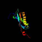
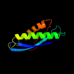
1 c3bk6C_
99.9
25
PDB header: membrane proteinChain: C: PDB Molecule: ph stomatin;PDBTitle: crystal structure of a core domain of stomatin from2 pyrococcus horikoshii
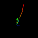
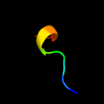
2 c2rpbA_
99.8
25
PDB header: membrane proteinChain: A: PDB Molecule: hypothetical membrane protein;PDBTitle: the solution structure of membrane protein
3 d1wina_
99.7
14
Fold: EF-Ts domain-likeSuperfamily: Band 7/SPFH domainFamily: Band 7/SPFH domain4 c2zv4O_
98.1
20
PDB header: structural proteinChain: O: PDB Molecule: major vault protein;PDBTitle: the structure of rat liver vault at 3.5 angstrom resolution
5 c2kk7A_
80.5
26
PDB header: hydrolaseChain: A: PDB Molecule: v-type atp synthase subunit e;PDBTitle: nmr solution structure of the n terminal domain of subunit e2 (e1-52) of a1ao atp synthase from methanocaldococcus3 jannaschii
6 c2k88A_
51.3
29
PDB header: hydrolaseChain: A: PDB Molecule: vacuolar proton pump subunit g;PDBTitle: association of subunit d (vma6p) and e (vma4p) with g2 (vma10p) and the nmr solution structure of subunit g (g1-3 59) of the saccharomyces cerevisiae v1vo atpase
7 d1l2pa_
49.5
28
Fold: Single transmembrane helixSuperfamily: F1F0 ATP synthase subunit B, membrane domainFamily: F1F0 ATP synthase subunit B, membrane domain8 c2kepA_
44.4
41
PDB header: transport proteinChain: A: PDB Molecule: general secretion pathway protein g;PDBTitle: solution structure of xcpt, the main component of the type 22 secretion system of pseudomonas aeruginosa
9 c3k5bE_
43.5
13
PDB header: hydrolaseChain: E: PDB Molecule: v-type atp synthase subunit e;PDBTitle: crystal structure of the peripheral stalk of thermus thermophilus h+-2 atpase/synthase
10 c3u5gB_
33.8
17
PDB header: ribosomeChain: B: PDB Molecule: 40s ribosomal protein s1-a;PDBTitle: the structure of the eukaryotic ribosome at 3.0 a resolution
11 c1wd6B_
27.9
42
PDB header: structural genomics, unknown functionChain: B: PDB Molecule: protein ydhr;PDBTitle: crystal structure of jw1657 from escherichia coli
12 d1t92a_
25.9
29
Fold: Pili subunitsSuperfamily: Pili subunitsFamily: Pseudopilin13 c2i7uA_
25.0
36
PDB header: de novo protein/ligand binding proteinChain: A: PDB Molecule: four-alpha-helix bundle;PDBTitle: structural and dynamical analysis of a four-alpha-helix2 bundle with designed anesthetic binding pockets
14 c3fu1B_
19.2
24
PDB header: protein transportChain: B: PDB Molecule: general secretion pathway protein g;PDBTitle: crystal structure of the major pseudopilin from the type 2 secretion2 system of vibrio cholerae
15 c2xzm4_
18.9
14
PDB header: ribosomeChain: 4: PDB Molecule: 40s ribosomal protein s3a;PDBTitle: crystal structure of the eukaryotic 40s ribosomal2 subunit in complex with initiation factor 1. this file3 contains the 40s subunit and initiation factor for4 molecule 1
16 c2vgpD_
12.8
33
PDB header: transferaseChain: D: PDB Molecule: inner centromere protein a;PDBTitle: crystal structure of aurora b kinase in complex with a2 aminothiazole inhibitor
17 d2hiqa1
12.6
42
Fold: Ferredoxin-likeSuperfamily: Dimeric alpha+beta barrelFamily: Hypothetical protein YdhR18 d1tf5a1
11.3
23
Fold: Pre-protein crosslinking domain of SecASuperfamily: Pre-protein crosslinking domain of SecAFamily: Pre-protein crosslinking domain of SecA19 c3k5bB_
11.0
22
PDB header: hydrolaseChain: B: PDB Molecule: v-type atp synthase, subunit (vapc-therm);PDBTitle: crystal structure of the peripheral stalk of thermus thermophilus h+-2 atpase/synthase
20 c1tt9B_
10.9
14
PDB header: transferase, lyaseChain: B: PDB Molecule: formimidoyltransferase-cyclodeaminasePDBTitle: structure of the bifunctional and golgi associated2 formiminotransferase cyclodeaminase octamer
21 c3kdpD_
not modelled
10.5
18
PDB header: hydrolaseChain: D: PDB Molecule: sodium/potassium-transporting atpase subunit beta-1;PDBTitle: crystal structure of the sodium-potassium pump
22 d1htjf_
not modelled
9.1
15
Fold: Regulator of G-protein signaling, RGSSuperfamily: Regulator of G-protein signaling, RGSFamily: Regulator of G-protein signaling, RGS23 c1htjF_
not modelled
9.1
15
PDB header: signaling proteinChain: F: PDB Molecule: kiaa0380;PDBTitle: structure of the rgs-like domain from pdz-rhogef
24 c3swfA_
not modelled
8.2
40
PDB header: transport proteinChain: A: PDB Molecule: cgmp-gated cation channel alpha-1;PDBTitle: cnga1 621-690 containing clz domain
25 d5ruba2
not modelled
7.3
25
Fold: Ferredoxin-likeSuperfamily: RuBisCO, large subunit, small (N-terminal) domainFamily: Ribulose 1,5-bisphosphate carboxylase-oxygenase26 c1jsuC_
not modelled
7.1
32
PDB header: complex (transferase/cyclin/inhibitor)Chain: C: PDB Molecule: p27;PDBTitle: p27(kip1)/cyclin a/cdk2 complex
27 c2w2hD_
not modelled
6.8
32
PDB header: rna-binding proteinChain: D: PDB Molecule: protein tat;PDBTitle: structural basis of transcription activation by the cyclin2 t1-tat-tar rna complex from eiav
28 d1wuua1
not modelled
6.8
10
Fold: Ribosomal protein S5 domain 2-likeSuperfamily: Ribosomal protein S5 domain 2-likeFamily: GHMP Kinase, N-terminal domain29 c3s93B_
not modelled
6.5
11
PDB header: transcriptionChain: B: PDB Molecule: tudor domain-containing protein 5;PDBTitle: crystal structure of conserved motif in tdrd5
30 d1wmda1
not modelled
6.4
11
Fold: Galactose-binding domain-likeSuperfamily: Galactose-binding domain-likeFamily: Proprotein convertase P-domain31 c2jp3A_
not modelled
6.4
36
PDB header: transcriptionChain: A: PDB Molecule: fxyd domain-containing ion transport regulator 4;PDBTitle: solution structure of the human fxyd4 (chif) protein in sds2 micelles
32 c2ptmA_
not modelled
6.4
10
PDB header: transport proteinChain: A: PDB Molecule: hyperpolarization-activated (ih) channel;PDBTitle: structure and rearrangements in the carboxy-terminal region of spih2 channels
33 c2jo1A_
not modelled
6.3
24
PDB header: hydrolase regulatorChain: A: PDB Molecule: phospholemman;PDBTitle: structure of the na,k-atpase regulatory protein fxyd1 in2 micelles
34 d2o4ta1
not modelled
6.3
6
Fold: Left-handed superhelixSuperfamily: BH3980-likeFamily: BH3980-like35 d1o5ha_
not modelled
6.1
19
Fold: Methenyltetrahydrofolate cyclohydrolase-likeSuperfamily: Methenyltetrahydrofolate cyclohydrolase-likeFamily: Methenyltetrahydrofolate cyclohydrolase-like36 d2ozla1
not modelled
6.0
20
Fold: Thiamin diphosphate-binding fold (THDP-binding)Superfamily: Thiamin diphosphate-binding fold (THDP-binding)Family: Branched-chain alpha-keto acid dehydrogenase PP module37 c1y4cA_
not modelled
5.8
15
PDB header: de novo proteinChain: A: PDB Molecule: maltose binding protein fused with designedPDBTitle: designed helical protein fusion mbp
38 c2kncB_
not modelled
5.7
13
PDB header: cell adhesionChain: B: PDB Molecule: integrin beta-3;PDBTitle: platelet integrin alfaiib-beta3 transmembrane-cytoplasmic2 heterocomplex
39 d1fftb2
not modelled
5.7
7
Fold: Transmembrane helix hairpinSuperfamily: Cytochrome c oxidase subunit II-like, transmembrane regionFamily: Cytochrome c oxidase subunit II-like, transmembrane region40 d1nkta1
not modelled
5.5
14
Fold: Pre-protein crosslinking domain of SecASuperfamily: Pre-protein crosslinking domain of SecAFamily: Pre-protein crosslinking domain of SecA41 c1o7fA_
not modelled
5.5
17
PDB header: regulationChain: A: PDB Molecule: camp-dependent rap1 guanine-nucleotide exchangePDBTitle: crystal structure of the regulatory domain of epac2
42 c2k6iA_
not modelled
5.5
34
PDB header: structural proteinChain: A: PDB Molecule: uncharacterized protein mj0223;PDBTitle: the domain features of the peripheral stalk subunit h of the2 methanogenic a1ao atp synthase and the nmr solution3 structure of h1-47
43 c2zxeB_
not modelled
5.4
21
PDB header: hydrolase/transport proteinChain: B: PDB Molecule: na+,k+-atpase beta subunit;PDBTitle: crystal structure of the sodium - potassium pump in the e2.2k+.pi2 state
44 d1ykwa2
not modelled
5.4
21
Fold: Ferredoxin-likeSuperfamily: RuBisCO, large subunit, small (N-terminal) domainFamily: Ribulose 1,5-bisphosphate carboxylase-oxygenase45 c3nngA_
not modelled
5.3
21
PDB header: structural genomics, unknown functionChain: A: PDB Molecule: uncharacterized protein;PDBTitle: crystal structure of the f5/8 type c domain of q5lfr2_bacfn protein2 from bacteroides fragilis. northeast structural genomics consortium3 target bfr258e
46 d1e3pa2
not modelled
5.3
39
Fold: OB-foldSuperfamily: Nucleic acid-binding proteinsFamily: Cold shock DNA-binding domain-like47 c9paiB_
not modelled
5.3
15
PDB header: hydrolase inhibitorChain: B: PDB Molecule: protein (plasminogen activator inhibitor-1) residues 365-PDBTitle: cleaved substrate variant of plasminogen activator inhibitor-1
48 c2axcA_
not modelled
5.3
18
PDB header: hydrolaseChain: A: PDB Molecule: colicin e7;PDBTitle: crystal structure of cole7 translocation domain
49 c3i24B_
not modelled
5.1
17
PDB header: hydrolaseChain: B: PDB Molecule: hit family hydrolase;PDBTitle: crystal structure of a hit family hydrolase protein from2 vibrio fischeri. northeast structural genomics consortium3 target id vfr176
50 d2hsga1
not modelled
5.1
21
Fold: lambda repressor-like DNA-binding domainsSuperfamily: lambda repressor-like DNA-binding domainsFamily: GalR/LacI-like bacterial regulator