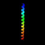
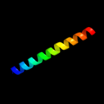
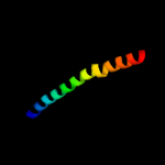
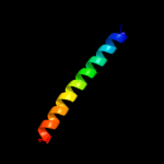
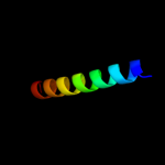
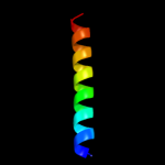
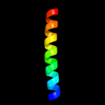
1 c2xzrA_
80.8
5
PDB header: cell adhesionChain: A: PDB Molecule: immunoglobulin-binding protein eibd;PDBTitle: escherichia coli immunoglobulin-binding protein eibd 391-438 fused2 to gcn4 adaptors
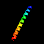
2 c1ci6B_
78.6
22
PDB header: transcriptionChain: B: PDB Molecule: transcription factor c/ebp beta;PDBTitle: transcription factor atf4-c/ebp beta bzip heterodimer
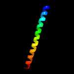
3 c2yy0D_
77.5
14
PDB header: transcriptionChain: D: PDB Molecule: c-myc-binding protein;PDBTitle: crystal structure of ms0802, c-myc-1 binding protein domain2 from homo sapiens
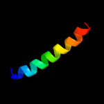
4 c2e43A_
74.6
23
PDB header: transcription/dnaChain: A: PDB Molecule: ccaat/enhancer-binding protein beta;PDBTitle: crystal structure of c/ebpbeta bzip homodimer k269a mutant2 bound to a high affinity dna fragment
5 c1fosF_
72.5
21
PDB header: transcription/dnaChain: F: PDB Molecule: c-jun proto-oncogene protein;PDBTitle: two human c-fos:c-jun:dna complexes
6 c1ci6A_
72.3
16
PDB header: transcriptionChain: A: PDB Molecule: transcription factor atf-4;PDBTitle: transcription factor atf4-c/ebp beta bzip heterodimer
7 c2xdjF_
71.8
9
PDB header: unknown functionChain: F: PDB Molecule: uncharacterized protein ybgf;PDBTitle: crystal structure of the n-terminal domain of e.coli ybgf
8 c1fosE_
69.8
28
PDB header: transcription/dnaChain: E: PDB Molecule: p55-c-fos proto-oncogene protein;PDBTitle: two human c-fos:c-jun:dna complexes
9 c1u2uA_
69.7
38
PDB header: transcriptionChain: A: PDB Molecule: general control protein gcn4;PDBTitle: nmr solution structure of a designed heterodimeric leucine2 zipper
10 c3mudA_
69.7
15
PDB header: contractile proteinChain: A: PDB Molecule: dna repair protein xrcc4, tropomyosin alpha-1 chain;PDBTitle: structure of the tropomyosin overlap complex from chicken smooth2 muscle
11 c1t2kD_
67.5
15
PDB header: transcription/dnaChain: D: PDB Molecule: cyclic-amp-dependent transcription factor atf-2;PDBTitle: structure of the dna binding domains of irf3, atf-2 and jun2 bound to dna
12 c1gk7A_
67.4
28
PDB header: vimentinChain: A: PDB Molecule: vimentin;PDBTitle: human vimentin coil 1a fragment (1a)
13 c1ce0B_
64.9
14
PDB header: hiv-1 envelope proteinChain: B: PDB Molecule: protein (leucine zipper model h38-p1);PDBTitle: trimerization specificity in hiv-1 gp41: analysis with a2 gcn4 leucine zipper model
14 c3m9bK_
63.0
18
PDB header: chaperoneChain: K: PDB Molecule: proteasome-associated atpase;PDBTitle: crystal structure of the amino terminal coiled coil domain and the2 inter domain of the mycobacterium tuberculosis proteasomal atpase mpa
15 c1dh3A_
62.3
22
PDB header: transcription/dnaChain: A: PDB Molecule: transcription factor creb;PDBTitle: crystal structure of a creb bzip-cre complex reveals the2 basis for creb faimly selective dimerization and dna3 binding
16 c1cz7C_
60.6
23
PDB header: contractile proteinChain: C: PDB Molecule: microtubule motor protein ncd;PDBTitle: the crystal structure of a minus-end directed microtubule2 motor protein ncd reveals variable dimer conformations
17 d1nkpa_
58.6
21
Fold: HLH-likeSuperfamily: HLH, helix-loop-helix DNA-binding domainFamily: HLH, helix-loop-helix DNA-binding domain18 c1ij3C_
57.1
15
PDB header: transcriptionChain: C: PDB Molecule: general control protein gcn4;PDBTitle: gcn4-pvsl coiled-coil trimer with serine at the a(16)2 position
19 c1ij3B_
57.1
15
PDB header: transcriptionChain: B: PDB Molecule: general control protein gcn4;PDBTitle: gcn4-pvsl coiled-coil trimer with serine at the a(16)2 position
20 c1ij2C_
56.9
15
PDB header: transcriptionChain: C: PDB Molecule: general control protein gcn4;PDBTitle: gcn4-pvtl coiled-coil trimer with threonine at the a(16)2 position
21 c1rb1B_
not modelled
56.6
15
PDB header: dna binding proteinChain: B: PDB Molecule: general control protein gcn4;PDBTitle: gcn4-leucine zipper core mutant as n16a trigonal automatic2 solution
22 c1swiA_
not modelled
56.6
15
PDB header: leucine zipperChain: A: PDB Molecule: gcn4p1;PDBTitle: gcn4-leucine zipper core mutant as n16a complexed with2 benzene
23 c1rb6C_
not modelled
56.6
15
PDB header: dna binding proteinChain: C: PDB Molecule: general control protein gcn4;PDBTitle: antiparallel trimer of gcn4-leucine zipper core mutant as2 n16a tetragonal form
24 c3k7zB_
not modelled
56.6
15
PDB header: dna binding proteinChain: B: PDB Molecule: general control protein gcn4;PDBTitle: gcn4-leucine zipper core mutant as n16a trigonal automatic2 solution
25 c1rb1A_
not modelled
56.6
15
PDB header: dna binding proteinChain: A: PDB Molecule: general control protein gcn4;PDBTitle: gcn4-leucine zipper core mutant as n16a trigonal automatic2 solution
26 c3k7zA_
not modelled
56.6
15
PDB header: dna binding proteinChain: A: PDB Molecule: general control protein gcn4;PDBTitle: gcn4-leucine zipper core mutant as n16a trigonal automatic2 solution
27 c1gd2G_
not modelled
56.0
18
PDB header: transcription/dnaChain: G: PDB Molecule: transcription factor pap1;PDBTitle: crystal structure of bzip transcription factor pap1 bound2 to dna
28 d1ivsa1
not modelled
55.8
17
Fold: Long alpha-hairpinSuperfamily: tRNA-binding armFamily: Valyl-tRNA synthetase (ValRS) C-terminal domain29 c1fmhA_
not modelled
55.5
35
PDB header: transcriptionChain: A: PDB Molecule: general control protein gcn4;PDBTitle: nmr solution structure of a designed heterodimeric leucine2 zipper
30 d1nkpb_
not modelled
55.1
18
Fold: HLH-likeSuperfamily: HLH, helix-loop-helix DNA-binding domainFamily: HLH, helix-loop-helix DNA-binding domain31 c2gd7B_
not modelled
54.5
25
PDB header: transcriptionChain: B: PDB Molecule: hexim1 protein;PDBTitle: the structure of the cyclin t-binding domain of hexim12 reveals the molecular basis for regulation of3 transcription elongation
32 c1ij2B_
not modelled
54.0
15
PDB header: transcriptionChain: B: PDB Molecule: general control protein gcn4;PDBTitle: gcn4-pvtl coiled-coil trimer with threonine at the a(16)2 position
33 c2w6bA_
not modelled
53.9
20
PDB header: signaling proteinChain: A: PDB Molecule: rho guanine nucleotide exchange factor 7;PDBTitle: crystal structure of the trimeric beta-pix coiled-coil2 domain
34 c2o7hF_
not modelled
53.0
15
PDB header: transcriptionChain: F: PDB Molecule: general control protein gcn4;PDBTitle: crystal structure of trimeric coiled coil gcn4 leucine zipper
35 d2azeb1
not modelled
52.4
11
Fold: E2F-DP heterodimerization regionSuperfamily: E2F-DP heterodimerization regionFamily: E2F dimerization segment36 c2wt7B_
not modelled
52.1
14
PDB header: transcriptionChain: B: PDB Molecule: transcription factor mafb;PDBTitle: crystal structure of the bzip heterodimeric complex2 mafb:cfos bound to dna
37 c1wpaA_
not modelled
52.1
12
PDB header: cell adhesionChain: A: PDB Molecule: occludin;PDBTitle: 1.5 angstrom crystal structure of human occludin fragment2 413-522
38 c2j5uB_
not modelled
51.9
18
PDB header: cell shape regulationChain: B: PDB Molecule: mrec protein;PDBTitle: mrec lysteria monocytogenes
39 c2wvrB_
not modelled
51.7
15
PDB header: replicationChain: B: PDB Molecule: geminin;PDBTitle: human cdt1:geminin complex
40 d1r05a_
not modelled
50.8
24
Fold: HLH-likeSuperfamily: HLH, helix-loop-helix DNA-binding domainFamily: HLH, helix-loop-helix DNA-binding domain41 c1ztaA_
not modelled
49.8
20
PDB header: dna-binding motifChain: A: PDB Molecule: leucine zipper monomer;PDBTitle: the solution structure of a leucine-zipper motif peptide
42 c2oqqB_
not modelled
49.2
14
PDB header: transcriptionChain: B: PDB Molecule: transcription factor hy5;PDBTitle: crystal structure of hy5 leucine zipper homodimer from2 arabidopsis thaliana
43 c2jeeA_
not modelled
48.8
14
PDB header: cell cycleChain: A: PDB Molecule: yiiu;PDBTitle: xray structure of e. coli yiiu
44 c3n4xB_
not modelled
46.2
16
PDB header: replicationChain: B: PDB Molecule: monopolin complex subunit csm1;PDBTitle: structure of csm1 full-length
45 c2wg6L_
not modelled
45.6
17
PDB header: transcription,hydrolaseChain: L: PDB Molecule: general control protein gcn4,PDBTitle: proteasome-activating nucleotidase (pan) n-domain (57-134)2 from archaeoglobus fulgidus fused to gcn4, p61a mutant
46 c3a5tB_
not modelled
45.3
23
PDB header: transcription regulator/dnaChain: B: PDB Molecule: transcription factor mafg;PDBTitle: crystal structure of mafg-dna complex
47 c1wt6B_
not modelled
44.6
21
PDB header: transferaseChain: B: PDB Molecule: myotonin-protein kinase;PDBTitle: coiled-coil domain of dmpk
48 c1xawA_
not modelled
44.3
12
PDB header: cell adhesionChain: A: PDB Molecule: occludin;PDBTitle: crystal structure of the cytoplasmic distal c-terminal2 domain of occludin
49 c3mkxC_
not modelled
41.2
14
PDB header: antiviral proteinChain: C: PDB Molecule: bone marrow stromal antigen 2;PDBTitle: crystal structure of bst2/tetherin
50 d1an2a_
not modelled
40.1
18
Fold: HLH-likeSuperfamily: HLH, helix-loop-helix DNA-binding domainFamily: HLH, helix-loop-helix DNA-binding domain51 c3kinB_
not modelled
39.4
16
PDB header: motor proteinChain: B: PDB Molecule: kinesin heavy chain;PDBTitle: kinesin (dimeric) from rattus norvegicus
52 c3q4fG_
not modelled
39.0
32
PDB header: dna binding protein/protein bindingChain: G: PDB Molecule: dna repair protein xrcc4;PDBTitle: crystal structure of xrcc4/xlf-cernunnos complex
53 c3oa7A_
not modelled
38.5
16
PDB header: structural proteinChain: A: PDB Molecule: head morphogenesis protein, chaotic nuclear migrationPDBTitle: structure of the c-terminal domain of cnm67, a core component of the2 spindle pole body of saccharomyces cerevisiae
54 c2w6aB_
not modelled
32.8
22
PDB header: signaling proteinChain: B: PDB Molecule: arf gtpase-activating protein git1;PDBTitle: x-ray structure of the dimeric git1 coiled-coil domain
55 c1junB_
not modelled
32.6
21
PDB header: transcription regulationChain: B: PDB Molecule: c-jun homodimer;PDBTitle: nmr study of c-jun homodimer
56 c3he5A_
not modelled
32.3
24
PDB header: de novo proteinChain: A: PDB Molecule: synzip1;PDBTitle: heterospecific coiled-coil pair synzip2:synzip1
57 c2wukD_
not modelled
32.2
15
PDB header: cell cycleChain: D: PDB Molecule: septum site-determining protein diviva;PDBTitle: diviva n-terminal domain, f17a mutant
58 c2zxxA_
not modelled
31.8
15
PDB header: cell cycle/replicationChain: A: PDB Molecule: geminin;PDBTitle: crystal structure of cdt1/geminin complex
59 d1nlwe_
not modelled
31.8
17
Fold: HLH-likeSuperfamily: HLH, helix-loop-helix DNA-binding domainFamily: HLH, helix-loop-helix DNA-binding domain60 c1dipA_
not modelled
31.5
15
PDB header: acetylationChain: A: PDB Molecule: delta-sleep-inducing peptide immunoreactivePDBTitle: the solution structure of porcine delta-sleep-inducing2 peptide immunoreactive peptide, nmr, 10 structures
61 c2x7aB_
not modelled
29.7
12
PDB header: immune systemChain: B: PDB Molecule: bone marrow stromal antigen 2;PDBTitle: structural basis of hiv-1 tethering to membranes by the2 bst2-tetherin ectodomain
62 d1uklc_
not modelled
29.4
25
Fold: HLH-likeSuperfamily: HLH, helix-loop-helix DNA-binding domainFamily: HLH, helix-loop-helix DNA-binding domain63 d1fxkc_
not modelled
29.2
12
Fold: Long alpha-hairpinSuperfamily: PrefoldinFamily: Prefoldin64 c1x4qA_
not modelled
27.3
25
PDB header: rna binding proteinChain: A: PDB Molecule: u4/u6 small nuclear ribonucleoprotein prp3;PDBTitle: solution structure of pwi domain in u4/u6 small nuclear2 ribonucleoprotein prp3(hprp3)
65 c1t6fA_
not modelled
26.9
21
PDB header: cell cycleChain: A: PDB Molecule: geminin;PDBTitle: crystal structure of the coiled-coil dimerization motif of2 geminin
66 c2a93B_
not modelled
26.8
21
PDB header: leucine zippersChain: B: PDB Molecule: c-myc-max heterodimeric leucine zipper;PDBTitle: nmr solution structure of the c-myc-max heterodimeric2 leucine zipper, 40 structures
67 c2v4hA_
not modelled
26.6
17
PDB header: transcriptionChain: A: PDB Molecule: nf-kappa-b essential modulator;PDBTitle: nemo cc2-lz domain - 1d5 darpin complex
68 d2e74h1
not modelled
25.7
29
Fold: Single transmembrane helixSuperfamily: PetN subunit of the cytochrome b6f complexFamily: PetN subunit of the cytochrome b6f complex69 c2e74H_
not modelled
25.0
29
PDB header: photosynthesisChain: H: PDB Molecule: cytochrome b6-f complex subunit 8;PDBTitle: crystal structure of the cytochrome b6f complex from m.laminosus
70 c2efrB_
not modelled
24.2
18
PDB header: contractile proteinChain: B: PDB Molecule: general control protein gcn4 and tropomyosin 1 alpha chain;PDBTitle: crystal structure of the c-terminal tropomyosin fragment with n- and2 c-terminal extensions of the leucine zipper at 1.8 angstroms3 resolution
71 c3tlxA_
not modelled
24.0
19
PDB header: transferaseChain: A: PDB Molecule: adenylate kinase 2;PDBTitle: crystal structure of pf10_0086, adenylate kinase from plasmodium2 falciparum
72 c2e75H_
not modelled
23.6
29
PDB header: photosynthesisChain: H: PDB Molecule: cytochrome b6-f complex subunit 8;PDBTitle: crystal structure of the cytochrome b6f complex with 2-nonyl-4-2 hydroxyquinoline n-oxide (nqno) from m.laminosus
73 c2e76H_
not modelled
23.6
29
PDB header: photosynthesisChain: H: PDB Molecule: cytochrome b6-f complex subunit 8;PDBTitle: crystal structure of the cytochrome b6f complex with tridecyl-2 stigmatellin (tds) from m.laminosus
74 d2oa5a1
not modelled
23.5
18
Fold: BLRF2-likeSuperfamily: BLRF2-likeFamily: BLRF2-like75 c1deqO_
not modelled
23.3
9
PDB header: PDB COMPND: 76 d1nlwa_
not modelled
23.1
10
Fold: HLH-likeSuperfamily: HLH, helix-loop-helix DNA-binding domainFamily: HLH, helix-loop-helix DNA-binding domain77 c3he4F_
not modelled
22.8
24
PDB header: de novo proteinChain: F: PDB Molecule: synzip5;PDBTitle: heterospecific coiled-coil pair synzip5:synzip6
78 c3hnwB_
not modelled
22.5
15
PDB header: structural genomics, unknown functionChain: B: PDB Molecule: uncharacterized protein;PDBTitle: crystal structure of a basic coiled-coil protein of unknown function2 from eubacterium eligens atcc 27750
79 c3ol1A_
not modelled
22.4
8
PDB header: structural proteinChain: A: PDB Molecule: vimentin;PDBTitle: crystal structure of vimentin (fragment 144-251) from homo sapiens,2 northeast structural genomics consortium target hr4796b
80 c3hizB_
not modelled
22.0
10
PDB header: transferase/oncoproteinChain: B: PDB Molecule: phosphatidylinositol 3-kinase regulatory subunitPDBTitle: crystal structure of p110alpha h1047r mutant in complex with2 nish2 of p85alpha
81 c1jccC_
not modelled
21.9
13
PDB header: membrane proteinChain: C: PDB Molecule: major outer membrane lipoprotein;PDBTitle: crystal structure of a novel alanine-zipper trimer at 1.7 a2 resolution, v13a,l16a,v20a,l23a,v27a,m30a,v34a mutations
82 c1vf5H_
not modelled
21.6
29
PDB header: photosynthesisChain: H: PDB Molecule: protein pet n;PDBTitle: crystal structure of cytochrome b6f complex from m.laminosus
83 c1vf5U_
not modelled
21.6
29
PDB header: photosynthesisChain: U: PDB Molecule: protein pet n;PDBTitle: crystal structure of cytochrome b6f complex from m.laminosus
84 c1ihqA_
not modelled
21.3
23
PDB header: de novo proteinChain: A: PDB Molecule: chimeric peptide glytm1bzip: tropomyosin alphaPDBTitle: glytm1bzip: a chimeric peptide model of the n-terminus of a2 rat short alpha tropomyosin with the n-terminus encoded by3 exon 1b
85 c1l8dB_
not modelled
20.7
24
PDB header: replicationChain: B: PDB Molecule: dna double-strand break repair rad50 atpase;PDBTitle: rad50 coiled-coil zn hook
86 c2l5gA_
not modelled
20.7
13
PDB header: transcription regulatorChain: A: PDB Molecule: g protein pathway suppressor 2;PDBTitle: co-ordinates and 1h, 13c and 15n chemical shift assignments for the2 complex of gps2 53-90 and smrt 167-207
87 c1hf9B_
not modelled
20.4
9
PDB header: atpase inhibitorChain: B: PDB Molecule: atpase inhibitor (mitochondrial);PDBTitle: c-terminal coiled-coil domain from bovine if1
88 c3qh9A_
not modelled
19.2
18
PDB header: structural proteinChain: A: PDB Molecule: liprin-beta-2;PDBTitle: human liprin-beta2 coiled-coil
89 c1ik9B_
not modelled
18.2
15
PDB header: gene regulation/ligaseChain: B: PDB Molecule: dna repair protein xrcc4;PDBTitle: crystal structure of a xrcc4-dna ligase iv complex
90 c2zt9H_
not modelled
17.5
29
PDB header: photosynthesisChain: H: PDB Molecule: cytochrome b6-f complex subunit 8;PDBTitle: crystal structure of the cytochrome b6f complex from nostoc sp. pcc2 7120
91 c1p9iA_
not modelled
17.1
33
PDB header: unknown functionChain: A: PDB Molecule: cortexillin i/gcn4 hybrid peptide;PDBTitle: coiled-coil x-ray structure at 1.17 a resolution
92 c3he4A_
not modelled
16.9
26
PDB header: de novo proteinChain: A: PDB Molecule: synzip6;PDBTitle: heterospecific coiled-coil pair synzip5:synzip6
93 c1u2uB_
not modelled
16.9
33
PDB header: transcriptionChain: B: PDB Molecule: general control protein gcn4;PDBTitle: nmr solution structure of a designed heterodimeric leucine2 zipper
94 c2d2cU_
not modelled
16.8
29
PDB header: photosynthesisChain: U: PDB Molecule: cytochrome b6-f complex subunit viii;PDBTitle: crystal structure of cytochrome b6f complex with dbmib from2 m. laminosus
95 c2d2cH_
not modelled
16.8
29
PDB header: photosynthesisChain: H: PDB Molecule: cytochrome b6-f complex subunit viii;PDBTitle: crystal structure of cytochrome b6f complex with dbmib from2 m. laminosus
96 c1u0iA_
not modelled
16.7
25
PDB header: de novo proteinChain: A: PDB Molecule: iaal-e3;PDBTitle: iaal-e3/k3 heterodimer
97 c2zvnF_
not modelled
16.4
17
PDB header: signaling protein/transcriptionChain: F: PDB Molecule: nf-kappa-b essential modulator;PDBTitle: nemo cozi domain incomplex with diubiquitin in p2121212 space group
98 c3a7pB_
not modelled
15.8
9
PDB header: protein transportChain: B: PDB Molecule: autophagy protein 16;PDBTitle: the crystal structure of saccharomyces cerevisiae atg16
99 c2ke4A_
not modelled
14.9
10
PDB header: membrane proteinChain: A: PDB Molecule: cdc42-interacting protein 4;PDBTitle: the nmr structure of the tc10 and cdc42 interacting domain2 of cip4


