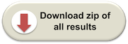
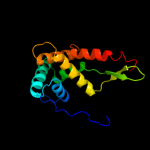
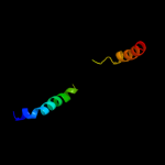
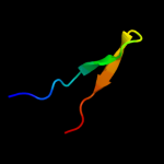
PDB header:chaperone/ribosomal protein Chain: S: PDB Molecule:30s ribosomal protein s7; PDBTitle: promiscuous substrate recognition in folding and assembly activities2 of the trigger factor chaperone
Confidence and coverage
Confidence:
100.0%
Coverage:
82%
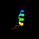
147 residues ( 82% of your sequence) have been modelled with 100.0% confidence by the single highest scoring template.
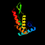
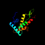
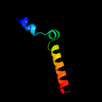
Region: 3 - 152 Aligned: 150 Modelled: 150 Confidence: 100.0% Identity: 100% PDB header:ribosome Chain: G: PDB Molecule:30s ribosomal protein s7; PDBTitle: crystal structure of the bacterial ribosome from2 escherichia coli in complex with the antibiotic kasugamyin3 at 3.5a resolution. this file contains the 30s subunit of4 one 70s ribosome. the entire crystal structure contains5 two 70s ribosomes and is described in remark 400.
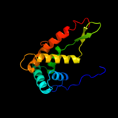
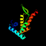
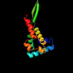
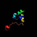
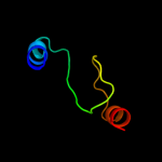
Region: 13 - 149 Aligned: 137 Modelled: 137 Confidence: 100.0% Identity: 29% PDB header:ribosome Chain: G: PDB Molecule:40s ribosomal protein s5; PDBTitle: structure of the ribosomal 80s-eef2-sordarin complex from2 yeast obtained by docking atomic models for rna and protein3 components into a 11.7 a cryo-em map. this file, 1s1h,4 contains 40s subunit. the 60s ribosomal subunit is in file5 1s1i.
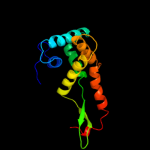
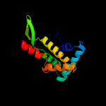
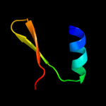
Region: 19 - 149 Aligned: 130 Modelled: 131 Confidence: 100.0% Identity: 28% PDB header:ribosomal protein/rna Chain: G: PDB Molecule:rna helix; PDBTitle: structure of a mammalian ribosomal 40s subunit within an2 80s complex obtained by docking homology models of the rna3 and proteins into an 8.7 a cryo-em map
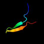
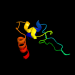
Region: 29 - 73 Aligned: 45 Modelled: 45 Confidence: 20.0% Identity: 11% PDB header:protein binding, dna binding protein Chain: A: PDB Molecule:dna cleavage and packaging protein large subunit, ul89; PDBTitle: nmr structure of the c-terminal domain of pul89
Region: 62 - 101 Aligned: 40 Modelled: 40 Confidence: 11.4% Identity: 15% PDB header:transport protein Chain: A: PDB Molecule:appears to be functionally related to snf7; PDBTitle: structure of the escrt-ii endosomal trafficking complex
Phyre2
21
22
23
24
25
26
27
28
29
30
31
32
33
34
35
Detailed template information
Binding site prediction
Due to computational demand, binding site predictions are not run for batch jobs
If you want to predict binding sites, please manually submit your model of choice to 3DLigandSite
Phyre is for academic use only
Please cite: Protein structure prediction on
the web: a case study using the Phyre server
Kelley LA and Sternberg MJE. Nature Protocols
4, 363 - 371 (2009) [pdf] [Import into BibTeX]
If you use the binding site
predictions from 3DLigandSite, please also cite:
3DLigandSite: predicting ligand-binding sites using similar structures.
Wass MN, Kelley LA and Sternberg
MJ Nucleic Acids Research 38, W469-73 (2010) [PubMed]