| 1 | c2gejA_
|
|
 |
98.2 |
12 |
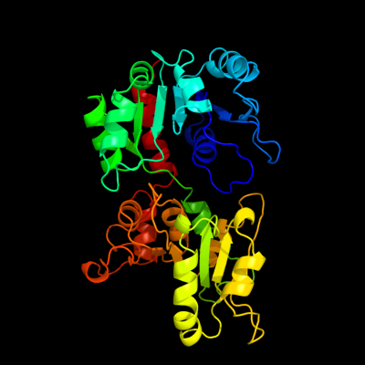
PDB header:transferase
Chain: A: PDB Molecule:phosphatidylinositol mannosyltransferase (pima);
PDBTitle: crystal structure of phosphatidylinositol mannosyltransferase (pima)2 from mycobacterium smegmatis in complex with gdp-man
|
| 2 | c3ot5D_
|
|
 |
97.5 |
12 |
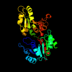
PDB header:isomerase
Chain: D: PDB Molecule:udp-n-acetylglucosamine 2-epimerase;
PDBTitle: 2.2 angstrom resolution crystal structure of putative udp-n-2 acetylglucosamine 2-epimerase from listeria monocytogenes
|
| 3 | c2x0dA_
|
|
 |
96.9 |
13 |
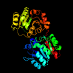
PDB header:transferase
Chain: A: PDB Molecule:wsaf;
PDBTitle: apo structure of wsaf
|
| 4 | d1v4va_
|
|
 |
96.8 |
14 |
Fold:UDP-Glycosyltransferase/glycogen phosphorylase
Superfamily:UDP-Glycosyltransferase/glycogen phosphorylase
Family:UDP-N-acetylglucosamine 2-epimerase |
| 5 | c2xmpB_
|
|
 |
96.5 |
14 |
PDB header:sugar binding protein
Chain: B: PDB Molecule:trehalose-synthase tret;
PDBTitle: crystal structure of trehalose synthase tret mutant e326a2 from p.horishiki in complex with udp
|
| 6 | c2vsnB_
|
|
 |
96.5 |
12 |
PDB header:transferase
Chain: B: PDB Molecule:xcogt;
PDBTitle: structure and topological arrangement of an o-glcnac2 transferase homolog: insight into molecular control of3 intracellular glycosylation
|
| 7 | c3c4vB_
|
|
 |
96.0 |
8 |
PDB header:transferase
Chain: B: PDB Molecule:predicted glycosyltransferases;
PDBTitle: structure of the retaining glycosyltransferase msha:the2 first step in mycothiol biosynthesis. organism:3 corynebacterium glutamicum : complex with udp and 1l-ins-1-4 p.
|
| 8 | c3oy2A_
|
|
 |
96.0 |
10 |
PDB header:viral protein,transferase
Chain: A: PDB Molecule:glycosyltransferase b736l;
PDBTitle: crystal structure of a putative glycosyltransferase from paramecium2 bursaria chlorella virus ny2a
|
| 9 | c2jjmH_
|
|
 |
95.9 |
13 |
PDB header:transferase
Chain: H: PDB Molecule:glycosyl transferase, group 1 family protein;
PDBTitle: crystal structure of a family gt4 glycosyltransferase from2 bacillus anthracis orf ba1558.
|
| 10 | d1f6da_
|
|
 |
95.7 |
13 |
Fold:UDP-Glycosyltransferase/glycogen phosphorylase
Superfamily:UDP-Glycosyltransferase/glycogen phosphorylase
Family:UDP-N-acetylglucosamine 2-epimerase |
| 11 | c2x6rA_
|
|
 |
95.6 |
12 |
PDB header:isomerase
Chain: A: PDB Molecule:trehalose-synthase tret;
PDBTitle: crystal structure of trehalose synthase tret from p.2 horikoshi produced by soaking in trehalose
|
| 12 | d1o6ca_
|
|
 |
95.4 |
14 |
Fold:UDP-Glycosyltransferase/glycogen phosphorylase
Superfamily:UDP-Glycosyltransferase/glycogen phosphorylase
Family:UDP-N-acetylglucosamine 2-epimerase |
| 13 | c1uquB_
|
|
 |
95.2 |
14 |
PDB header:synthase
Chain: B: PDB Molecule:alpha, alpha-trehalose-phosphate synthase;
PDBTitle: trehalose-6-phosphate from e. coli bound with udp-glucose.
|
| 14 | c3pe3D_
|
|
 |
95.2 |
13 |
PDB header:transferase
Chain: D: PDB Molecule:udp-n-acetylglucosamine--peptide n-
PDBTitle: structure of human o-glcnac transferase and its complex with a peptide2 substrate
|
| 15 | c2r60A_
|
|
 |
94.7 |
14 |
PDB header:transferase
Chain: A: PDB Molecule:glycosyl transferase, group 1;
PDBTitle: structure of apo sucrose phosphate synthase (sps) of2 halothermothrix orenii
|
| 16 | c3okaA_
|
|
 |
92.5 |
12 |
PDB header:transferase
Chain: A: PDB Molecule:gdp-mannose-dependent alpha-(1-6)-phosphatidylinositol
PDBTitle: crystal structure of corynebacterium glutamicum pimb' in complex with2 gdp-man (triclinic crystal form)
|
| 17 | c3dzcA_
|
|
 |
91.9 |
13 |
PDB header:isomerase
Chain: A: PDB Molecule:udp-n-acetylglucosamine 2-epimerase;
PDBTitle: 2.35 angstrom resolution structure of wecb (vc0917), a udp-n-2 acetylglucosamine 2-epimerase from vibrio cholerae.
|
| 18 | c2xcuC_
|
|
 |
87.5 |
12 |
PDB header:transferase
Chain: C: PDB Molecule:3-deoxy-d-manno-2-octulosonic acid transferase;
PDBTitle: membrane-embedded monofunctional glycosyltransferase waaa of aquifex2 aeolicus, comlex with cmp
|
| 19 | d2iw1a1
|
|
 |
85.7 |
11 |
Fold:UDP-Glycosyltransferase/glycogen phosphorylase
Superfamily:UDP-Glycosyltransferase/glycogen phosphorylase
Family:Glycosyl transferases group 1 |
| 20 | d2f9fa1
|
|
 |
83.7 |
14 |
Fold:UDP-Glycosyltransferase/glycogen phosphorylase
Superfamily:UDP-Glycosyltransferase/glycogen phosphorylase
Family:Glycosyl transferases group 1 |
| 21 | d2bfwa1 |
|
not modelled |
76.2 |
10 |
Fold:UDP-Glycosyltransferase/glycogen phosphorylase
Superfamily:UDP-Glycosyltransferase/glycogen phosphorylase
Family:Glycosyl transferases group 1 |
| 22 | d1duvg2 |
|
not modelled |
69.1 |
20 |
Fold:ATC-like
Superfamily:Aspartate/ornithine carbamoyltransferase
Family:Aspartate/ornithine carbamoyltransferase |
| 23 | d1uqta_ |
|
not modelled |
66.3 |
12 |
Fold:UDP-Glycosyltransferase/glycogen phosphorylase
Superfamily:UDP-Glycosyltransferase/glycogen phosphorylase
Family:Trehalose-6-phosphate synthase, OtsA |
| 24 | d2bisa1 |
|
not modelled |
64.8 |
12 |
Fold:UDP-Glycosyltransferase/glycogen phosphorylase
Superfamily:UDP-Glycosyltransferase/glycogen phosphorylase
Family:Glycosyl transferases group 1 |
| 25 | d1pswa_ |
|
not modelled |
60.5 |
13 |
Fold:UDP-Glycosyltransferase/glycogen phosphorylase
Superfamily:UDP-Glycosyltransferase/glycogen phosphorylase
Family:ADP-heptose LPS heptosyltransferase II |
| 26 | c3oneA_ |
|
not modelled |
55.1 |
9 |
PDB header:hydrolase/hydrolase substrate
Chain: A: PDB Molecule:adenosylhomocysteinase;
PDBTitle: crystal structure of lupinus luteus s-adenosyl-l-homocysteine2 hydrolase in complex with adenine
|
| 27 | c2w37A_ |
|
not modelled |
53.7 |
18 |
PDB header:transferase
Chain: A: PDB Molecule:ornithine carbamoyltransferase, catabolic;
PDBTitle: crystal structure of the hexameric catabolic ornithine2 transcarbamylase from lactobacillus hilgardii
|
| 28 | c1v8bA_ |
|
not modelled |
49.2 |
13 |
PDB header:hydrolase
Chain: A: PDB Molecule:adenosylhomocysteinase;
PDBTitle: crystal structure of a hydrolase
|
| 29 | c3tovB_ |
|
not modelled |
48.5 |
17 |
PDB header:transferase
Chain: B: PDB Molecule:glycosyl transferase family 9;
PDBTitle: the crystal structure of the glycosyl transferase family 9 from2 veillonella parvula dsm 2008
|
| 30 | d1qs0b1 |
|
not modelled |
43.7 |
16 |
Fold:Thiamin diphosphate-binding fold (THDP-binding)
Superfamily:Thiamin diphosphate-binding fold (THDP-binding)
Family:Branched-chain alpha-keto acid dehydrogenase Pyr module |
| 31 | d1dxha2 |
|
not modelled |
38.8 |
14 |
Fold:ATC-like
Superfamily:Aspartate/ornithine carbamoyltransferase
Family:Aspartate/ornithine carbamoyltransferase |
| 32 | d1vlva2 |
|
not modelled |
38.0 |
16 |
Fold:ATC-like
Superfamily:Aspartate/ornithine carbamoyltransferase
Family:Aspartate/ornithine carbamoyltransferase |
| 33 | c3dhyC_ |
|
not modelled |
36.4 |
18 |
PDB header:hydrolase
Chain: C: PDB Molecule:adenosylhomocysteinase;
PDBTitle: crystal structures of mycobacterium tuberculosis s-adenosyl-l-2 homocysteine hydrolase in ternary complex with substrate and3 inhibitors
|
| 34 | c2h1fB_ |
|
not modelled |
33.1 |
12 |
PDB header:transferase
Chain: B: PDB Molecule:lipopolysaccharide heptosyltransferase-1;
PDBTitle: e. coli heptosyltransferase waac with adp
|
| 35 | d1li4a2 |
|
not modelled |
32.9 |
13 |
Fold:Flavodoxin-like
Superfamily:Formate/glycerate dehydrogenase catalytic domain-like
Family:S-adenosylhomocystein hydrolase |
| 36 | d1pvva2 |
|
not modelled |
27.9 |
24 |
Fold:ATC-like
Superfamily:Aspartate/ornithine carbamoyltransferase
Family:Aspartate/ornithine carbamoyltransferase |
| 37 | c1d4fD_ |
|
not modelled |
27.5 |
14 |
PDB header:hydrolase
Chain: D: PDB Molecule:s-adenosylhomocysteine hydrolase;
PDBTitle: crystal structure of recombinant rat-liver d244e mutant s-2 adenosylhomocysteine hydrolase
|
| 38 | c3r1fO_ |
|
not modelled |
27.5 |
26 |
PDB header:transcription
Chain: O: PDB Molecule:esx-1 secretion-associated regulator espr;
PDBTitle: crystal structure of a key regulator of virulence in mycobacterium2 tuberculosis
|
| 39 | c1ortD_ |
|
not modelled |
27.3 |
14 |
PDB header:transferase
Chain: D: PDB Molecule:ornithine transcarbamoylase;
PDBTitle: ornithine transcarbamoylase from pseudomonas aeruginosa
|
| 40 | c3updA_ |
|
not modelled |
26.6 |
19 |
PDB header:transferase
Chain: A: PDB Molecule:ornithine carbamoyltransferase;
PDBTitle: 2.9 angstrom crystal structure of ornithine carbamoyltransferase2 (argf) from vibrio vulnificus
|
| 41 | d1iyjb5 |
|
not modelled |
22.6 |
21 |
Fold:OB-fold
Superfamily:Nucleic acid-binding proteins
Family:Single strand DNA-binding domain, SSB |
| 42 | d1xmba1 |
|
not modelled |
22.3 |
8 |
Fold:Phosphorylase/hydrolase-like
Superfamily:Zn-dependent exopeptidases
Family:Bacterial dinuclear zinc exopeptidases |
| 43 | c2otcA_ |
|
not modelled |
22.1 |
17 |
PDB header:transferase
Chain: A: PDB Molecule:ornithine carbamoyltransferase;
PDBTitle: ornithine transcarbamoylase complexed with n-2 (phosphonacetyl)-l-ornithine
|
| 44 | d2bfdb1 |
|
not modelled |
21.9 |
14 |
Fold:Thiamin diphosphate-binding fold (THDP-binding)
Superfamily:Thiamin diphosphate-binding fold (THDP-binding)
Family:Branched-chain alpha-keto acid dehydrogenase Pyr module |
| 45 | c1olsB_ |
|
not modelled |
20.7 |
16 |
PDB header:oxidoreductase
Chain: B: PDB Molecule:2-oxoisovalerate dehydrogenase beta subunit;
PDBTitle: roles of his291-alpha and his146-beta' in the reductive2 acylation reaction catalyzed by human branched-chain3 alpha-ketoacid dehydrogenase
|
| 46 | c3en0A_ |
|
not modelled |
18.9 |
13 |
PDB header:hydrolase
Chain: A: PDB Molecule:cyanophycinase;
PDBTitle: the structure of cyanophycinase
|
| 47 | c3rhzB_ |
|
not modelled |
17.8 |
10 |
PDB header:transferase
Chain: B: PDB Molecule:nucleotide sugar synthetase-like protein;
PDBTitle: structure and functional analysis of a new subfamily of2 glycosyltransferases required for glycosylation of serine-rich3 streptococcal adhesions
|
| 48 | c3cseA_ |
|
not modelled |
15.6 |
14 |
PDB header:oxidoreductase
Chain: A: PDB Molecule:dihydrofolate reductase;
PDBTitle: candida glabrata dihydrofolate reductase complexed with2 nadph and 2,4-diamino-5-(3-(2,5-dimethoxyphenyl)prop-1-3 ynyl)-6-ethylpyrimidine (ucp120b)
|
| 49 | c1vlvA_ |
|
not modelled |
15.3 |
13 |
PDB header:transferase
Chain: A: PDB Molecule:ornithine carbamoyltransferase;
PDBTitle: crystal structure of ornithine carbamoyltransferase (tm1097) from2 thermotoga maritima at 2.25 a resolution
|
| 50 | c2kzkA_ |
|
not modelled |
15.2 |
37 |
PDB header:protein transport
Chain: A: PDB Molecule:uncharacterized protein yol083w;
PDBTitle: solution structure of alpha-mannosidase binding domain of atg34
|
| 51 | c1jb9A_ |
|
not modelled |
15.1 |
9 |
PDB header:oxidoreductase
Chain: A: PDB Molecule:ferredoxin-nadp reductase;
PDBTitle: crystal structure of the ferredoxin:nadp+ reductase from maize root at2 1.7 angstroms
|
| 52 | c3q3hA_ |
|
not modelled |
14.7 |
13 |
PDB header:transferase
Chain: A: PDB Molecule:hmw1c-like glycosyltransferase;
PDBTitle: crystal structure of the actinobacillus pleuropneumoniae hmw1c2 glycosyltransferase in complex with udp-glc
|
| 53 | d1x6va1 |
|
not modelled |
14.5 |
20 |
Fold:PUA domain-like
Superfamily:PUA domain-like
Family:ATP sulfurylase N-terminal domain |
| 54 | d1otha2 |
|
not modelled |
14.3 |
20 |
Fold:ATC-like
Superfamily:Aspartate/ornithine carbamoyltransferase
Family:Aspartate/ornithine carbamoyltransferase |
| 55 | c2qzsA_ |
|
not modelled |
14.2 |
12 |
PDB header:transferase
Chain: A: PDB Molecule:glycogen synthase;
PDBTitle: crystal structure of wild-type e.coli gs in complex with adp2 and glucose(wtgsb)
|
| 56 | d1rzua_ |
|
not modelled |
14.1 |
12 |
Fold:UDP-Glycosyltransferase/glycogen phosphorylase
Superfamily:UDP-Glycosyltransferase/glycogen phosphorylase
Family:Glycosyl transferases group 1 |
| 57 | c2kzbA_ |
|
not modelled |
14.0 |
18 |
PDB header:protein transport
Chain: A: PDB Molecule:autophagy-related protein 19;
PDBTitle: solution structure of alpha-mannosidase binding domain of atg19
|
| 58 | d2nx2a1 |
|
not modelled |
13.4 |
14 |
Fold:MCP/YpsA-like
Superfamily:MCP/YpsA-like
Family:YpsA-like |
| 59 | c2jzcA_ |
|
not modelled |
13.2 |
18 |
PDB header:transferase
Chain: A: PDB Molecule:udp-n-acetylglucosamine transferase subunit
PDBTitle: nmr solution structure of alg13: the sugar donor subunit of2 a yeast n-acetylglucosamine transferase. northeast3 structural genomics consortium target yg1
|
| 60 | c3jrnA_ |
|
not modelled |
13.0 |
16 |
PDB header:plant protein
Chain: A: PDB Molecule:at1g72930 protein;
PDBTitle: crystal structure of tir domain from arabidopsis thaliana
|
| 61 | d1u9ya1 |
|
not modelled |
12.9 |
8 |
Fold:PRTase-like
Superfamily:PRTase-like
Family:Phosphoribosylpyrophosphate synthetase-like |
| 62 | c3d64A_ |
|
not modelled |
12.5 |
13 |
PDB header:hydrolase
Chain: A: PDB Molecule:adenosylhomocysteinase;
PDBTitle: crystal structure of s-adenosyl-l-homocysteine hydrolase from2 burkholderia pseudomallei
|
| 63 | d1pj3a1 |
|
not modelled |
12.2 |
17 |
Fold:NAD(P)-binding Rossmann-fold domains
Superfamily:NAD(P)-binding Rossmann-fold domains
Family:Aminoacid dehydrogenase-like, C-terminal domain |
| 64 | c1dkrB_ |
|
not modelled |
12.1 |
27 |
PDB header:transferase
Chain: B: PDB Molecule:phosphoribosyl pyrophosphate synthetase;
PDBTitle: crystal structures of bacillus subtilis phosphoribosylpyrophosphate2 synthetase: molecular basis of allosteric inhibition and activation.
|
| 65 | d2f62a1 |
|
not modelled |
12.0 |
11 |
Fold:Flavodoxin-like
Superfamily:N-(deoxy)ribosyltransferase-like
Family:N-deoxyribosyltransferase |
| 66 | d1jhda1 |
|
not modelled |
11.9 |
7 |
Fold:PUA domain-like
Superfamily:PUA domain-like
Family:ATP sulfurylase N-terminal domain |
| 67 | d1wdds_ |
|
not modelled |
11.3 |
33 |
Fold:RuBisCO, small subunit
Superfamily:RuBisCO, small subunit
Family:RuBisCO, small subunit |
| 68 | d1s2da_ |
|
not modelled |
11.1 |
15 |
Fold:Flavodoxin-like
Superfamily:N-(deoxy)ribosyltransferase-like
Family:N-deoxyribosyltransferase |
| 69 | d2v6ai1 |
|
not modelled |
11.1 |
33 |
Fold:RuBisCO, small subunit
Superfamily:RuBisCO, small subunit
Family:RuBisCO, small subunit |
| 70 | c3c7cB_ |
|
not modelled |
11.1 |
9 |
PDB header:oxidoreductase
Chain: B: PDB Molecule:octopine dehydrogenase;
PDBTitle: a structural basis for substrate and stereo selectivity in2 octopine dehydrogenase (odh-nadh-l-arginine)
|
| 71 | c3q98A_ |
|
not modelled |
10.9 |
13 |
PDB header:transferase
Chain: A: PDB Molecule:transcarbamylase;
PDBTitle: structure of ygew encoded protein from e. coli
|
| 72 | c2z5bB_ |
|
not modelled |
10.6 |
21 |
PDB header:chaperone
Chain: B: PDB Molecule:uncharacterized protein ylr021w;
PDBTitle: crystal structure of a novel chaperone complex for yeast2 20s proteasome assembly
|
| 73 | d1miua5 |
|
not modelled |
10.5 |
19 |
Fold:OB-fold
Superfamily:Nucleic acid-binding proteins
Family:Single strand DNA-binding domain, SSB |
| 74 | d1g8fa1 |
|
not modelled |
10.5 |
21 |
Fold:PUA domain-like
Superfamily:PUA domain-like
Family:ATP sulfurylase N-terminal domain |
| 75 | c3q6nF_ |
|
not modelled |
10.4 |
16 |
PDB header:chaperone
Chain: F: PDB Molecule:heat shock protein hsp 90-alpha;
PDBTitle: crystal structure of human mc-hsp90 in p21 space group
|
| 76 | d1uzdc1 |
|
not modelled |
10.3 |
33 |
Fold:RuBisCO, small subunit
Superfamily:RuBisCO, small subunit
Family:RuBisCO, small subunit |
| 77 | c2dgdD_ |
|
not modelled |
10.3 |
10 |
PDB header:lyase
Chain: D: PDB Molecule:223aa long hypothetical arylmalonate decarboxylase;
PDBTitle: crystal structure of st0656, a function unknown protein from2 sulfolobus tokodaii
|
| 78 | d1v47a1 |
|
not modelled |
10.3 |
10 |
Fold:PUA domain-like
Superfamily:PUA domain-like
Family:ATP sulfurylase N-terminal domain |
| 79 | d8ruci_ |
|
not modelled |
10.3 |
33 |
Fold:RuBisCO, small subunit
Superfamily:RuBisCO, small subunit
Family:RuBisCO, small subunit |
| 80 | d1ej7s_ |
|
not modelled |
10.2 |
33 |
Fold:RuBisCO, small subunit
Superfamily:RuBisCO, small subunit
Family:RuBisCO, small subunit |
| 81 | d1gk8i_ |
|
not modelled |
9.8 |
33 |
Fold:RuBisCO, small subunit
Superfamily:RuBisCO, small subunit
Family:RuBisCO, small subunit |
| 82 | d2pmra1 |
|
not modelled |
9.7 |
25 |
Fold:immunoglobulin/albumin-binding domain-like
Superfamily:AF1782-like
Family:AF1782-like |
| 83 | c2r5wA_ |
|
not modelled |
9.0 |
13 |
PDB header:hydrolase, transferase
Chain: A: PDB Molecule:nicotinamide-nucleotide adenylyltransferase;
PDBTitle: crystal structure of a bifunctional nmn2 adenylyltransferase/adp ribose pyrophosphatase from3 francisella tularensis
|
| 84 | d2e74g1 |
|
not modelled |
8.8 |
44 |
Fold:Single transmembrane helix
Superfamily:PetG subunit of the cytochrome b6f complex
Family:PetG subunit of the cytochrome b6f complex |
| 85 | c1ml4A_ |
|
not modelled |
8.7 |
18 |
PDB header:transferase
Chain: A: PDB Molecule:aspartate transcarbamoylase;
PDBTitle: the pala-liganded aspartate transcarbamoylase catalytic subunit from2 pyrococcus abyssi
|
| 86 | d1m8pa1 |
|
not modelled |
8.6 |
17 |
Fold:PUA domain-like
Superfamily:PUA domain-like
Family:ATP sulfurylase N-terminal domain |
| 87 | c1zh8B_ |
|
not modelled |
8.6 |
20 |
PDB header:oxidoreductase
Chain: B: PDB Molecule:oxidoreductase;
PDBTitle: crystal structure of oxidoreductase (tm0312) from thermotoga maritima2 at 2.50 a resolution
|
| 88 | c3nv9A_ |
|
not modelled |
8.4 |
20 |
PDB header:oxidoreductase
Chain: A: PDB Molecule:malic enzyme;
PDBTitle: crystal structure of entamoeba histolytica malic enzyme
|
| 89 | d1f8ya_ |
|
not modelled |
8.3 |
15 |
Fold:Flavodoxin-like
Superfamily:N-(deoxy)ribosyltransferase-like
Family:N-deoxyribosyltransferase |
| 90 | c3oziB_ |
|
not modelled |
8.1 |
21 |
PDB header:plant protein
Chain: B: PDB Molecule:l6tr;
PDBTitle: crystal structure of the tir domain from the flax disease resistance2 protein l6
|
| 91 | c1zcoA_ |
|
not modelled |
8.1 |
15 |
PDB header:lyase
Chain: A: PDB Molecule:2-dehydro-3-deoxyphosphoheptonate aldolase;
PDBTitle: crystal structure of pyrococcus furiosus 3-deoxy-d-arabino-2 heptulosonate 7-phosphate synthase
|
| 92 | c3mtqA_ |
|
not modelled |
7.6 |
11 |
PDB header:transferase
Chain: A: PDB Molecule:putative phosphoenolpyruvate-dependent sugar
PDBTitle: crystal structure of a putative phosphoenolpyruvate-dependent sugar2 phosphotransferase system (pts) permease (kpn_04802) from klebsiella3 pneumoniae subsp. pneumoniae mgh 78578 at 1.70 a resolution
|
| 93 | d1ir1s_ |
|
not modelled |
7.6 |
33 |
Fold:RuBisCO, small subunit
Superfamily:RuBisCO, small subunit
Family:RuBisCO, small subunit |
| 94 | c1qr6A_ |
|
not modelled |
7.5 |
18 |
PDB header:oxidoreductase
Chain: A: PDB Molecule:malic enzyme 2;
PDBTitle: human mitochondrial nad(p)-dependent malic enzyme
|
| 95 | c3hftA_ |
|
not modelled |
7.5 |
16 |
PDB header:hydrolase
Chain: A: PDB Molecule:wbms, polysaccharide deacetylase involved in o-antigen
PDBTitle: crystal structure of a putative polysaccharide deacetylase involved in2 o-antigen biosynthesis (wbms, bb0128) from bordetella bronchiseptica3 at 1.90 a resolution
|
| 96 | c3clbA_ |
|
not modelled |
7.3 |
21 |
PDB header:oxidoreductase, transferase
Chain: A: PDB Molecule:dhfr-ts;
PDBTitle: structure of bifunctional tcdhfr-ts in complex with tmq
|
| 97 | c2z5cA_ |
|
not modelled |
7.3 |
19 |
PDB header:chaperone/hydrolase
Chain: A: PDB Molecule:protein ypl144w;
PDBTitle: crystal structure of a novel chaperone complex for yeast2 20s proteasome assembly
|
| 98 | d1gq2a1 |
|
not modelled |
7.2 |
14 |
Fold:NAD(P)-binding Rossmann-fold domains
Superfamily:NAD(P)-binding Rossmann-fold domains
Family:Aminoacid dehydrogenase-like, C-terminal domain |
| 99 | c1a1sA_ |
|
not modelled |
7.2 |
24 |
PDB header:transcarbamylase
Chain: A: PDB Molecule:ornithine carbamoyltransferase;
PDBTitle: ornithine carbamoyltransferase from pyrococcus furiosus
|