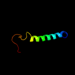
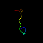
| 1 |
|
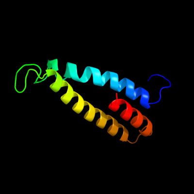
PDB 1jb0 chain L
Region: 214 - 299
Aligned: 81
Modelled: 86
Confidence: 31.8%
Identity: 14%
Fold: Photosystem I reaction center subunit XI, PsaL
Superfamily: Photosystem I reaction center subunit XI, PsaL
Family: Photosystem I reaction center subunit XI, PsaL
Phyre2
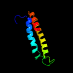
| 2 |
|
PDB 2wvm chain A
Region: 86 - 122
Aligned: 37
Modelled: 37
Confidence: 11.5%
Identity: 24%
PDB header:transferase
Chain: A: PDB Molecule:mannosyl-3-phosphoglycerate synthase;
PDBTitle: h309a mutant of mannosyl-3-phosphoglycerate synthase from2 thermus thermophilus hb27 in complex with3 gdp-alpha-d-mannose and mg(ii)
Phyre2
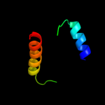
| 3 |
|
PDB 2zu8 chain A
Region: 86 - 122
Aligned: 37
Modelled: 34
Confidence: 9.1%
Identity: 19%
PDB header:transferase
Chain: A: PDB Molecule:mannosyl-3-phosphoglycerate synthase;
PDBTitle: crystal structure of mannosyl-3-phosphoglycerate synthase2 from pyrococcus horikoshii
Phyre2
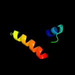
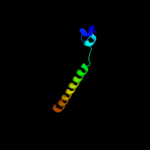
| 4 |
|
PDB 3hfx chain A
Region: 356 - 398
Aligned: 43
Modelled: 43
Confidence: 8.6%
Identity: 9%
PDB header:transport protein
Chain: A: PDB Molecule:l-carnitine/gamma-butyrobetaine antiporter;
PDBTitle: crystal structure of carnitine transporter
Phyre2
| 5 |
|
PDB 2y69 chain Q
Region: 234 - 288
Aligned: 55
Modelled: 55
Confidence: 8.1%
Identity: 9%
PDB header:electron transport
Chain: Q: PDB Molecule:cytochrome c oxidase subunit 4 isoform 1;
PDBTitle: bovine heart cytochrome c oxidase re-refined with molecular2 oxygen
Phyre2
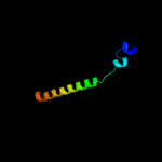
| 6 |
|
PDB 2vv5 chain A domain 3
Region: 149 - 236
Aligned: 82
Modelled: 88
Confidence: 7.3%
Identity: 16%
Fold: Mechanosensitive channel protein MscS (YggB), transmembrane region
Superfamily: Mechanosensitive channel protein MscS (YggB), transmembrane region
Family: Mechanosensitive channel protein MscS (YggB), transmembrane region
Phyre2
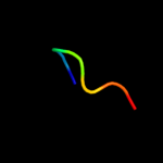
| 7 |
|
PDB 2b5k chain A
Region: 236 - 242
Aligned: 7
Modelled: 7
Confidence: 7.0%
Identity: 29%
PDB header:antimicrobial protein
Chain: A: PDB Molecule:polyphemusin-1;
PDBTitle: pv5 nmr solution structure in dpc micelles
Phyre2
| 8 |
|
PDB 1v54 chain D
Region: 234 - 288
Aligned: 55
Modelled: 55
Confidence: 6.7%
Identity: 9%
Fold: Single transmembrane helix
Superfamily: Mitochondrial cytochrome c oxidase subunit IV
Family: Mitochondrial cytochrome c oxidase subunit IV
Phyre2
| 9 |
|
PDB 3pm7 chain A
Region: 2 - 17
Aligned: 16
Modelled: 16
Confidence: 6.0%
Identity: 31%
PDB header:structural genomics, unknown function
Chain: A: PDB Molecule:uncharacterized protein;
PDBTitle: crystal structure of ef_3132 protein from enterococcus faecalis at the2 resolution 2a, northeast structural genomics consortium target efr184
Phyre2
| 10 |
|
PDB 2w2e chain A
Region: 221 - 347
Aligned: 122
Modelled: 127
Confidence: 5.4%
Identity: 17%
PDB header:membrane protein
Chain: A: PDB Molecule:aquaporin;
PDBTitle: 1.15 angstrom crystal structure of p.pastoris aquaporin,2 aqy1, in a closed conformation at ph 3.5
Phyre2