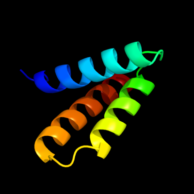
Phyre Home Retrieve Phyre Job Id
Emaill.a.kelley@imperial.ac.uk
DescriptionP75993
DateThu Jan 5 12:17:06 GMT 2012
Unique Job IDdffc6475b4c10124

Summary 

Top model
Image coloured by rainbow N → C terminus
Model (left) based on template c2oxlA_
Top template information
PDB header:gene regulation
Chain: A: PDB Molecule:hypothetical protein ymgb;
PDBTitle: structure and function of the e. coli protein ymgb: a protein critical2 for biofilm formation and acid resistance
Confidence and coverage
Confidence: 99.9% Coverage: 70%
62 residues ( 70% of your sequence) have been modelled with 99.9% confidence by the single highest scoring template.
You may wish to submit your sequence to Phyrealarm. This will automatically scan your sequence every week for new potential templates as they appear in the Phyre2 library.
3D viewing
Interactive 3D view in Jmol

Sequence analysis 

Secondary structure and disorder prediction 

   1........10.........20.........30.........40.........50.........60
Sequence  MLEDTTIHNAITDKALASYFRSSGNLLEEESAVLGQAVTNLMLSGDNVNNKNIILSLIHS
Secondary structure 


SS confidence 


Disorder  ?????????????


?

?


???????


Disorder confidence 


   .........70.........80........
Sequence  LETTSDILKADVIRKTLEIVLRYTADDM
Secondary structure 


SS confidence 


Disorder 


?????
Disorder confidence 


Confidence Key
High(9)                    Low (0)
?Disordered
Alpha helix
Beta strand

Domain analysis 

Hover over an aligned region to see model and summary info

Please note, only up to the top 20 hits are modelled to reduce computer load

RankAligned region

PDB 2oxl chain A

3D model

Region: 25 - 86
Aligned: 62
Modelled: 62
Confidence: 99.9%
Identity: 98%
PDB header:gene regulation
Chain: A: PDB Molecule:hypothetical protein ymgb;
PDBTitle: structure and function of the e. coli protein ymgb: a protein critical2 for biofilm formation and acid resistance

Phyre2

PDB 1stz chain B

3D model

Region: 30 - 53
Aligned: 24
Modelled: 24
Confidence: 25.1%
Identity: 17%
PDB header:transcription
Chain: B: PDB Molecule:heat-inducible transcription repressor hrca homolog;
PDBTitle: crystal structure of a hypothetical protein at 2.2 a resolution

Phyre2

PDB 1v92 chain A

3D model

Region: 13 - 24
Aligned: 12
Modelled: 12
Confidence: 15.5%
Identity: 42%
Fold: RuvA C-terminal domain-like
Superfamily: UBA-like
Family: TAP-C domain-like

Phyre2

PDB 3ct5 chain A

3D model

Region: 14 - 40
Aligned: 27
Modelled: 27
Confidence: 13.4%
Identity: 19%
PDB header:hydrolase
Chain: A: PDB Molecule:morphogenesis protein 1;
PDBTitle: crystal and cryoem structural studies of a cell wall degrading enzyme2 in the bacteriophage phi29 tail

Phyre2

PDB 2bn5 chain A

3D model

Region: 13 - 31
Aligned: 19
Modelled: 19
Confidence: 11.2%
Identity: 37%
PDB header:nuclear protein
Chain: A: PDB Molecule:psi;
PDBTitle: p-element somatic inhibitor protein complex with u1-70k2 proline-rich peptide

Phyre2

PDB 1stz chain A domain 1

3D model

Region: 32 - 53
Aligned: 22
Modelled: 22
Confidence: 10.7%
Identity: 18%
Fold: DNA/RNA-binding 3-helical bundle
Superfamily: "Winged helix" DNA-binding domain
Family: Heat-inducible transcription repressor HrcA, N-terminal domain

Phyre2

PDB 3gxv chain A

3D model

Region: 18 - 87
Aligned: 60
Modelled: 70
Confidence: 10.3%
Identity: 20%
PDB header:hydrolase/replication
Chain: A: PDB Molecule:replicative dna helicase;
PDBTitle: three-dimensional structure of n-terminal domain of dnab2 helicase from helicobacter pylori and its interactions with3 primase

Phyre2

PDB 2j8a chain A

3D model

Region: 4 - 25
Aligned: 22
Modelled: 22
Confidence: 9.8%
Identity: 23%
PDB header:transferase
Chain: A: PDB Molecule:histone-lysine n-methyltransferase, h3 lysine-4
PDBTitle: x-ray structure of the n-terminus rrm domain of set1

Phyre2

PDB 3eyy chain A

3D model

Region: 13 - 80
Aligned: 61
Modelled: 68
Confidence: 9.5%
Identity: 18%
PDB header:transport
Chain: A: PDB Molecule:putative iron uptake regulatory protein;
PDBTitle: structural basis for the specialization of nur, a nickel-2 specific fur homologue, in metal sensing and dna3 recognition

Phyre2

PDB 1eiy chain A domain 1

3D model

Region: 15 - 41
Aligned: 27
Modelled: 27
Confidence: 8.9%
Identity: 22%
Fold: Long alpha-hairpin
Superfamily: tRNA-binding arm
Family: Phenylalanyl-tRNA synthetase (PheRS)

Phyre2

PDB 2izp chain B

3D model

Region: 14 - 44
Aligned: 31
Modelled: 31
Confidence: 8.9%
Identity: 16%
PDB header:toxin
Chain: B: PDB Molecule:putative membrane antigen;
PDBTitle: bipd - an invasion prtein associated with the type-iii2 secretion system of burkholderia pseudomallei.

Phyre2

PDB 2b3y chain A domain 2

3D model

Region: 48 - 61
Aligned: 14
Modelled: 14
Confidence: 8.6%
Identity: 29%
Fold: Aconitase iron-sulfur domain
Superfamily: Aconitase iron-sulfur domain
Family: Aconitase iron-sulfur domain

Phyre2

PDB 1ugp chain A

3D model

Region: 20 - 76
Aligned: 56
Modelled: 57
Confidence: 8.0%
Identity: 20%
Fold: Nitrile hydratase alpha chain
Superfamily: Nitrile hydratase alpha chain
Family: Nitrile hydratase alpha chain

Phyre2

PDB 1got chain G

3D model

Region: 48 - 82
Aligned: 35
Modelled: 35
Confidence: 7.8%
Identity: 11%
Fold: Non-globular all-alpha subunits of globular proteins
Superfamily: Transducin (heterotrimeric G protein), gamma chain
Family: Transducin (heterotrimeric G protein), gamma chain

Phyre2

PDB 3h8d chain E

3D model

Region: 12 - 22
Aligned: 11
Modelled: 11
Confidence: 7.7%
Identity: 45%
PDB header:motor protein/signaling protein
Chain: E: PDB Molecule:disabled homolog 2;
PDBTitle: crystal structure of myosin vi in complex with dab2 peptide

Phyre2

PDB 3f1i chain H

3D model

Region: 32 - 61
Aligned: 30
Modelled: 30
Confidence: 7.6%
Identity: 20%
PDB header:protein binding
Chain: H: PDB Molecule:hepatocyte growth factor-regulated tyrosine kinase
PDBTitle: human escrt-0 core complex

Phyre2

PDB 1aco chain A domain 2

3D model

Region: 48 - 61
Aligned: 14
Modelled: 14
Confidence: 7.3%
Identity: 29%
Fold: Aconitase iron-sulfur domain
Superfamily: Aconitase iron-sulfur domain
Family: Aconitase iron-sulfur domain

Phyre2

PDB 2dzl chain A

3D model

Region: 13 - 24
Aligned: 12
Modelled: 12
Confidence: 6.9%
Identity: 25%
PDB header:structural genomics unknown function
Chain: A: PDB Molecule:protein fam100b;
PDBTitle: solution structure of the uba domain in human protein2 fam100b

Phyre2

PDB 1q5z chain A

3D model

Region: 58 - 78
Aligned: 21
Modelled: 21
Confidence: 6.8%
Identity: 38%
Fold: Invasion protein A (SipA) , C-terminal actin binding domain
Superfamily: Invasion protein A (SipA) , C-terminal actin binding domain
Family: Invasion protein A (SipA) , C-terminal actin binding domain

Phyre2

PDB 1q5z chain A

3D model

Region: 58 - 78
Aligned: 21
Modelled: 21
Confidence: 6.8%
Identity: 38%
PDB header:cell invasion
Chain: A: PDB Molecule:sipa;
PDBTitle: crystal structure of the c-terminal actin binding domain of2 salmonella invasion protein a (sipa)

Phyre2
1

c2oxlA_
2

c1stzB_
3

d1v92a_
4

c3ct5A_
5

c2bn5A_
6

d1stza1
7

c3gxvA_
8

c2j8aA_
9

c3eyyA_
10

d1eiya1
11

c2izpB_
12

d2b3ya2
13

d1ugpa_
14

d1gotg_
15

c3h8dE_
16

c3f1iH_
17

d1acoa2
18

c2dzlA_
19

d1q5za_
20

c1q5zA_
21


22


23


24


25


26


27


Detailed template information 

#
Template Alignment Coverage3D Model Confidence
% i.d. Template Information
1c2oxlA_


99.9 98 PDB header:gene regulation
Chain: A: PDB Molecule:hypothetical protein ymgb;
PDBTitle: structure and function of the e. coli protein ymgb: a protein critical2 for biofilm formation and acid resistance
2c1stzB_


25.1 17 PDB header:transcription
Chain: B: PDB Molecule:heat-inducible transcription repressor hrca homolog;
PDBTitle: crystal structure of a hypothetical protein at 2.2 a resolution
3d1v92a_


15.5 42 Fold:RuvA C-terminal domain-like
Superfamily:UBA-like
Family:TAP-C domain-like
4c3ct5A_


13.4 19 PDB header:hydrolase
Chain: A: PDB Molecule:morphogenesis protein 1;
PDBTitle: crystal and cryoem structural studies of a cell wall degrading enzyme2 in the bacteriophage phi29 tail
5c2bn5A_


11.2 37 PDB header:nuclear protein
Chain: A: PDB Molecule:psi;
PDBTitle: p-element somatic inhibitor protein complex with u1-70k2 proline-rich peptide
6d1stza1


10.7 18 Fold:DNA/RNA-binding 3-helical bundle
Superfamily:"Winged helix" DNA-binding domain
Family:Heat-inducible transcription repressor HrcA, N-terminal domain
7c3gxvA_


10.3 20 PDB header:hydrolase/replication
Chain: A: PDB Molecule:replicative dna helicase;
PDBTitle: three-dimensional structure of n-terminal domain of dnab2 helicase from helicobacter pylori and its interactions with3 primase
8c2j8aA_


9.8 23 PDB header:transferase
Chain: A: PDB Molecule:histone-lysine n-methyltransferase, h3 lysine-4
PDBTitle: x-ray structure of the n-terminus rrm domain of set1
9c3eyyA_


9.5 18 PDB header:transport
Chain: A: PDB Molecule:putative iron uptake regulatory protein;
PDBTitle: structural basis for the specialization of nur, a nickel-2 specific fur homologue, in metal sensing and dna3 recognition
10d1eiya1


8.9 22 Fold:Long alpha-hairpin
Superfamily:tRNA-binding arm
Family:Phenylalanyl-tRNA synthetase (PheRS)
11c2izpB_


8.9 16 PDB header:toxin
Chain: B: PDB Molecule:putative membrane antigen;
PDBTitle: bipd - an invasion prtein associated with the type-iii2 secretion system of burkholderia pseudomallei.
12d2b3ya2


8.6 29 Fold:Aconitase iron-sulfur domain
Superfamily:Aconitase iron-sulfur domain
Family:Aconitase iron-sulfur domain
13d1ugpa_


8.0 20 Fold:Nitrile hydratase alpha chain
Superfamily:Nitrile hydratase alpha chain
Family:Nitrile hydratase alpha chain
14d1gotg_


7.8 11 Fold:Non-globular all-alpha subunits of globular proteins
Superfamily:Transducin (heterotrimeric G protein), gamma chain
Family:Transducin (heterotrimeric G protein), gamma chain
15c3h8dE_


7.7 45 PDB header:motor protein/signaling protein
Chain: E: PDB Molecule:disabled homolog 2;
PDBTitle: crystal structure of myosin vi in complex with dab2 peptide
16c3f1iH_


7.6 20 PDB header:protein binding
Chain: H: PDB Molecule:hepatocyte growth factor-regulated tyrosine kinase
PDBTitle: human escrt-0 core complex
17d1acoa2


7.3 29 Fold:Aconitase iron-sulfur domain
Superfamily:Aconitase iron-sulfur domain
Family:Aconitase iron-sulfur domain
18c2dzlA_


6.9 25 PDB header:structural genomics unknown function
Chain: A: PDB Molecule:protein fam100b;
PDBTitle: solution structure of the uba domain in human protein2 fam100b
19d1q5za_


6.8 38 Fold:Invasion protein A (SipA) , C-terminal actin binding domain
Superfamily:Invasion protein A (SipA) , C-terminal actin binding domain
Family:Invasion protein A (SipA) , C-terminal actin binding domain
20c1q5zA_


6.8 38 PDB header:cell invasion
Chain: A: PDB Molecule:sipa;
PDBTitle: crystal structure of the c-terminal actin binding domain of2 salmonella invasion protein a (sipa)
21d2iy5a1


not modelled 6.5 22 Fold:Long alpha-hairpin
Superfamily:tRNA-binding arm
Family:Phenylalanyl-tRNA synthetase (PheRS)
22d1mzba_


not modelled 6.2 18 Fold:DNA/RNA-binding 3-helical bundle
Superfamily:"Winged helix" DNA-binding domain
Family:FUR-like
23c2b3yB_


not modelled 6.1 29 PDB header:lyase
Chain: B: PDB Molecule:iron-responsive element binding protein 1;
PDBTitle: structure of a monoclinic crystal form of human cytosolic aconitase2 (irp1)
24c3csqC_


not modelled 5.7 18 PDB header:hydrolase
Chain: C: PDB Molecule:morphogenesis protein 1;
PDBTitle: crystal and cryoem structural studies of a cell wall2 degrading enzyme in the bacteriophage phi29 tail
25c2o8sA_


not modelled 5.5 12 PDB header:structural genomics, unknown function
Chain: A: PDB Molecule:agr_c_984p;
PDBTitle: x-ray crystal structure of protein agr_c_984 from agrobacterium2 tumefaciens. northeast structural genomics consortium target atr120.
26d1l5ja3


not modelled 5.4 0 Fold:Aconitase iron-sulfur domain
Superfamily:Aconitase iron-sulfur domain
Family:Aconitase iron-sulfur domain
27d2f76x1


not modelled 5.2 17 Fold:Retroviral matrix proteins
Superfamily:Retroviral matrix proteins
Family:Mason-Pfizer monkey virus matrix protein

Binding site prediction 

Due to computational demand, binding site predictions are not run for batch jobs

If you want to predict binding sites, please manually submit your model of choice to 3DLigandSite


Phyre is for academic use only

Please cite: Protein structure prediction on the web: a case study using the Phyre server
Kelley LA and Sternberg MJE. Nature Protocols 4, 363 - 371 (2009) [pdf] [Import into BibTeX]
 
If you use the binding site predictions from 3DLigandSite, please also cite:
3DLigandSite: predicting ligand-binding sites using similar structures.
Wass MN, Kelley LA and Sternberg MJ Nucleic Acids Research 38, W469-73 (2010) [PubMed]
 
© Structural Bioinformatics Group
Imperial College London
Lawrence Kelley, Benjamin Jefferys
Disclaimer
Terms and Conditions
Component software
Template detection: HHpred 1.51
Secondary structure prediction: Psi-pred 2.5
Disorder prediction: Disopred 2.4
Transmembrane prediction: Memsat_SVM
Multi-template modelling and ab initio: Poing 1.0