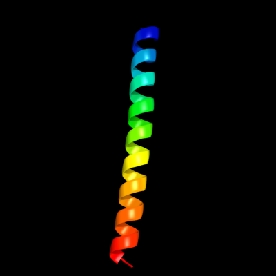
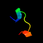
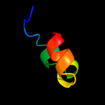
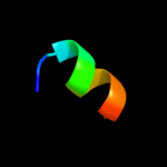
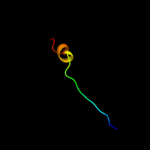
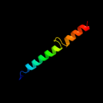
PDB header:structural protein Chain: A: PDB Molecule:adenomatous polyposis coli protein; PDBTitle: crystal structure of the n-terminal coiled coil domain from2 apc
Confidence and coverage
Confidence:
48.0%
Coverage:
24%
35 residues ( 24% of your sequence) have been modelled with 48.0% confidence by the single highest scoring template.
You may wish to submit your sequence to Phyrealarm. This will automatically scan your sequence every week for new potential templates as they appear in the Phyre2 library.
Please note: You must be registered and logged in to use Phyrealarm.
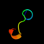
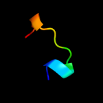
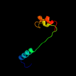
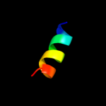
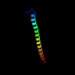
Region: 23 - 46 Aligned: 24 Modelled: 24 Confidence: 17.6% Identity: 33% PDB header:rna binding protein Chain: A: PDB Molecule:protein vts1; PDBTitle: solution structure of the vts1 sam domain in the presence2 of rna
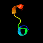
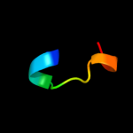
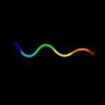
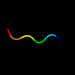
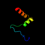
Region: 35 - 44 Aligned: 10 Modelled: 10 Confidence: 16.7% Identity: 40% PDB header:dna-binding protein Chain: K: PDB Molecule:terminase small subunit; PDBTitle: crystal structure of the small terminase oligomerization2 core domain from a spp1-like bacteriophage (crystal form 3)
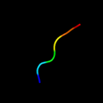
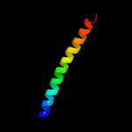
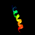
Region: 105 - 124 Aligned: 20 Modelled: 20 Confidence: 11.6% Identity: 45% PDB header:virus Chain: A: PDB Molecule:capsid protein; PDBTitle: backbone trace of the capsid protein dimer of a fungal partitivirus2 from electron cryomicroscopy and homology modeling