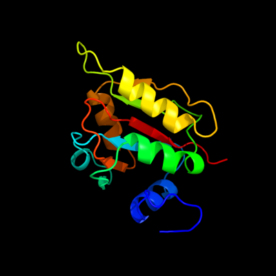
Phyre Home Retrieve Phyre Job Id
Emaill.a.kelley@imperial.ac.uk
DescriptionP0AEN8
DateThu Jan 5 11:23:48 GMT 2012
Unique Job IDd9e31f5a62f9b50a

Summary 

Top model
Image coloured by rainbow N → C terminus
Model (left) based on template c2wcvI_
Top template information
PDB header:isomerase
Chain: I: PDB Molecule:l-fucose mutarotase;
PDBTitle: crystal structure of bacterial fucu
Confidence and coverage
Confidence:100.0% Coverage:100%
140 residues (100% of your sequence) have been modelled with 100.0% confidence by the single highest scoring template.
3D viewing
Interactive 3D view in Jmol

Sequence analysis 

Secondary structure and disorder prediction 

   1........10.........20.........30.........40.........50.........60
Sequence  MLKTISPLISPELLKVLAEMGHGDEIIFSDAHFPAHSMGPQVIRADGLLVSDLLQAIIPL
Secondary structure 


SS confidence 


Disorder  ???????


Disorder confidence 


   .........70.........80.........90.........100.........110.........120
Sequence  FELDSYAPPLVMMAAVEGDTLDPEVERRYRNALSLQAPCPDIIRINRFAFYERAQKAFAI
Secondary structure 


SS confidence 


Disorder 


?


?


Disorder confidence 


   .........130.........140
Sequence  VITGERAKYGNILLKKGVTP
Secondary structure 


SS confidence 


Disorder 


???
Disorder confidence 


Confidence Key
High(9)                    Low (0)
?Disordered
Alpha helix
Beta strand

Domain analysis 

Hover over an aligned region to see model and summary info

Please note, only up to the top 20 hits are modelled to reduce computer load

RankAligned region

PDB 2wcv chain I

3D model

Region: 1 - 140
Aligned: 140
Modelled: 140
Confidence: 100.0%
Identity: 100%
PDB header:isomerase
Chain: I: PDB Molecule:l-fucose mutarotase;
PDBTitle: crystal structure of bacterial fucu

Phyre2

PDB 2ob5 chain A domain 1

3D model

Region: 1 - 140
Aligned: 140
Modelled: 139
Confidence: 100.0%
Identity: 33%
Fold: RbsD-like
Superfamily: RbsD-like
Family: RbsD-like

Phyre2

PDB 2wcu chain B

3D model

Region: 2 - 139
Aligned: 138
Modelled: 138
Confidence: 100.0%
Identity: 47%
PDB header:isomerase
Chain: B: PDB Molecule:protein fucu homolog;
PDBTitle: crystal structure of mammalian fucu

Phyre2

PDB 3mvk chain A

3D model

Region: 5 - 139
Aligned: 135
Modelled: 134
Confidence: 100.0%
Identity: 41%
PDB header:isomerase
Chain: A: PDB Molecule:protein fucu;
PDBTitle: the crystal structure of fucu from bifidobacterium longum to 1.65a

Phyre2

PDB 3e7n chain B

3D model

Region: 2 - 139
Aligned: 135
Modelled: 138
Confidence: 100.0%
Identity: 18%
PDB header:transport protein
Chain: B: PDB Molecule:d-ribose high-affinity transport system;
PDBTitle: crystal structure of d-ribose high-affinity transport system from2 salmonella typhimurium lt2

Phyre2

PDB 1ogd chain A

3D model

Region: 1 - 139
Aligned: 130
Modelled: 139
Confidence: 100.0%
Identity: 21%
Fold: RbsD-like
Superfamily: RbsD-like
Family: RbsD-like

Phyre2

PDB 3p13 chain B

3D model

Region: 7 - 139
Aligned: 129
Modelled: 133
Confidence: 100.0%
Identity: 16%
PDB header:isomerase
Chain: B: PDB Molecule:d-ribose pyranase;
PDBTitle: complex structure of d-ribose pyranase sa240 with d-ribose

Phyre2

PDB 1qki chain A domain 1

3D model

Region: 51 - 119
Aligned: 65
Modelled: 69
Confidence: 26.7%
Identity: 15%
Fold: NAD(P)-binding Rossmann-fold domains
Superfamily: NAD(P)-binding Rossmann-fold domains
Family: Glyceraldehyde-3-phosphate dehydrogenase-like, N-terminal domain

Phyre2

PDB 1e1c chain A

3D model

Region: 11 - 64
Aligned: 54
Modelled: 54
Confidence: 22.2%
Identity: 20%
PDB header:isomerase
Chain: A: PDB Molecule:methylmalonyl-coa mutase alpha chain;
PDBTitle: methylmalonyl-coa mutase h244a mutant

Phyre2

PDB 1e3d chain A

3D model

Region: 108 - 132
Aligned: 25
Modelled: 25
Confidence: 18.9%
Identity: 36%
Fold: HydA/Nqo6-like
Superfamily: HydA/Nqo6-like
Family: Nickel-iron hydrogenase, small subunit

Phyre2

PDB 2dgb chain A

3D model

Region: 1 - 23
Aligned: 21
Modelled: 23
Confidence: 17.6%
Identity: 33%
PDB header:structural genomics, unknown function
Chain: A: PDB Molecule:hypothetical protein purs;
PDBTitle: structure of thermus thermophilus purs in the p21 form

Phyre2

PDB 1t4a chain A

3D model

Region: 1 - 24
Aligned: 22
Modelled: 24
Confidence: 16.3%
Identity: 18%
Fold: PurS-like
Superfamily: PurS-like
Family: PurS subunit of FGAM synthetase

Phyre2

PDB 1frf chain S

3D model

Region: 108 - 132
Aligned: 25
Modelled: 25
Confidence: 15.4%
Identity: 28%
Fold: HydA/Nqo6-like
Superfamily: HydA/Nqo6-like
Family: Nickel-iron hydrogenase, small subunit

Phyre2

PDB 1cc1 chain S

3D model

Region: 108 - 132
Aligned: 25
Modelled: 25
Confidence: 15.1%
Identity: 32%
Fold: HydA/Nqo6-like
Superfamily: HydA/Nqo6-like
Family: Nickel-iron hydrogenase, small subunit

Phyre2

PDB 2yx5 chain A

3D model

Region: 1 - 24
Aligned: 22
Modelled: 24
Confidence: 14.4%
Identity: 27%
PDB header:structural genomics, unknown function
Chain: A: PDB Molecule:upf0062 protein mj1593;
PDBTitle: crystal structure of methanocaldococcus jannaschii purs, one of the2 subunits of formylglycinamide ribonucleotide amidotransferase in the3 purine biosynthetic pathway

Phyre2

PDB 3myr chain E

3D model

Region: 108 - 132
Aligned: 25
Modelled: 25
Confidence: 14.0%
Identity: 24%
PDB header:oxidoreductase
Chain: E: PDB Molecule:hydrogenase (nife) small subunit hyda;
PDBTitle: crystal structure of [nife] hydrogenase from allochromatium vinosum in2 its ni-a state

Phyre2

PDB 1wui chain S domain 1

3D model

Region: 108 - 132
Aligned: 25
Modelled: 25
Confidence: 13.8%
Identity: 24%
Fold: HydA/Nqo6-like
Superfamily: HydA/Nqo6-like
Family: Nickel-iron hydrogenase, small subunit

Phyre2

PDB 1h2a chain S

3D model

Region: 108 - 132
Aligned: 25
Modelled: 25
Confidence: 13.7%
Identity: 24%
PDB header:oxidoreductase
Chain: S: PDB Molecule:hydrogenase;
PDBTitle: single crystals of hydrogenase from desulfovibrio vulgaris

Phyre2

PDB 3iym chain A

3D model

Region: 60 - 110
Aligned: 50
Modelled: 51
Confidence: 13.0%
Identity: 24%
PDB header:virus
Chain: A: PDB Molecule:capsid protein;
PDBTitle: backbone trace of the capsid protein dimer of a fungal partitivirus2 from electron cryomicroscopy and homology modeling

Phyre2

PDB 1guz chain A domain 1

3D model

Region: 13 - 32
Aligned: 20
Modelled: 20
Confidence: 11.4%
Identity: 25%
Fold: NAD(P)-binding Rossmann-fold domains
Superfamily: NAD(P)-binding Rossmann-fold domains
Family: LDH N-terminal domain-like

Phyre2
1

c2wcvI_
2

d2ob5a1
3

c2wcuB_
4

c3mvkA_
5

c3e7nB_
6

d1ogda_
7

c3p13B_
8

d1qkia1
9

c1e1cA_
10

d1e3da_
11

c2dgbA_
12

d1t4aa_
13

d1frfs_
14

d1cc1s_
15

c2yx5A_
16

c3myrE_
17

d1wuis1
18

c1h2aS_
19

c3iymA_
20

d1guza1
21


22


23


24


25


26


27


28


29


30


31


32


33


34


35


36


37


38


39


40


41


42


43


44


45


46


47


48


49


Detailed template information 

#
Template Alignment Coverage3D Model Confidence
% i.d. Template Information
1c2wcvI_


100.0 100 PDB header:isomerase
Chain: I: PDB Molecule:l-fucose mutarotase;
PDBTitle: crystal structure of bacterial fucu
2d2ob5a1


100.0 33 Fold:RbsD-like
Superfamily:RbsD-like
Family:RbsD-like
3c2wcuB_


100.0 47 PDB header:isomerase
Chain: B: PDB Molecule:protein fucu homolog;
PDBTitle: crystal structure of mammalian fucu
4c3mvkA_


100.0 41 PDB header:isomerase
Chain: A: PDB Molecule:protein fucu;
PDBTitle: the crystal structure of fucu from bifidobacterium longum to 1.65a
5c3e7nB_


100.0 18 PDB header:transport protein
Chain: B: PDB Molecule:d-ribose high-affinity transport system;
PDBTitle: crystal structure of d-ribose high-affinity transport system from2 salmonella typhimurium lt2
6d1ogda_


100.0 21 Fold:RbsD-like
Superfamily:RbsD-like
Family:RbsD-like
7c3p13B_


100.0 16 PDB header:isomerase
Chain: B: PDB Molecule:d-ribose pyranase;
PDBTitle: complex structure of d-ribose pyranase sa240 with d-ribose
8d1qkia1


26.7 15 Fold:NAD(P)-binding Rossmann-fold domains
Superfamily:NAD(P)-binding Rossmann-fold domains
Family:Glyceraldehyde-3-phosphate dehydrogenase-like, N-terminal domain
9c1e1cA_


22.2 20 PDB header:isomerase
Chain: A: PDB Molecule:methylmalonyl-coa mutase alpha chain;
PDBTitle: methylmalonyl-coa mutase h244a mutant
10d1e3da_


18.9 36 Fold:HydA/Nqo6-like
Superfamily:HydA/Nqo6-like
Family:Nickel-iron hydrogenase, small subunit
11c2dgbA_


17.6 33 PDB header:structural genomics, unknown function
Chain: A: PDB Molecule:hypothetical protein purs;
PDBTitle: structure of thermus thermophilus purs in the p21 form
12d1t4aa_


16.3 18 Fold:PurS-like
Superfamily:PurS-like
Family:PurS subunit of FGAM synthetase
13d1frfs_


15.4 28 Fold:HydA/Nqo6-like
Superfamily:HydA/Nqo6-like
Family:Nickel-iron hydrogenase, small subunit
14d1cc1s_


15.1 32 Fold:HydA/Nqo6-like
Superfamily:HydA/Nqo6-like
Family:Nickel-iron hydrogenase, small subunit
15c2yx5A_


14.4 27 PDB header:structural genomics, unknown function
Chain: A: PDB Molecule:upf0062 protein mj1593;
PDBTitle: crystal structure of methanocaldococcus jannaschii purs, one of the2 subunits of formylglycinamide ribonucleotide amidotransferase in the3 purine biosynthetic pathway
16c3myrE_


14.0 24 PDB header:oxidoreductase
Chain: E: PDB Molecule:hydrogenase (nife) small subunit hyda;
PDBTitle: crystal structure of [nife] hydrogenase from allochromatium vinosum in2 its ni-a state
17d1wuis1


13.8 24 Fold:HydA/Nqo6-like
Superfamily:HydA/Nqo6-like
Family:Nickel-iron hydrogenase, small subunit
18c1h2aS_


13.7 24 PDB header:oxidoreductase
Chain: S: PDB Molecule:hydrogenase;
PDBTitle: single crystals of hydrogenase from desulfovibrio vulgaris
19c3iymA_


13.0 24 PDB header:virus
Chain: A: PDB Molecule:capsid protein;
PDBTitle: backbone trace of the capsid protein dimer of a fungal partitivirus2 from electron cryomicroscopy and homology modeling
20d1guza1


11.4 25 Fold:NAD(P)-binding Rossmann-fold domains
Superfamily:NAD(P)-binding Rossmann-fold domains
Family:LDH N-terminal domain-like
21d1gtda_


not modelled 10.8 35 Fold:PurS-like
Superfamily:PurS-like
Family:PurS subunit of FGAM synthetase
22d176la_


not modelled 10.7 27 Fold:Lysozyme-like
Superfamily:Lysozyme-like
Family:Phage lysozyme
23c3rgwS_


not modelled 10.3 28 PDB header:oxidoreductase/oxidoreductase
Chain: S: PDB Molecule:membrane-bound hydrogenase (nife) small subunit hoxk;
PDBTitle: crystal structure at 1.5 a resolution of an h2-reduced, o2-tolerant2 hydrogenase from ralstonia eutropha unmasks a novel iron-sulfur3 cluster
24c2wpnA_


not modelled 10.1 36 PDB header:oxidoreductase
Chain: A: PDB Molecule:periplasmic [nifese] hydrogenase, small subunit;
PDBTitle: structure of the oxidised, as-isolated nifese hydrogenase2 from d. vulgaris hildenborough
25d5ldha1


not modelled 9.6 14 Fold:NAD(P)-binding Rossmann-fold domains
Superfamily:NAD(P)-binding Rossmann-fold domains
Family:LDH N-terminal domain-like
26d1yq9a1


not modelled 8.8 35 Fold:HydA/Nqo6-like
Superfamily:HydA/Nqo6-like
Family:Nickel-iron hydrogenase, small subunit
27d1vs6z1


not modelled 8.0 36 Fold:L28p-like
Superfamily:L28p-like
Family:Ribosomal protein L31p
28d1llda1


not modelled 8.0 30 Fold:NAD(P)-binding Rossmann-fold domains
Superfamily:NAD(P)-binding Rossmann-fold domains
Family:LDH N-terminal domain-like
29c1w1wF_


not modelled 7.7 20 PDB header:cell adhesion
Chain: F: PDB Molecule:sister chromatid cohesion protein 1;
PDBTitle: sc smc1hd:scc1-c complex, atpgs
30d1w1we_


not modelled 7.7 20 Fold:DNA/RNA-binding 3-helical bundle
Superfamily:"Winged helix" DNA-binding domain
Family:Rad21/Rec8-like
31d1vpka2


not modelled 7.7 9 Fold:DNA clamp
Superfamily:DNA clamp
Family:DNA polymerase III, beta subunit
32d189la_


not modelled 7.6 27 Fold:Lysozyme-like
Superfamily:Lysozyme-like
Family:Phage lysozyme
33d1ldna1


not modelled 6.9 35 Fold:NAD(P)-binding Rossmann-fold domains
Superfamily:NAD(P)-binding Rossmann-fold domains
Family:LDH N-terminal domain-like
34d1p37a_


not modelled 6.9 27 Fold:Lysozyme-like
Superfamily:Lysozyme-like
Family:Phage lysozyme
35d2ldxa1


not modelled 6.7 25 Fold:NAD(P)-binding Rossmann-fold domains
Superfamily:NAD(P)-binding Rossmann-fold domains
Family:LDH N-terminal domain-like
36d1swya_


not modelled 6.4 27 Fold:Lysozyme-like
Superfamily:Lysozyme-like
Family:Phage lysozyme
37d1yjma1


not modelled 6.2 23 Fold:SMAD/FHA domain
Superfamily:SMAD/FHA domain
Family:FHA domain
38d1gpma3


not modelled 5.9 30 Fold:Alpha-lytic protease prodomain-like
Superfamily:GMP synthetase C-terminal dimerisation domain
Family:GMP synthetase C-terminal dimerisation domain
39d1hyha1


not modelled 5.9 30 Fold:NAD(P)-binding Rossmann-fold domains
Superfamily:NAD(P)-binding Rossmann-fold domains
Family:LDH N-terminal domain-like
40d1vq3a_


not modelled 5.8 27 Fold:PurS-like
Superfamily:PurS-like
Family:PurS subunit of FGAM synthetase
41d1cp2a_


not modelled 5.8 26 Fold:P-loop containing nucleoside triphosphate hydrolases
Superfamily:P-loop containing nucleoside triphosphate hydrolases
Family:Nitrogenase iron protein-like
42c2b664_


not modelled 5.6 36 PDB header:ribosome
Chain: 4: PDB Molecule:50s ribosomal protein l31;
PDBTitle: 50s ribosomal subunit from a crystal structure of release factor rf1,2 trnas and mrna bound to the ribosome. this file contains the 50s3 subunit from a crystal structure of release factor rf1, trnas and4 mrna bound to the ribosome and is described in remark 400
43c2xd4A_


not modelled 5.6 9 PDB header:ligase
Chain: A: PDB Molecule:phosphoribosylamine--glycine ligase;
PDBTitle: nucleotide-bound structures of bacillus subtilis glycinamide2 ribonucleotide synthetase
44d1jkea_


not modelled 5.5 25 Fold:DTD-like
Superfamily:DTD-like
Family:DTD-like
45c1yj5C_


not modelled 5.5 23 PDB header:transferase
Chain: C: PDB Molecule:5' polynucleotide kinase-3' phosphatase fha domain;
PDBTitle: molecular architecture of mammalian polynucleotide kinase, a dna2 repair enzyme
46d1llca1


not modelled 5.5 20 Fold:NAD(P)-binding Rossmann-fold domains
Superfamily:NAD(P)-binding Rossmann-fold domains
Family:LDH N-terminal domain-like
47c2zw2B_


not modelled 5.4 18 PDB header:ligase
Chain: B: PDB Molecule:putative uncharacterized protein sts178;
PDBTitle: crystal structure of formylglycinamide ribonucleotide amidotransferase2 iii from sulfolobus tokodaii (stpurs)
48c3hl4B_


not modelled 5.2 10 PDB header:transferase
Chain: B: PDB Molecule:choline-phosphate cytidylyltransferase a;
PDBTitle: crystal structure of a mammalian ctp:phosphocholine2 cytidylyltransferase with cdp-choline
49c2j034_


not modelled 5.1 36 PDB header:ribosome
Chain: 4: PDB Molecule:50s ribosomal protein l31;
PDBTitle: structure of the thermus thermophilus 70s ribosome2 complexed with mrna, trna and paromomycin (part 4 of 4).3 this file contains the 50s subunit from molecule ii.

Binding site prediction 

Due to computational demand, binding site predictions are not run for batch jobs

If you want to predict binding sites, please manually submit your model of choice to 3DLigandSite


Phyre is for academic use only

Please cite: Protein structure prediction on the web: a case study using the Phyre server
Kelley LA and Sternberg MJE. Nature Protocols 4, 363 - 371 (2009) [pdf] [Import into BibTeX]
 
If you use the binding site predictions from 3DLigandSite, please also cite:
3DLigandSite: predicting ligand-binding sites using similar structures.
Wass MN, Kelley LA and Sternberg MJ Nucleic Acids Research 38, W469-73 (2010) [PubMed]
 
© Structural Bioinformatics Group
Imperial College London
Lawrence Kelley, Benjamin Jefferys
Disclaimer
Terms and Conditions
Component software
Template detection: HHpred 1.51
Secondary structure prediction: Psi-pred 2.5
Disorder prediction: Disopred 2.4
Transmembrane prediction: Memsat_SVM
Multi-template modelling and ab initio: Poing 1.0