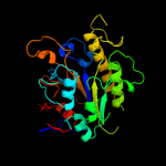
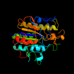
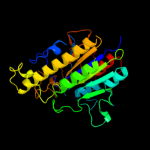
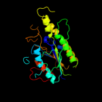
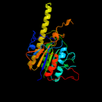
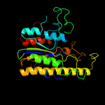
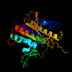
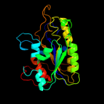
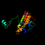
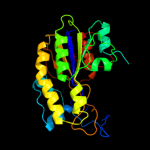
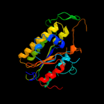
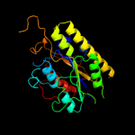
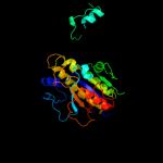
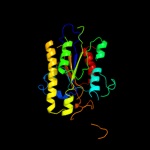
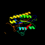
1 c2w8dB_
100.0
12
PDB header: transferaseChain: B: PDB Molecule: processed glycerol phosphate lipoteichoic acid synthase 2;PDBTitle: distinct and essential morphogenic functions for wall- and2 lipo-teichoic acids in bacillus subtilis
2 c2w5tA_
100.0
12
PDB header: transferaseChain: A: PDB Molecule: processed glycerol phosphate lipoteichoic acidPDBTitle: structure-based mechanism of lipoteichoic acid synthesis by2 staphylococcus aureus ltas.
3 c2qzuA_
100.0
13
PDB header: hydrolaseChain: A: PDB Molecule: putative sulfatase yidj;PDBTitle: crystal structure of the putative sulfatase yidj from bacteroides2 fragilis. northeast structural genomics consortium target bfr123
4 c3ed4A_
100.0
18
PDB header: transferaseChain: A: PDB Molecule: arylsulfatase;PDBTitle: crystal structure of putative arylsulfatase from escherichia coli
5 c3lxqB_
100.0
15
PDB header: structural genomics, unknown functionChain: B: PDB Molecule: uncharacterized protein vp1736;PDBTitle: the crystal structure of a protein in the alkaline2 phosphatase superfamily from vibrio parahaemolyticus to3 1.95a
6 d1auka_
100.0
15
Fold: Alkaline phosphatase-likeSuperfamily: Alkaline phosphatase-likeFamily: Arylsulfatase7 d1fsua_
100.0
14
Fold: Alkaline phosphatase-likeSuperfamily: Alkaline phosphatase-likeFamily: Arylsulfatase8 c3b5qB_
100.0
12
PDB header: hydrolaseChain: B: PDB Molecule: putative sulfatase yidj;PDBTitle: crystal structure of a putative sulfatase (np_810509.1)2 from bacteroides thetaiotaomicron vpi-5482 at 2.40 a3 resolution
9 d1hdha_
100.0
19
Fold: Alkaline phosphatase-likeSuperfamily: Alkaline phosphatase-likeFamily: Arylsulfatase10 c2vqrA_
100.0
16
PDB header: hydrolaseChain: A: PDB Molecule: putative sulfatase;PDBTitle: crystal structure of a phosphonate monoester hydrolase2 from rhizobium leguminosarum: a new member of the3 alkaline phosphatase superfamily
11 d1p49a_
100.0
15
Fold: Alkaline phosphatase-likeSuperfamily: Alkaline phosphatase-likeFamily: Arylsulfatase12 d1o98a2
100.0
9
Fold: Alkaline phosphatase-likeSuperfamily: Alkaline phosphatase-likeFamily: 2,3-Bisphosphoglycerate-independent phosphoglycerate mutase, catalytic domain13 c2zktB_
100.0
16
PDB header: isomeraseChain: B: PDB Molecule: 2,3-bisphosphoglycerate-independent phosphoglyceratePDBTitle: structure of ph0037 protein from pyrococcus horikoshii
14 c3m8yC_
100.0
14
PDB header: isomeraseChain: C: PDB Molecule: phosphopentomutase;PDBTitle: phosphopentomutase from bacillus cereus after glucose-1,6-bisphosphate2 activation
15 d2i09a1
100.0
14
Fold: Alkaline phosphatase-likeSuperfamily: Alkaline phosphatase-likeFamily: DeoB catalytic domain-like16 c3q3qA_
100.0
16
PDB header: hydrolaseChain: A: PDB Molecule: alkaline phosphatase;PDBTitle: crystal structure of spap: an novel alkaline phosphatase from2 bacterium sphingomonas sp. strain bsar-1
17 c2gsoB_
100.0
14
PDB header: hydrolaseChain: B: PDB Molecule: phosphodiesterase-nucleotide pyrophosphatase;PDBTitle: structure of xac nucleotide2 pyrophosphatase/phosphodiesterase in complex with vanadate
18 c2i09A_
99.9
11
PDB header: isomeraseChain: A: PDB Molecule: phosphopentomutase;PDBTitle: crystal structure of putative phosphopentomutase from streptococcus2 mutans
19 c2xrgA_
99.9
15
PDB header: hydrolaseChain: A: PDB Molecule: ectonucleotide pyrophosphatase/phosphodiesterase familyPDBTitle: crystal structure of autotaxin (enpp2) in complex with the2 ha155 boronic acid inhibitor
20 c2xr9A_
99.9
16
PDB header: hydrolaseChain: A: PDB Molecule: ectonucleotide pyrophosphatase/phosphodiesterase familyPDBTitle: crystal structure of autotaxin (enpp2)
21 c3szzA_
not modelled
99.9
18
PDB header: hydrolaseChain: A: PDB Molecule: phosphonoacetate hydrolase;PDBTitle: crystal structure of phosphonoacetate hydrolase from sinorhizobium2 meliloti 1021 in complex with acetate
22 d1ei6a_
not modelled
99.9
16
Fold: Alkaline phosphatase-likeSuperfamily: Alkaline phosphatase-likeFamily: Phosphonoacetate hydrolase23 c1o98A_
not modelled
99.7
13
PDB header: isomeraseChain: A: PDB Molecule: 2,3-bisphosphoglycerate-independentPDBTitle: 1.4a crystal structure of phosphoglycerate mutase from2 bacillus stearothermophilus complexed with3 2-phosphoglycerate
24 c3igzB_
not modelled
99.6
12
PDB header: isomeraseChain: B: PDB Molecule: cofactor-independent phosphoglycerate mutase;PDBTitle: crystal structures of leishmania mexicana phosphoglycerate2 mutase at low cobalt concentration
25 c2d1gB_
not modelled
99.6
17
PDB header: hydrolaseChain: B: PDB Molecule: acid phosphatase;PDBTitle: structure of francisella tularensis acid phosphatase a (acpa) bound to2 orthovanadate
26 c2iucB_
not modelled
99.4
16
PDB header: hydrolaseChain: B: PDB Molecule: alkaline phosphatase;PDBTitle: structure of alkaline phosphatase from the antarctic2 bacterium tab5
27 d1y6va1
not modelled
99.3
12
Fold: Alkaline phosphatase-likeSuperfamily: Alkaline phosphatase-likeFamily: Alkaline phosphatase28 c1ew2A_
not modelled
99.2
14
PDB header: hydrolaseChain: A: PDB Molecule: phosphatase;PDBTitle: crystal structure of a human phosphatase
29 d1zeda1
not modelled
99.2
14
Fold: Alkaline phosphatase-likeSuperfamily: Alkaline phosphatase-likeFamily: Alkaline phosphatase30 c2w0yB_
not modelled
99.1
12
PDB header: hydrolaseChain: B: PDB Molecule: alkaline phosphatase;PDBTitle: h.salinarum alkaline phosphatase
31 d1k7ha_
not modelled
99.1
13
Fold: Alkaline phosphatase-likeSuperfamily: Alkaline phosphatase-likeFamily: Alkaline phosphatase32 c2x98A_
not modelled
99.1
13
PDB header: hydrolaseChain: A: PDB Molecule: alkaline phosphatase;PDBTitle: h.salinarum alkaline phosphatase
33 c3a52A_
not modelled
99.0
14
PDB header: hydrolaseChain: A: PDB Molecule: cold-active alkaline phosphatase;PDBTitle: crystal structure of cold-active alkailne phosphatase from2 psychrophile shewanella sp.
34 c3e2dB_
not modelled
98.7
15
PDB header: hydrolaseChain: B: PDB Molecule: alkaline phosphatase;PDBTitle: the 1.4 a crystal structure of the large and cold-active2 vibrio sp. alkaline phosphatase
35 c3iddA_
not modelled
96.7
20
PDB header: isomeraseChain: A: PDB Molecule: 2,3-bisphosphoglycerate-independentPDBTitle: cofactor-independent phosphoglycerate mutase from2 thermoplasma acidophilum dsm 1728
36 d1b4ub_
not modelled
42.4
5
Fold: Phosphorylase/hydrolase-likeSuperfamily: LigB-likeFamily: LigB-like37 c3bijC_
not modelled
38.2
18
PDB header: structural genomics, unknown functionChain: C: PDB Molecule: uncharacterized protein gsu0716;PDBTitle: crystal structure of protein gsu0716 from geobacter2 sulfurreducens. northeast structural genomics target gsr13
38 d1j33a_
not modelled
28.7
13
Fold: Nicotinate mononucleotide:5,6-dimethylbenzimidazole phosphoribosyltransferase (CobT)Superfamily: Nicotinate mononucleotide:5,6-dimethylbenzimidazole phosphoribosyltransferase (CobT)Family: Nicotinate mononucleotide:5,6-dimethylbenzimidazole phosphoribosyltransferase (CobT)39 d1l5oa_
not modelled
24.9
7
Fold: Nicotinate mononucleotide:5,6-dimethylbenzimidazole phosphoribosyltransferase (CobT)Superfamily: Nicotinate mononucleotide:5,6-dimethylbenzimidazole phosphoribosyltransferase (CobT)Family: Nicotinate mononucleotide:5,6-dimethylbenzimidazole phosphoribosyltransferase (CobT)40 c3ib7A_
not modelled
22.6
23
PDB header: hydrolaseChain: A: PDB Molecule: icc protein;PDBTitle: crystal structure of full length rv0805
41 d1xo1a2
not modelled
22.6
12
Fold: PIN domain-likeSuperfamily: PIN domain-likeFamily: 5' to 3' exonuclease catalytic domain42 d3c9fa2
not modelled
22.0
18
Fold: Metallo-dependent phosphatasesSuperfamily: Metallo-dependent phosphatasesFamily: 5'-nucleotidase (syn. UDP-sugar hydrolase), N-terminal domain43 c2xokG_
not modelled
21.3
11
PDB header: hydrolaseChain: G: PDB Molecule: atp synthase subunit gamma, mitochondrial;PDBTitle: refined structure of yeast f1c10 atpase complex to 3 a2 resolution
44 c2hy1A_
not modelled
20.6
31
PDB header: hydrolaseChain: A: PDB Molecule: rv0805;PDBTitle: crystal structure of rv0805
45 d2hy1a1
not modelled
20.6
31
Fold: Metallo-dependent phosphatasesSuperfamily: Metallo-dependent phosphatasesFamily: GpdQ-like46 c3e4cB_
not modelled
19.1
17
PDB header: hydrolaseChain: B: PDB Molecule: caspase-1;PDBTitle: procaspase-1 zymogen domain crystal strucutre
47 d1s1qa_
not modelled
19.0
12
Fold: UBC-likeSuperfamily: UBC-likeFamily: UEV domain48 d1usha2
not modelled
16.9
11
Fold: Metallo-dependent phosphatasesSuperfamily: Metallo-dependent phosphatasesFamily: 5'-nucleotidase (syn. UDP-sugar hydrolase), N-terminal domain49 d1uzdc1
not modelled
14.7
9
Fold: RuBisCO, small subunitSuperfamily: RuBisCO, small subunitFamily: RuBisCO, small subunit50 d1yj5a1
not modelled
13.1
26
Fold: HAD-likeSuperfamily: HAD-likeFamily: phosphatase domain of polynucleotide kinase51 d2p0va1
not modelled
12.2
11
Fold: alpha/alpha toroidSuperfamily: Six-hairpin glycosidasesFamily: CPF0428-like52 c2p0vA_
not modelled
12.2
11
PDB header: structural genomics, unknown functionChain: A: PDB Molecule: hypothetical protein bt3781;PDBTitle: crystal structure of bt3781 protein from bacteroides2 thetaiotaomicron, northeast structural genomics target3 btr58
53 d1ej7s_
not modelled
12.0
11
Fold: RuBisCO, small subunitSuperfamily: RuBisCO, small subunitFamily: RuBisCO, small subunit54 d2z1aa2
not modelled
11.9
8
Fold: Metallo-dependent phosphatasesSuperfamily: Metallo-dependent phosphatasesFamily: 5'-nucleotidase (syn. UDP-sugar hydrolase), N-terminal domain55 d1tfra2
not modelled
11.8
15
Fold: PIN domain-likeSuperfamily: PIN domain-likeFamily: 5' to 3' exonuclease catalytic domain56 d2hrca1
not modelled
11.0
7
Fold: Chelatase-likeSuperfamily: ChelataseFamily: Ferrochelatase57 c3uoaB_
not modelled
10.6
18
PDB header: hydrolase/hydrolase inhibitorChain: B: PDB Molecule: mucosa-associated lymphoid tissue lymphoma translocationPDBTitle: crystal structure of the malt1 paracaspase (p21 form)
58 c2jcmA_
not modelled
9.1
14
PDB header: hydrolaseChain: A: PDB Molecule: cytosolic purine 5'-nucleotidase;PDBTitle: crystal structure of human cytosolic 5'-nucleotidase ii in2 complex with beryllium trifluoride
59 d8ruci_
not modelled
8.9
11
Fold: RuBisCO, small subunitSuperfamily: RuBisCO, small subunitFamily: RuBisCO, small subunit60 c1oidA_
not modelled
8.9
10
PDB header: hydrolaseChain: A: PDB Molecule: protein usha;PDBTitle: 5'-nucleotidase (e. coli) with an engineered disulfide2 bridge (s228c, p513c)
61 c2dfjA_
not modelled
8.3
8
PDB header: hydrolaseChain: A: PDB Molecule: diadenosinetetraphosphatase;PDBTitle: crystal structure of the diadenosine tetraphosphate2 hydrolase from shigella flexneri 2a
62 d2jdig1
not modelled
7.7
17
Fold: Pyruvate kinase C-terminal domain-likeSuperfamily: ATP synthase (F1-ATPase), gamma subunitFamily: ATP synthase (F1-ATPase), gamma subunit63 d1jb0i_
not modelled
7.2
23
Fold: Single transmembrane helixSuperfamily: Subunit VIII of photosystem I reaction centre, PsaIFamily: Subunit VIII of photosystem I reaction centre, PsaI64 d2hk6a1
not modelled
7.2
11
Fold: Chelatase-likeSuperfamily: ChelataseFamily: Ferrochelatase65 c3a0hk_
not modelled
7.2
38
PDB header: electron transportChain: K: PDB Molecule: photosystem ii reaction center protein k;PDBTitle: crystal structure of i-substituted photosystem ii complex
66 d1yp2a2
not modelled
7.1
3
Fold: Nucleotide-diphospho-sugar transferasesSuperfamily: Nucleotide-diphospho-sugar transferasesFamily: glucose-1-phosphate thymidylyltransferase67 d1cmwa2
not modelled
6.4
13
Fold: PIN domain-likeSuperfamily: PIN domain-likeFamily: 5' to 3' exonuclease catalytic domain68 d2nxfa1
not modelled
6.1
14
Fold: Metallo-dependent phosphatasesSuperfamily: Metallo-dependent phosphatasesFamily: ADPRibase-Mn-like69 c3c9fB_
not modelled
6.0
18
PDB header: hydrolaseChain: B: PDB Molecule: 5'-nucleotidase;PDBTitle: crystal structure of 5'-nucleotidase from candida albicans sc5314
70 d1fs0g_
not modelled
5.8
20
Fold: Pyruvate kinase C-terminal domain-likeSuperfamily: ATP synthase (F1-ATPase), gamma subunitFamily: ATP synthase (F1-ATPase), gamma subunit71 c3a0bK_
not modelled
5.7
38
PDB header: electron transportChain: K: PDB Molecule: photosystem ii reaction center protein k;PDBTitle: crystal structure of br-substituted photosystem ii complex
72 c3a0bk_
not modelled
5.7
38
PDB header: electron transportChain: K: PDB Molecule: photosystem ii reaction center protein k;PDBTitle: crystal structure of br-substituted photosystem ii complex
73 c2w6jG_
not modelled
5.7
17
PDB header: hydrolaseChain: G: PDB Molecule: atp synthase subunit gamma, mitochondrial;PDBTitle: low resolution structures of bovine mitochondrial f1-atpase2 during controlled dehydration: hydration state 5.
74 c1oy8A_
not modelled
5.7
10
PDB header: membrane proteinChain: A: PDB Molecule: acriflavine resistance protein b;PDBTitle: structural basis of multiple drug binding capacity of the acrb2 multidrug efflux pump
75 c2hbzA_
not modelled
5.6
16
PDB header: hydrolase/hydrolase inhibitorChain: A: PDB Molecule: caspase-1;PDBTitle: crystal structure of human caspase-1 (arg286->ala, glu390->ala) in2 complex with 3-[2-(2-benzyloxycarbonylamino-3-methyl-butyrylamino)-3 propionylamino]-4-oxo-pentanoic acid (z-vad-fmk)
76 c3e20C_
not modelled
5.6
12
PDB header: translationChain: C: PDB Molecule: eukaryotic peptide chain release factor subunit 1;PDBTitle: crystal structure of s.pombe erf1/erf3 complex
77 d1a9xa3
not modelled
5.5
17
Fold: PreATP-grasp domainSuperfamily: PreATP-grasp domainFamily: BC N-terminal domain-like78 c3oaaO_
not modelled
5.2
12
PDB header: hydrolase/transport proteinChain: O: PDB Molecule: atp synthase gamma chain;PDBTitle: structure of the e.coli f1-atp synthase inhibited by subunit epsilon
79 d1szpb1
not modelled
5.2
17
Fold: SAM domain-likeSuperfamily: Rad51 N-terminal domain-likeFamily: DNA repair protein Rad51, N-terminal domain80 c3zvmA_
not modelled
5.1
26
PDB header: hydrolase/transferase/dnaChain: A: PDB Molecule: bifunctional polynucleotide phosphatase/kinase;PDBTitle: the structural basis for substrate recognition by mammalian2 polynucleotide kinase 3' phosphatase
81 d1dt9a3
not modelled
5.1
13
Fold: N-terminal domain of eukaryotic peptide chain release factor subunit 1, ERF1Superfamily: N-terminal domain of eukaryotic peptide chain release factor subunit 1, ERF1Family: N-terminal domain of eukaryotic peptide chain release factor subunit 1, ERF1


