Phyre Home Retrieve Phyre Job Id
Emaill.a.kelley@imperial.ac.uk
DescriptionP16685
DateThu Jan 5 11:35:31 GMT 2012
Unique Job IDd4d340cf27050f5b

Summary 

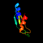
Top model
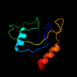
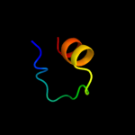
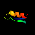
Image coloured by rainbow N → C terminus
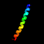
Model (left) based on template c2pc9B_
Top template information
PDB header:lyase
Chain: B: PDB Molecule:phosphoenolpyruvate carboxykinase [atp];
PDBTitle: crystal structure of atp-dependent phosphoenolpyruvate carboxykinase2 from thermus thermophilus hb8
Confidence and coverage
Confidence: 20.4% Coverage: 52%
78 residues ( 52% of your sequence) have been modelled with 20.4% confidence by the single highest scoring template.
You may wish to submit your sequence to Phyrealarm. This will automatically scan your sequence every week for new potential templates as they appear in the Phyre2 library.
Please note: You must be registered and logged in to use Phyrealarm.
3D viewing
Interactive 3D view in Jmol

Sequence analysis 

Secondary structure and disorder prediction 

   1........10.........20.........30.........40.........50.........60
Sequence  MHADTATRQHWMSVLAHSQPAELAARLNALNITADYEVIRAAETGLVQIQARMGGTGERF
Secondary structure 


SS confidence 


Disorder  ?????


?


??
??


Disorder confidence 


   .........70.........80.........90.........100.........110.........120
Sequence  FAGDATLTRAAVRLTDGTLGYSWVQGRDKQHAERCALIDALMQQSRHFQNLSETLIAPLD
Secondary structure 


SS confidence 


Disorder 


Disorder confidence 


   .........130.........140.........150
Sequence  ADRMARIAARQAEVNASRVDFFTMVRGDNA
Secondary structure 


SS confidence 


Disorder 


??


??????
Disorder confidence 


Confidence Key
High(9)                    Low (0)
?Disordered
Alpha helix
Beta strand

Domain analysis 

Hover over an aligned region to see model and summary info

Please note, only up to the top 20 hits are modelled to reduce computer load

RankAligned region

PDB 2pc9 chain B

3D model

Region: 28 - 106
Aligned: 78
Modelled: 79
Confidence: 20.4%
Identity: 18%
PDB header:lyase
Chain: B: PDB Molecule:phosphoenolpyruvate carboxykinase [atp];
PDBTitle: crystal structure of atp-dependent phosphoenolpyruvate carboxykinase2 from thermus thermophilus hb8

Phyre2

PDB 2h12 chain C

3D model

Region: 79 - 145
Aligned: 66
Modelled: 66
Confidence: 19.8%
Identity: 12%
PDB header:transferase
Chain: C: PDB Molecule:citrate synthase;
PDBTitle: structure of acetobacter aceti citrate synthase complexed2 with oxaloacetate and carboxymethyldethia coenzyme a (cmx)

Phyre2

PDB 1bqg chain A domain 2

3D model

Region: 68 - 115
Aligned: 48
Modelled: 48
Confidence: 17.2%
Identity: 23%
Fold: Enolase N-terminal domain-like
Superfamily: Enolase N-terminal domain-like
Family: Enolase N-terminal domain-like

Phyre2

PDB 2a9d chain A domain 1

3D model

Region: 37 - 77
Aligned: 41
Modelled: 41
Confidence: 16.2%
Identity: 17%
Fold: Immunoglobulin-like beta-sandwich
Superfamily: E set domains
Family: Molybdenum-containing oxidoreductases-like dimerisation domain

Phyre2

PDB 3f4y chain F

3D model

Region: 88 - 119
Aligned: 32
Modelled: 32
Confidence: 11.9%
Identity: 31%
PDB header:viral protein
Chain: F: PDB Molecule:mutant peptide derived from hiv gp41 chr domain;
PDBTitle: hiv gp41 six-helix bundle containing a mutant chr alpha-2 peptide sequence

Phyre2

PDB 1ogp chain A domain 1

3D model

Region: 37 - 77
Aligned: 41
Modelled: 41
Confidence: 10.9%
Identity: 17%
Fold: Immunoglobulin-like beta-sandwich
Superfamily: E set domains
Family: Molybdenum-containing oxidoreductases-like dimerisation domain

Phyre2

PDB 2eqj chain A

3D model

Region: 47 - 79
Aligned: 29
Modelled: 33
Confidence: 10.1%
Identity: 24%
PDB header:transcription
Chain: A: PDB Molecule:metal-response element-binding transcription
PDBTitle: solution structure of the tudor domain of metal-response2 element-binding transcription factor 2

Phyre2

PDB 2e5p chain A

3D model

Region: 47 - 79
Aligned: 29
Modelled: 33
Confidence: 9.5%
Identity: 24%
PDB header:transcription
Chain: A: PDB Molecule:phd finger protein 1;
PDBTitle: solution structure of the tudor domain of phd finger2 protein 1 (phf1 protein)

Phyre2

PDB 1z1b chain A domain 1

3D model

Region: 76 - 97
Aligned: 22
Modelled: 22
Confidence: 9.4%
Identity: 32%
Fold: DNA-binding domain
Superfamily: DNA-binding domain
Family: lambda integrase N-terminal domain

Phyre2

PDB 1kjk chain A

3D model

Region: 76 - 97
Aligned: 22
Modelled: 22
Confidence: 9.4%
Identity: 32%
PDB header:viral protein
Chain: A: PDB Molecule:integrase;
PDBTitle: solution structure of the lambda integrase amino-terminal2 domain

Phyre2

PDB 3c4t chain A

3D model

Region: 69 - 101
Aligned: 31
Modelled: 33
Confidence: 9.1%
Identity: 29%
PDB header:hydrolase
Chain: A: PDB Molecule:endoribonuclease dicer;
PDBTitle: structure of rnaseiiib and dsrna binding domains of mouse dicer

Phyre2

PDB 1o0w chain B

3D model

Region: 66 - 101
Aligned: 34
Modelled: 36
Confidence: 8.7%
Identity: 29%
PDB header:hydrolase
Chain: B: PDB Molecule:ribonuclease iii;
PDBTitle: crystal structure of ribonuclease iii (tm1102) from2 thermotoga maritima at 2.0 a resolution

Phyre2

PDB 1x47 chain A domain 1

3D model

Region: 69 - 101
Aligned: 31
Modelled: 33
Confidence: 8.2%
Identity: 26%
Fold: dsRBD-like
Superfamily: dsRNA-binding domain-like
Family: Double-stranded RNA-binding domain (dsRBD)

Phyre2

PDB 2enp chain A

3D model

Region: 51 - 68
Aligned: 18
Modelled: 18
Confidence: 8.0%
Identity: 17%
PDB header:transport protein
Chain: A: PDB Molecule:b/k protein;
PDBTitle: solution structure of the first c2 domain from human b/k2 protein

Phyre2

PDB 2e5q chain A

3D model

Region: 47 - 79
Aligned: 29
Modelled: 33
Confidence: 8.0%
Identity: 24%
PDB header:transcription
Chain: A: PDB Molecule:phd finger protein 19;
PDBTitle: solution structure of the tudor domain of phd finger2 protein 19, isoform b [homo sapiens]

Phyre2

PDB 2fd6 chain U domain 2

3D model

Region: 40 - 71
Aligned: 32
Modelled: 32
Confidence: 7.4%
Identity: 6%
Fold: Snake toxin-like
Superfamily: Snake toxin-like
Family: Extracellular domain of cell surface receptors

Phyre2

PDB 1stu chain A

3D model

Region: 19 - 101
Aligned: 62
Modelled: 64
Confidence: 7.3%
Identity: 19%
Fold: dsRBD-like
Superfamily: dsRNA-binding domain-like
Family: Double-stranded RNA-binding domain (dsRBD)

Phyre2

PDB 2bv3 chain A domain 4

3D model

Region: 29 - 51
Aligned: 23
Modelled: 23
Confidence: 6.9%
Identity: 26%
Fold: Ferredoxin-like
Superfamily: EF-G C-terminal domain-like
Family: EF-G/eEF-2 domains III and V

Phyre2

PDB 3ts3 chain D

3D model

Region: 39 - 85
Aligned: 38
Modelled: 47
Confidence: 6.9%
Identity: 26%
PDB header:viral protein
Chain: D: PDB Molecule:capsid polyprotein;
PDBTitle: crystal structure of the projection domain of the turkey astrovirus2 capsid protein at 1.5 angstrom resolution

Phyre2

PDB 1ef1 chain C

3D model

Region: 107 - 142
Aligned: 35
Modelled: 36
Confidence: 6.2%
Identity: 14%
Fold: Non-globular all-alpha subunits of globular proteins
Superfamily: Moesin tail domain
Family: Moesin tail domain

Phyre2
1

c2pc9B_
2

c2h12C_
3

d1bqga2
4

d2a9da1
5

c3f4yF_
6

d1ogpa1
7

c2eqjA_
8

c2e5pA_
9

d1z1ba1
10

c1kjkA_
11

c3c4tA_
12

c1o0wB_
13

d1x47a1
14

c2enpA_
15

c2e5qA_
16

d2fd6u2
17

d1stua_
18

d2bv3a4
19

c3ts3D_
20

d1ef1c_
21


22


23


24


25


26


27


28


29


30


Detailed template information 

#
Template Alignment Coverage3D Model Confidence
% i.d. Template Information
1c2pc9B_


20.4 18 PDB header:lyase
Chain: B: PDB Molecule:phosphoenolpyruvate carboxykinase [atp];
PDBTitle: crystal structure of atp-dependent phosphoenolpyruvate carboxykinase2 from thermus thermophilus hb8
2c2h12C_


19.8 12 PDB header:transferase
Chain: C: PDB Molecule:citrate synthase;
PDBTitle: structure of acetobacter aceti citrate synthase complexed2 with oxaloacetate and carboxymethyldethia coenzyme a (cmx)
3d1bqga2


17.2 23 Fold:Enolase N-terminal domain-like
Superfamily:Enolase N-terminal domain-like
Family:Enolase N-terminal domain-like
4d2a9da1


16.2 17 Fold:Immunoglobulin-like beta-sandwich
Superfamily:E set domains
Family:Molybdenum-containing oxidoreductases-like dimerisation domain
5c3f4yF_


11.9 31 PDB header:viral protein
Chain: F: PDB Molecule:mutant peptide derived from hiv gp41 chr domain;
PDBTitle: hiv gp41 six-helix bundle containing a mutant chr alpha-2 peptide sequence
6d1ogpa1


10.9 17 Fold:Immunoglobulin-like beta-sandwich
Superfamily:E set domains
Family:Molybdenum-containing oxidoreductases-like dimerisation domain
7c2eqjA_


10.1 24 PDB header:transcription
Chain: A: PDB Molecule:metal-response element-binding transcription
PDBTitle: solution structure of the tudor domain of metal-response2 element-binding transcription factor 2
8c2e5pA_


9.5 24 PDB header:transcription
Chain: A: PDB Molecule:phd finger protein 1;
PDBTitle: solution structure of the tudor domain of phd finger2 protein 1 (phf1 protein)
9d1z1ba1


9.4 32 Fold:DNA-binding domain
Superfamily:DNA-binding domain
Family:lambda integrase N-terminal domain
10c1kjkA_


9.4 32 PDB header:viral protein
Chain: A: PDB Molecule:integrase;
PDBTitle: solution structure of the lambda integrase amino-terminal2 domain
11c3c4tA_


9.1 29 PDB header:hydrolase
Chain: A: PDB Molecule:endoribonuclease dicer;
PDBTitle: structure of rnaseiiib and dsrna binding domains of mouse dicer
12c1o0wB_


8.7 29 PDB header:hydrolase
Chain: B: PDB Molecule:ribonuclease iii;
PDBTitle: crystal structure of ribonuclease iii (tm1102) from2 thermotoga maritima at 2.0 a resolution
13d1x47a1


8.2 26 Fold:dsRBD-like
Superfamily:dsRNA-binding domain-like
Family:Double-stranded RNA-binding domain (dsRBD)
14c2enpA_


8.0 17 PDB header:transport protein
Chain: A: PDB Molecule:b/k protein;
PDBTitle: solution structure of the first c2 domain from human b/k2 protein
15c2e5qA_


8.0 24 PDB header:transcription
Chain: A: PDB Molecule:phd finger protein 19;
PDBTitle: solution structure of the tudor domain of phd finger2 protein 19, isoform b [homo sapiens]
16d2fd6u2


7.4 6 Fold:Snake toxin-like
Superfamily:Snake toxin-like
Family:Extracellular domain of cell surface receptors
17d1stua_


7.3 19 Fold:dsRBD-like
Superfamily:dsRNA-binding domain-like
Family:Double-stranded RNA-binding domain (dsRBD)
18d2bv3a4


6.9 26 Fold:Ferredoxin-like
Superfamily:EF-G C-terminal domain-like
Family:EF-G/eEF-2 domains III and V
19c3ts3D_


6.9 26 PDB header:viral protein
Chain: D: PDB Molecule:capsid polyprotein;
PDBTitle: crystal structure of the projection domain of the turkey astrovirus2 capsid protein at 1.5 angstrom resolution
20d1ef1c_


6.2 14 Fold:Non-globular all-alpha subunits of globular proteins
Superfamily:Moesin tail domain
Family:Moesin tail domain
21d2gl5a2


not modelled 6.1 15 Fold:Enolase N-terminal domain-like
Superfamily:Enolase N-terminal domain-like
Family:Enolase N-terminal domain-like
22d2bo3a1


not modelled 5.9 16 Fold:HP0242-like
Superfamily:HP0242-like
Family:HP0242-like
23d1m06f_


not modelled 5.8 75 Fold:Nucleoplasmin-like/VP (viral coat and capsid proteins)
Superfamily:ssDNA viruses
Family:Microviridae-like VP
24d1x48a1


not modelled 5.7 23 Fold:dsRBD-like
Superfamily:dsRNA-binding domain-like
Family:Double-stranded RNA-binding domain (dsRBD)
25d1hi9a_


not modelled 5.5 10 Fold:Dipeptide transport protein
Superfamily:Dipeptide transport protein
Family:Dipeptide transport protein
26d1t0aa_


not modelled 5.5 16 Fold:Bacillus chorismate mutase-like
Superfamily:IpsF-like
Family:IpsF-like
27c1h2iG_


not modelled 5.4 35 PDB header:dna-binding protein
Chain: G: PDB Molecule:dna repair protein rad52 homolog;
PDBTitle: human rad52 protein, n-terminal domain
28d1w55a2


not modelled 5.2 16 Fold:Bacillus chorismate mutase-like
Superfamily:IpsF-like
Family:IpsF-like
29d1pvma3


not modelled 5.2 22 Fold:Rubredoxin-like
Superfamily:Hypothetical protein Ta0289 C-terminal domain
Family:Hypothetical protein Ta0289 C-terminal domain
30c3kysB_


not modelled 5.1 29 PDB header:transcription/protein binding
Chain: B: PDB Molecule:65 kda yes-associated protein;
PDBTitle: crystal structure of human yap and tead complex

Binding site prediction 

Due to computational demand, binding site predictions are not run for batch jobs

If you want to predict binding sites, please manually submit your model of choice to 3DLigandSite


Phyre is for academic use only

Please cite: Protein structure prediction on the web: a case study using the Phyre server
Kelley LA and Sternberg MJE. Nature Protocols 4, 363 - 371 (2009) [pdf] [Import into BibTeX]
 
If you use the binding site predictions from 3DLigandSite, please also cite:
3DLigandSite: predicting ligand-binding sites using similar structures.
Wass MN, Kelley LA and Sternberg MJ Nucleic Acids Research 38, W469-73 (2010) [PubMed]
 
© Structural Bioinformatics Group
Imperial College London
Lawrence Kelley, Benjamin Jefferys
Disclaimer
Terms and Conditions
Component software
Template detection: HHpred 1.51
Secondary structure prediction: Psi-pred 2.5
Disorder prediction: Disopred 2.4
Transmembrane prediction: Memsat_SVM
Multi-template modelling and ab initio: Poing 1.0