Phyre Home Retrieve Phyre Job Id
Emaill.a.kelley@imperial.ac.uk
DescriptionP67153
DateThu Jan 5 12:10:31 GMT 2012
Unique Job IDd3ccacbdcc188ef8

Summary 

Top model
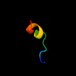
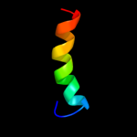
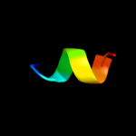
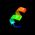
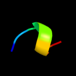
Image coloured by rainbow N → C terminus
Model (left) based on template d1xl3c1
Top template information
Fold:Type III secretion system domain
Superfamily:Type III secretion system domain
Family:TyeA-like
Confidence and coverage
Confidence: 25.7% Coverage: 6%
13 residues ( 6% of your sequence) have been modelled with 25.7% confidence by the single highest scoring template.
You may wish to submit your sequence to Phyrealarm. This will automatically scan your sequence every week for new potential templates as they appear in the Phyre2 library.
Please note: You must be registered and logged in to use Phyrealarm.
3D viewing
Interactive 3D view in Jmol

Sequence analysis 

Secondary structure and disorder prediction 

   1........10.........20.........30.........40.........50.........60
Sequence  MVQKPLIKQGYSLAEEIANSVSHGIGLVFGIVGLVLLLVQAVDLNASATAITSYSLYGGS
Secondary structure 


SS confidence 


Disorder  ??????????


?????


Disorder confidence 


   .........70.........80.........90.........100.........110.........120
Sequence  MILLFLASTLYHAIPHQRAKMWLKKFDHCAIYLLIAGTYTPFLLVGLDSPLARGLMIVIW
Secondary structure 


SS confidence 


Disorder 


Disorder confidence 


   .........130.........140.........150.........160.........170.........180
Sequence  SLALLGILFKLTIAHRFKILSLVTYLAMGWLSLVVIYEMAVKLAAGSVTLLAVGGVVYSL
Secondary structure 


SS confidence 


Disorder 


Disorder confidence 


   .........190.........200.........210.........
Sequence  GVIFYVCKRIPYNHAIWHGFVLGGSVCHFLAIYLYIGQA
Secondary structure 


SS confidence 


Disorder 


??
Disorder confidence 


Confidence Key
High(9)                    Low (0)
?Disordered
Alpha helix
Beta strand

Domain analysis 

Hover over an aligned region to see model and summary info

Please note, only up to the top 20 hits are modelled to reduce computer load

RankAligned region

PDB 1xl3 chain C domain 1

3D model

Region: 188 - 200
Aligned: 13
Modelled: 13
Confidence: 25.7%
Identity: 31%
Fold: Type III secretion system domain
Superfamily: Type III secretion system domain
Family: TyeA-like

Phyre2

PDB 2e74 chain G domain 1

3D model

Region: 172 - 186
Aligned: 15
Modelled: 15
Confidence: 20.3%
Identity: 40%
Fold: Single transmembrane helix
Superfamily: PetG subunit of the cytochrome b6f complex
Family: PetG subunit of the cytochrome b6f complex

Phyre2

PDB 1vf5 chain G

3D model

Region: 172 - 186
Aligned: 15
Modelled: 15
Confidence: 20.0%
Identity: 40%
Fold: Single transmembrane helix
Superfamily: PetG subunit of the cytochrome b6f complex
Family: PetG subunit of the cytochrome b6f complex

Phyre2

PDB 1vf5 chain G

3D model

Region: 172 - 186
Aligned: 15
Modelled: 15
Confidence: 20.0%
Identity: 40%
PDB header:photosynthesis
Chain: G: PDB Molecule:protein pet g;
PDBTitle: crystal structure of cytochrome b6f complex from m.laminosus

Phyre2

PDB 2jv7 chain A

3D model

Region: 4 - 24
Aligned: 21
Modelled: 21
Confidence: 19.9%
Identity: 10%
PDB header:metal binding protein
Chain: A: PDB Molecule:calcium-binding protein;
PDBTitle: nmr solution structure of histoplasma capsulatum cbp2 homodimer

Phyre2

PDB 1srb chain A

3D model

Region: 1 - 7
Aligned: 7
Modelled: 7
Confidence: 12.1%
Identity: 29%
PDB header:toxin
Chain: A: PDB Molecule:sarafotoxins precursor;
PDBTitle: conformational studies on srtb, a non-selective endothelin2 receptor agonist, and on irl 1620, an etb receptor3 specific agonist

Phyre2

PDB 1ik7 chain A

3D model

Region: 188 - 214
Aligned: 27
Modelled: 27
Confidence: 12.0%
Identity: 26%
Fold: DEATH domain
Superfamily: DEATH domain
Family: DEATH domain, DD

Phyre2

PDB 2khs chain B

3D model

Region: 3 - 22
Aligned: 20
Modelled: 20
Confidence: 9.4%
Identity: 25%
PDB header:hydrolase
Chain: B: PDB Molecule:nuclease;
PDBTitle: solution structure of snase121:snase(111-143) complex

Phyre2

PDB 1rkc chain B

3D model

Region: 2 - 10
Aligned: 9
Modelled: 9
Confidence: 9.3%
Identity: 33%
PDB header:cell adhesion, structural protein
Chain: B: PDB Molecule:talin;
PDBTitle: human vinculin head (1-258) in complex with talin's2 vinculin binding site 3 (residues 1944-1969)

Phyre2

PDB 2kkt chain A

3D model

Region: 2 - 10
Aligned: 9
Modelled: 9
Confidence: 8.9%
Identity: 11%
PDB header:transcription
Chain: A: PDB Molecule:ataxin-7-like protein 3;
PDBTitle: solution structure of the sca7 domain of human ataxin-7-l3 protein

Phyre2

PDB 1mul chain A

3D model

Region: 1 - 33
Aligned: 33
Modelled: 33
Confidence: 6.8%
Identity: 15%
Fold: IHF-like DNA-binding proteins
Superfamily: IHF-like DNA-binding proteins
Family: Prokaryotic DNA-bending protein

Phyre2

PDB 2xse chain A

3D model

Region: 1 - 48
Aligned: 48
Modelled: 48
Confidence: 6.7%
Identity: 8%
PDB header:oxidoreductase
Chain: A: PDB Molecule:thymine dioxygenase jbp1;
PDBTitle: the structural basis for recognition of j-base containing2 dna by a novel dna-binding domain in jbp1

Phyre2

PDB 1owf chain B

3D model

Region: 1 - 10
Aligned: 10
Modelled: 10
Confidence: 6.6%
Identity: 30%
Fold: IHF-like DNA-binding proteins
Superfamily: IHF-like DNA-binding proteins
Family: Prokaryotic DNA-bending protein

Phyre2

PDB 2o97 chain B domain 1

3D model

Region: 1 - 10
Aligned: 10
Modelled: 10
Confidence: 6.6%
Identity: 30%
Fold: IHF-like DNA-binding proteins
Superfamily: IHF-like DNA-binding proteins
Family: Prokaryotic DNA-bending protein

Phyre2

PDB 1p71 chain A

3D model

Region: 1 - 10
Aligned: 10
Modelled: 10
Confidence: 6.5%
Identity: 20%
Fold: IHF-like DNA-binding proteins
Superfamily: IHF-like DNA-binding proteins
Family: Prokaryotic DNA-bending protein

Phyre2

PDB 3kb4 chain D

3D model

Region: 185 - 201
Aligned: 17
Modelled: 17
Confidence: 6.4%
Identity: 24%
PDB header:structural genomics, unknown function
Chain: D: PDB Molecule:alr8543 protein;
PDBTitle: crystal structure of the alr8543 protein in complex with2 geranylgeranyl monophosphate and magnesium ion from nostoc sp. pcc3 7120, northeast structural genomics consortium target nsr141

Phyre2

PDB 2ldf chain A

3D model

Region: 1 - 7
Aligned: 7
Modelled: 7
Confidence: 6.1%
Identity: 14%
PDB header:toxin
Chain: A: PDB Molecule:sarafotoxin-m;
PDBTitle: solution structure of the long sarafotoxin srtx-m

Phyre2

PDB 2koe chain A

3D model

Region: 56 - 68
Aligned: 13
Modelled: 13
Confidence: 5.9%
Identity: 23%
PDB header:membrane protein, signaling protein
Chain: A: PDB Molecule:human cannabinoid receptor 1 - helix 7/8 peptide;
PDBTitle: human cannabinoid receptor 1 - helix 7/8 peptide

Phyre2

PDB 3msq chain C

3D model

Region: 185 - 201
Aligned: 17
Modelled: 17
Confidence: 5.7%
Identity: 24%
PDB header:biosynthetic protein
Chain: C: PDB Molecule:putative ubiquinone biosynthesis protein;
PDBTitle: crystal structure of a putative ubiquinone biosynthesis protein2 (npun02000094) from nostoc punctiforme pcc 73102 at 2.85 a resolution

Phyre2

PDB 3s90 chain D

3D model

Region: 3 - 19
Aligned: 17
Modelled: 17
Confidence: 5.3%
Identity: 18%
PDB header:cell adhesion
Chain: D: PDB Molecule:talin-1;
PDBTitle: human vinculin head domain vh1 (residues 1-252) in complex with murine2 talin (vbs33; residues 1512-1546)

Phyre2
1

d1xl3c1
2

d2e74g1
3

d1vf5g_
4

c1vf5G_
5

c2jv7A_
6

c1srbA_
7

d1ik7a_
8

c2khsB_
9

c1rkcB_
10

c2kktA_
11

d1mula_
12

c2xseA_
13

d1owfb_
14

d2o97b1
15

d1p71a_
16

c3kb4D_
17

c2ldfA_
18

c2koeA_
19

c3msqC_
20

c3s90D_
21


22


23


24


Detailed template information 

#
Template Alignment Coverage3D Model Confidence
% i.d. Template Information
1d1xl3c1


25.7 31 Fold:Type III secretion system domain
Superfamily:Type III secretion system domain
Family:TyeA-like
2d2e74g1


20.3 40 Fold:Single transmembrane helix
Superfamily:PetG subunit of the cytochrome b6f complex
Family:PetG subunit of the cytochrome b6f complex
3d1vf5g_


20.0 40 Fold:Single transmembrane helix
Superfamily:PetG subunit of the cytochrome b6f complex
Family:PetG subunit of the cytochrome b6f complex
4c1vf5G_


20.0 40 PDB header:photosynthesis
Chain: G: PDB Molecule:protein pet g;
PDBTitle: crystal structure of cytochrome b6f complex from m.laminosus
5c2jv7A_


19.9 10 PDB header:metal binding protein
Chain: A: PDB Molecule:calcium-binding protein;
PDBTitle: nmr solution structure of histoplasma capsulatum cbp2 homodimer
6c1srbA_


12.1 29 PDB header:toxin
Chain: A: PDB Molecule:sarafotoxins precursor;
PDBTitle: conformational studies on srtb, a non-selective endothelin2 receptor agonist, and on irl 1620, an etb receptor3 specific agonist
7d1ik7a_


12.0 26 Fold:DEATH domain
Superfamily:DEATH domain
Family:DEATH domain, DD
8c2khsB_


9.4 25 PDB header:hydrolase
Chain: B: PDB Molecule:nuclease;
PDBTitle: solution structure of snase121:snase(111-143) complex
9c1rkcB_


9.3 33 PDB header:cell adhesion, structural protein
Chain: B: PDB Molecule:talin;
PDBTitle: human vinculin head (1-258) in complex with talin's2 vinculin binding site 3 (residues 1944-1969)
10c2kktA_


8.9 11 PDB header:transcription
Chain: A: PDB Molecule:ataxin-7-like protein 3;
PDBTitle: solution structure of the sca7 domain of human ataxin-7-l3 protein
11d1mula_


6.8 15 Fold:IHF-like DNA-binding proteins
Superfamily:IHF-like DNA-binding proteins
Family:Prokaryotic DNA-bending protein
12c2xseA_


6.7 8 PDB header:oxidoreductase
Chain: A: PDB Molecule:thymine dioxygenase jbp1;
PDBTitle: the structural basis for recognition of j-base containing2 dna by a novel dna-binding domain in jbp1
13d1owfb_


6.6 30 Fold:IHF-like DNA-binding proteins
Superfamily:IHF-like DNA-binding proteins
Family:Prokaryotic DNA-bending protein
14d2o97b1


6.6 30 Fold:IHF-like DNA-binding proteins
Superfamily:IHF-like DNA-binding proteins
Family:Prokaryotic DNA-bending protein
15d1p71a_


6.5 20 Fold:IHF-like DNA-binding proteins
Superfamily:IHF-like DNA-binding proteins
Family:Prokaryotic DNA-bending protein
16c3kb4D_


6.4 24 PDB header:structural genomics, unknown function
Chain: D: PDB Molecule:alr8543 protein;
PDBTitle: crystal structure of the alr8543 protein in complex with2 geranylgeranyl monophosphate and magnesium ion from nostoc sp. pcc3 7120, northeast structural genomics consortium target nsr141
17c2ldfA_


6.1 14 PDB header:toxin
Chain: A: PDB Molecule:sarafotoxin-m;
PDBTitle: solution structure of the long sarafotoxin srtx-m
18c2koeA_


5.9 23 PDB header:membrane protein, signaling protein
Chain: A: PDB Molecule:human cannabinoid receptor 1 - helix 7/8 peptide;
PDBTitle: human cannabinoid receptor 1 - helix 7/8 peptide
19c3msqC_


5.7 24 PDB header:biosynthetic protein
Chain: C: PDB Molecule:putative ubiquinone biosynthesis protein;
PDBTitle: crystal structure of a putative ubiquinone biosynthesis protein2 (npun02000094) from nostoc punctiforme pcc 73102 at 2.85 a resolution
20c3s90D_


5.3 18 PDB header:cell adhesion
Chain: D: PDB Molecule:talin-1;
PDBTitle: human vinculin head domain vh1 (residues 1-252) in complex with murine2 talin (vbs33; residues 1512-1546)
21d1exea_


not modelled 5.2 27 Fold:IHF-like DNA-binding proteins
Superfamily:IHF-like DNA-binding proteins
Family:Prokaryotic DNA-bending protein
22c2wy3B_


not modelled 5.1 12 PDB header:immune system/viral protein
Chain: B: PDB Molecule:uncharacterized protein ul16;
PDBTitle: structure of the hcmv ul16-micb complex elucidates select2 binding of a viral immunoevasin to diverse nkg2d ligands
23c2kkrA_


not modelled 5.1 22 PDB header:transcription, protein binding
Chain: A: PDB Molecule:ataxin-7;
PDBTitle: solution structure of sca7 zinc finger domain from human ataxin-72 protein
24c1unvA_


not modelled 5.1 17 PDB header:four helix bundle
Chain: A: PDB Molecule:general control protein gcn4;
PDBTitle: structure based engineering of internal molecular surfaces2 of four helix bundles

Binding site prediction 

Due to computational demand, binding site predictions are not run for batch jobs

If you want to predict binding sites, please manually submit your model of choice to 3DLigandSite


Phyre is for academic use only

Please cite: Protein structure prediction on the web: a case study using the Phyre server
Kelley LA and Sternberg MJE. Nature Protocols 4, 363 - 371 (2009) [pdf] [Import into BibTeX]
 
If you use the binding site predictions from 3DLigandSite, please also cite:
3DLigandSite: predicting ligand-binding sites using similar structures.
Wass MN, Kelley LA and Sternberg MJ Nucleic Acids Research 38, W469-73 (2010) [PubMed]
 
© Structural Bioinformatics Group
Imperial College London
Lawrence Kelley, Benjamin Jefferys
Disclaimer
Terms and Conditions
Component software
Template detection: HHpred 1.51
Secondary structure prediction: Psi-pred 2.5
Disorder prediction: Disopred 2.4
Transmembrane prediction: Memsat_SVM
Multi-template modelling and ab initio: Poing 1.0