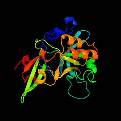
Phyre Home Retrieve Phyre Job Id
Emaill.a.kelley@imperial.ac.uk
DescriptionP76318
DateThu Jan 5 12:21:45 GMT 2012
Unique Job IDd3baa5c222a17b02

Summary 

Top model
Image coloured by rainbow N → C terminus
Model (left) based on template c2icuB_
Top template information
PDB header:structural genomics, unknown function
Chain: B: PDB Molecule:hypothetical protein yedk;
PDBTitle: crystal structure of hypothetical protein yedk from escherichia coli
Confidence and coverage
Confidence:100.0% Coverage: 96%
213 residues ( 96% of your sequence) have been modelled with 100.0% confidence by the single highest scoring template.
3D viewing
Interactive 3D view in Jmol

Sequence analysis 

Secondary structure and disorder prediction 

   1........10.........20.........30.........40.........50.........60
Sequence  MCGRFAQSQTREDYLALLAEDIERDIPYDPEPIGRYNVAPGTKVLLLSERDEHLHLDPVF
Secondary structure 


SS confidence 


Disorder  ????


?
??


Disorder confidence 


   .........70.........80.........90.........100.........110.........120
Sequence  WGYAPGWWDKPPLINARVETAATSRMFKPLWQHGRAICFADGWFEWKKEGDKKQPFFIYR
Secondary structure 


SS confidence 


Disorder 


??


Disorder confidence 


   .........130.........140.........150.........160.........170.........180
Sequence  ADGQPIFMAAIGSTPFERGDEAEGFLIVTAAADQGLVDIHDRRPLVLSPEAAREWMRQEI
Secondary structure 


SS confidence 


Disorder 


Disorder confidence 


   .........190.........200.........210.........220..
Sequence  SGKEASEIAASGCVPANQFSWHPVSRAVGNVKNQGAELIQPV
Secondary structure 


SS confidence 


Disorder 


?????


????
Disorder confidence 


Confidence Key
High(9)                    Low (0)
?Disordered
Alpha helix
Beta strand

Domain analysis 

Hover over an aligned region to see model and summary info

Please note, only up to the top 20 hits are modelled to reduce computer load

RankAligned region

PDB 2icu chain B

3D model

Region: 3 - 222
Aligned: 213
Modelled: 220
Confidence: 100.0%
Identity: 98%
PDB header:structural genomics, unknown function
Chain: B: PDB Molecule:hypothetical protein yedk;
PDBTitle: crystal structure of hypothetical protein yedk from escherichia coli

Phyre2

PDB 2f20 chain A domain 1

3D model

Region: 2 - 221
Aligned: 215
Modelled: 219
Confidence: 100.0%
Identity: 20%
Fold: BB1717-like
Superfamily: BB1717-like
Family: BB1717-like

Phyre2

PDB 2bdv chain A domain 1

3D model

Region: 2 - 217
Aligned: 208
Modelled: 216
Confidence: 100.0%
Identity: 38%
Fold: BB1717-like
Superfamily: BB1717-like
Family: BB1717-like

Phyre2

PDB 1zn6 chain A domain 1

3D model

Region: 2 - 204
Aligned: 191
Modelled: 197
Confidence: 100.0%
Identity: 20%
Fold: BB1717-like
Superfamily: BB1717-like
Family: BB1717-like

Phyre2

PDB 2aeg chain A

3D model

Region: 2 - 208
Aligned: 197
Modelled: 199
Confidence: 100.0%
Identity: 15%
PDB header:structural genomics, unknown function
Chain: A: PDB Molecule:hypothetical protein agr_pat_140;
PDBTitle: x-ray crystal structure of protein atu5096 from agrobacterium2 tumefaciens. northeast structural genomics consortium target atr63.

Phyre2

PDB 2aeg chain A domain 1

3D model

Region: 2 - 208
Aligned: 197
Modelled: 207
Confidence: 100.0%
Identity: 15%
Fold: BB1717-like
Superfamily: BB1717-like
Family: BB1717-like

Phyre2

PDB 3uks chain B

3D model

Region: 159 - 176
Aligned: 18
Modelled: 18
Confidence: 36.4%
Identity: 17%
PDB header:hydrolase
Chain: B: PDB Molecule:sedoheptulose-1,7 bisphosphatase, putative;
PDBTitle: 1.85 angstrom crystal structure of putative sedoheptulose-1,72 bisphosphatase from toxoplasma gondii

Phyre2

PDB 1d9q chain A

3D model

Region: 159 - 175
Aligned: 17
Modelled: 17
Confidence: 34.1%
Identity: 41%
Fold: Carbohydrate phosphatase
Superfamily: Carbohydrate phosphatase
Family: Inositol monophosphatase/fructose-1,6-bisphosphatase-like

Phyre2

PDB 1bk4 chain A

3D model

Region: 159 - 176
Aligned: 18
Modelled: 18
Confidence: 34.0%
Identity: 44%
Fold: Carbohydrate phosphatase
Superfamily: Carbohydrate phosphatase
Family: Inositol monophosphatase/fructose-1,6-bisphosphatase-like

Phyre2

PDB 1nuw chain A

3D model

Region: 159 - 176
Aligned: 18
Modelled: 18
Confidence: 33.2%
Identity: 50%
Fold: Carbohydrate phosphatase
Superfamily: Carbohydrate phosphatase
Family: Inositol monophosphatase/fructose-1,6-bisphosphatase-like

Phyre2

PDB 2gq1 chain A

3D model

Region: 159 - 175
Aligned: 17
Modelled: 17
Confidence: 32.9%
Identity: 18%
PDB header:hydrolase
Chain: A: PDB Molecule:fructose-1,6-bisphosphatase;
PDBTitle: crystal structure of recombinant type i fructose-1,6-bisphosphatase2 from escherichia coli complexed with sulfate ions

Phyre2

PDB 1fta chain A

3D model

Region: 159 - 176
Aligned: 18
Modelled: 18
Confidence: 32.0%
Identity: 44%
Fold: Carbohydrate phosphatase
Superfamily: Carbohydrate phosphatase
Family: Inositol monophosphatase/fructose-1,6-bisphosphatase-like

Phyre2

PDB 1spi chain A

3D model

Region: 159 - 177
Aligned: 19
Modelled: 19
Confidence: 30.4%
Identity: 37%
Fold: Carbohydrate phosphatase
Superfamily: Carbohydrate phosphatase
Family: Inositol monophosphatase/fructose-1,6-bisphosphatase-like

Phyre2

PDB 2fhy chain L

3D model

Region: 159 - 176
Aligned: 18
Modelled: 18
Confidence: 27.9%
Identity: 44%
PDB header:hydrolase
Chain: L: PDB Molecule:fructose-1,6-bisphosphatase 1;
PDBTitle: structure of human liver fpbase complexed with a novel2 benzoxazole as allosteric inhibitor

Phyre2

PDB 1v55 chain B

3D model

Region: 160 - 176
Aligned: 17
Modelled: 17
Confidence: 22.0%
Identity: 35%
PDB header:oxidoreductase
Chain: B: PDB Molecule:cytochrome c oxidase polypeptide ii;
PDBTitle: bovine heart cytochrome c oxidase at the fully reduced state

Phyre2

PDB 3dtu chain B domain 1

3D model

Region: 160 - 176
Aligned: 17
Modelled: 17
Confidence: 17.3%
Identity: 41%
Fold: Cupredoxin-like
Superfamily: Cupredoxins
Family: Periplasmic domain of cytochrome c oxidase subunit II

Phyre2

PDB 2hew chain F domain 1

3D model

Region: 97 - 104
Aligned: 8
Modelled: 8
Confidence: 17.0%
Identity: 25%
Fold: TNF-like
Superfamily: TNF-like
Family: TNF-like

Phyre2

PDB 2hew chain F

3D model

Region: 97 - 104
Aligned: 8
Modelled: 8
Confidence: 17.0%
Identity: 25%
PDB header:cytokine
Chain: F: PDB Molecule:tumor necrosis factor ligand superfamily member 4;
PDBTitle: the x-ray crystal structure of murine ox40l

Phyre2

PDB 2yqp chain A

3D model

Region: 1 - 6
Aligned: 6
Modelled: 6
Confidence: 16.9%
Identity: 50%
PDB header:gene regulation, hydrolase
Chain: A: PDB Molecule:probable atp-dependent rna helicase ddx59;
PDBTitle: solution structure of the zf-hit domain in dead (asp-glu-2 ala-asp) box polypeptide 59

Phyre2

PDB 3ehb chain B domain 1

3D model

Region: 160 - 176
Aligned: 17
Modelled: 17
Confidence: 15.9%
Identity: 29%
Fold: Cupredoxin-like
Superfamily: Cupredoxins
Family: Periplasmic domain of cytochrome c oxidase subunit II

Phyre2
1

c2icuB_
2

d2f20a1
3

d2bdva1
4

d1zn6a1
5

c2aegA_
6

d2aega1
7

c3uksB_
8

d1d9qa_
9

d1bk4a_
10

d1nuwa_
11

c2gq1A_
12

d1ftaa_
13

d1spia_
14

c2fhyL_
15

c1v55B_
16

d3dtub1
17

d2hewf1
18

c2hewF_
19

c2yqpA_
20

d3ehbb1
21


22


23


24


25


26


27


28


29


30


31


32


33


34


35


36


37


38


39


40


41


42


43


44


45


46


47


Detailed template information 

#
Template Alignment Coverage3D Model Confidence
% i.d. Template Information
1c2icuB_


100.0 98 PDB header:structural genomics, unknown function
Chain: B: PDB Molecule:hypothetical protein yedk;
PDBTitle: crystal structure of hypothetical protein yedk from escherichia coli
2d2f20a1


100.0 20 Fold:BB1717-like
Superfamily:BB1717-like
Family:BB1717-like
3d2bdva1


100.0 38 Fold:BB1717-like
Superfamily:BB1717-like
Family:BB1717-like
4d1zn6a1


100.0 20 Fold:BB1717-like
Superfamily:BB1717-like
Family:BB1717-like
5c2aegA_


100.0 15 PDB header:structural genomics, unknown function
Chain: A: PDB Molecule:hypothetical protein agr_pat_140;
PDBTitle: x-ray crystal structure of protein atu5096 from agrobacterium2 tumefaciens. northeast structural genomics consortium target atr63.
6d2aega1


100.0 15 Fold:BB1717-like
Superfamily:BB1717-like
Family:BB1717-like
7c3uksB_


36.4 17 PDB header:hydrolase
Chain: B: PDB Molecule:sedoheptulose-1,7 bisphosphatase, putative;
PDBTitle: 1.85 angstrom crystal structure of putative sedoheptulose-1,72 bisphosphatase from toxoplasma gondii
8d1d9qa_


34.1 41 Fold:Carbohydrate phosphatase
Superfamily:Carbohydrate phosphatase
Family:Inositol monophosphatase/fructose-1,6-bisphosphatase-like
9d1bk4a_


34.0 44 Fold:Carbohydrate phosphatase
Superfamily:Carbohydrate phosphatase
Family:Inositol monophosphatase/fructose-1,6-bisphosphatase-like
10d1nuwa_


33.2 50 Fold:Carbohydrate phosphatase
Superfamily:Carbohydrate phosphatase
Family:Inositol monophosphatase/fructose-1,6-bisphosphatase-like
11c2gq1A_


32.9 18 PDB header:hydrolase
Chain: A: PDB Molecule:fructose-1,6-bisphosphatase;
PDBTitle: crystal structure of recombinant type i fructose-1,6-bisphosphatase2 from escherichia coli complexed with sulfate ions
12d1ftaa_


32.0 44 Fold:Carbohydrate phosphatase
Superfamily:Carbohydrate phosphatase
Family:Inositol monophosphatase/fructose-1,6-bisphosphatase-like
13d1spia_


30.4 37 Fold:Carbohydrate phosphatase
Superfamily:Carbohydrate phosphatase
Family:Inositol monophosphatase/fructose-1,6-bisphosphatase-like
14c2fhyL_


27.9 44 PDB header:hydrolase
Chain: L: PDB Molecule:fructose-1,6-bisphosphatase 1;
PDBTitle: structure of human liver fpbase complexed with a novel2 benzoxazole as allosteric inhibitor
15c1v55B_


22.0 35 PDB header:oxidoreductase
Chain: B: PDB Molecule:cytochrome c oxidase polypeptide ii;
PDBTitle: bovine heart cytochrome c oxidase at the fully reduced state
16d3dtub1


17.3 41 Fold:Cupredoxin-like
Superfamily:Cupredoxins
Family:Periplasmic domain of cytochrome c oxidase subunit II
17d2hewf1


17.0 25 Fold:TNF-like
Superfamily:TNF-like
Family:TNF-like
18c2hewF_


17.0 25 PDB header:cytokine
Chain: F: PDB Molecule:tumor necrosis factor ligand superfamily member 4;
PDBTitle: the x-ray crystal structure of murine ox40l
19c2yqpA_


16.9 50 PDB header:gene regulation, hydrolase
Chain: A: PDB Molecule:probable atp-dependent rna helicase ddx59;
PDBTitle: solution structure of the zf-hit domain in dead (asp-glu-2 ala-asp) box polypeptide 59
20d3ehbb1


15.9 29 Fold:Cupredoxin-like
Superfamily:Cupredoxins
Family:Periplasmic domain of cytochrome c oxidase subunit II
21d2hevf1


not modelled 14.6 38 Fold:TNF-like
Superfamily:TNF-like
Family:TNF-like
22c2dhiA_


not modelled 14.4 11 PDB header:signaling protein
Chain: A: PDB Molecule:pleckstrin homology domain-containing family b
PDBTitle: solution structure of the ph domain of evectin-2 from mouse
23c1m57H_


not modelled 14.2 35 PDB header:oxidoreductase
Chain: H: PDB Molecule:cytochrome c oxidase;
PDBTitle: structure of cytochrome c oxidase from rhodobacter2 sphaeroides (eq(i-286) mutant))
24c1ar1B_


not modelled 13.7 29 PDB header:complex (oxidoreductase/antibody)
Chain: B: PDB Molecule:cytochrome c oxidase;
PDBTitle: structure at 2.7 angstrom resolution of the paracoccus2 denitrificans two-subunit cytochrome c oxidase complexed3 with an antibody fv fragment
25c1uijA_


not modelled 12.8 12 PDB header:sugar binding protein
Chain: A: PDB Molecule:beta subunit of beta conglycinin;
PDBTitle: crystal structure of soybean beta-conglycinin beta2 homotrimer (i122m/k124w)
26d1v54b1


not modelled 11.5 24 Fold:Cupredoxin-like
Superfamily:Cupredoxins
Family:Periplasmic domain of cytochrome c oxidase subunit II
27c3c3vA_


not modelled 10.9 10 PDB header:allergen
Chain: A: PDB Molecule:arachin arah3 isoform;
PDBTitle: crystal structure of peanut major allergen ara h 3
28c1fftG_


not modelled 9.4 12 PDB header:oxidoreductase
Chain: G: PDB Molecule:ubiquinol oxidase;
PDBTitle: the structure of ubiquinol oxidase from escherichia coli
29c2b3gB_


not modelled 9.0 42 PDB header:replication
Chain: B: PDB Molecule:cellular tumor antigen p53;
PDBTitle: p53n (fragment 33-60) bound to rpa70n
30d2k49a2


not modelled 8.7 20 Fold:YegP-like
Superfamily:YegP-like
Family:YegP-like
31c3kscD_


not modelled 8.1 8 PDB header:plant protein
Chain: D: PDB Molecule:lega class;
PDBTitle: crystal structure of pea prolegumin, an 11s seed globulin2 from pisum sativum l.
32d2k8ea1


not modelled 8.1 26 Fold:YegP-like
Superfamily:YegP-like
Family:YegP-like
33c1jsuC_


not modelled 8.0 27 PDB header:complex (transferase/cyclin/inhibitor)
Chain: C: PDB Molecule:p27;
PDBTitle: p27(kip1)/cyclin a/cdk2 complex
34d2gysa1


not modelled 7.8 9 Fold:Immunoglobulin-like beta-sandwich
Superfamily:Fibronectin type III
Family:Fibronectin type III
35c2l14B_


not modelled 7.6 26 PDB header:protein binding
Chain: B: PDB Molecule:cellular tumor antigen p53;
PDBTitle: structure of cbp nuclear coactivator binding domain in complex with2 p53 tad
36c2k8eA_


not modelled 7.5 30 PDB header:structural genomics, unknown function
Chain: A: PDB Molecule:upf0339 protein yegp;
PDBTitle: solution nmr structure of protein of unknown function yegp from e.2 coli. ontario center for structural proteomics target ec0640_1_1233 northeast structural genomics consortium target et102.
37c1t3bA_


not modelled 7.5 11 PDB header:isomerase
Chain: A: PDB Molecule:thiol:disulfide interchange protein dsbc;
PDBTitle: x-ray structure of dsbc from haemophilus influenzae
38c2gs0B_


not modelled 7.3 29 PDB header:transcription
Chain: B: PDB Molecule:cellular tumor antigen p53;
PDBTitle: nmr structure of the complex between the ph domain of the2 tfb1 subunit from tfiih and the activation domain of p53
39c3rbbA_


not modelled 6.9 44 PDB header:viral protein, protein binding
Chain: A: PDB Molecule:protein nef;
PDBTitle: hiv-1 nef protein in complex with engineered hck sh3 domain
40d1cyxa_


not modelled 6.6 18 Fold:Cupredoxin-like
Superfamily:Cupredoxins
Family:Periplasmic domain of cytochrome c oxidase subunit II
41c1cyxA_


not modelled 6.6 18 PDB header:electron transport
Chain: A: PDB Molecule:cyoa;
PDBTitle: quinol oxidase (periplasmic fragment of subunit ii with2 engineered cu-a binding site)(cyoa)
42c3qacA_


not modelled 5.6 10 PDB header:plant protein
Chain: A: PDB Molecule:11s globulin seed storage protein;
PDBTitle: structure of amaranth 11s proglobulin seed storage protein from2 amaranthus hypochondriacus l.
43d1kr4a_


not modelled 5.6 23 Fold:Ferredoxin-like
Superfamily:GlnB-like
Family:Divalent ion tolerance proteins CutA (CutA1)
44d2k57a1


not modelled 5.5 29 Fold:Sm-like fold
Superfamily:Sm-like ribonucleoproteins
Family:YgdI/YgdR-like
45c2b7fD_


not modelled 5.4 25 PDB header:hydrolase/hydrolase inhibitor
Chain: D: PDB Molecule:htlv protease;
PDBTitle: crystal structure of human t-cell leukemia virus protease, a novel2 target for anti-cancer design
46d1f0ya1


not modelled 5.4 29 Fold:6-phosphogluconate dehydrogenase C-terminal domain-like
Superfamily:6-phosphogluconate dehydrogenase C-terminal domain-like
Family:HCDH C-domain-like
47d1p1la_


not modelled 5.3 23 Fold:Ferredoxin-like
Superfamily:GlnB-like
Family:Divalent ion tolerance proteins CutA (CutA1)

Binding site prediction 

Due to computational demand, binding site predictions are not run for batch jobs

If you want to predict binding sites, please manually submit your model of choice to 3DLigandSite


Phyre is for academic use only

Please cite: Protein structure prediction on the web: a case study using the Phyre server
Kelley LA and Sternberg MJE. Nature Protocols 4, 363 - 371 (2009) [pdf] [Import into BibTeX]
 
If you use the binding site predictions from 3DLigandSite, please also cite:
3DLigandSite: predicting ligand-binding sites using similar structures.
Wass MN, Kelley LA and Sternberg MJ Nucleic Acids Research 38, W469-73 (2010) [PubMed]
 
© Structural Bioinformatics Group
Imperial College London
Lawrence Kelley, Benjamin Jefferys
Disclaimer
Terms and Conditions
Component software
Template detection: HHpred 1.51
Secondary structure prediction: Psi-pred 2.5
Disorder prediction: Disopred 2.4
Transmembrane prediction: Memsat_SVM
Multi-template modelling and ab initio: Poing 1.0