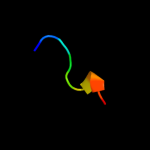
1 c3bk7A_
30.9
60
PDB header: hydrolyase/translationChain: A: PDB Molecule: abc transporter atp-binding protein;PDBTitle: structure of the complete abce1/rnaase-l inhibitor protein2 from pyrococcus abysii
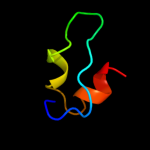
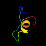
2 c2abyA_
17.5
40
PDB header: unknown functionChain: A: PDB Molecule: hypothetical protein ta0743;PDBTitle: solution structure of ta0743 from thermoplasma acidophilum
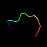
3 c2hlwA_
17.1
16
PDB header: ligase, signaling proteinChain: A: PDB Molecule: ubiquitin-conjugating enzyme e2 variant 1;PDBTitle: solution structure of the human ubiquitin-conjugating2 enzyme variant uev1a
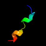
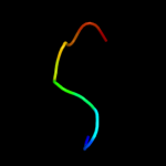
4 c1gthD_
15.6
23
PDB header: oxidoreductaseChain: D: PDB Molecule: dihydropyrimidine dehydrogenase;PDBTitle: dihydropyrimidine dehydrogenase (dpd) from pig, ternary2 complex with nadph and 5-iodouracil
5 c2kvoA_
15.4
32
PDB header: photosynthesisChain: A: PDB Molecule: photosystem ii reaction center psb28 protein;PDBTitle: solution nmr structure of photosystem ii reaction center psb28 protein2 from synechocystis sp.(strain pcc 6803), northeast structural3 genomics consortium target sgr171
6 c2gksB_
13.9
28
PDB header: transferaseChain: B: PDB Molecule: bifunctional sat/aps kinase;PDBTitle: crystal structure of the bi-functional atp sulfurylase-aps kinase from2 aquifex aeolicus, a chemolithotrophic thermophile
7 d3gata_
12.7
23
Fold: Glucocorticoid receptor-like (DNA-binding domain)Superfamily: Glucocorticoid receptor-like (DNA-binding domain)Family: Erythroid transcription factor GATA-18 c3u1xA_
10.7
39
PDB header: hydrolaseChain: A: PDB Molecule: putative glycosyl hydrolase;PDBTitle: crystal structure of a putative glycosyl hydrolase (bdi_1869) from2 parabacteroides distasonis atcc 8503 at 1.70 a resolution
9 d2bs2b1
9.8
24
Fold: Globin-likeSuperfamily: alpha-helical ferredoxinFamily: Fumarate reductase/Succinate dehydogenase iron-sulfur protein, C-terminal domain10 c2kslA_
9.8
38
PDB header: toxinChain: A: PDB Molecule: u1-agatoxin-ta1a;PDBTitle: structure of the insecticidal toxin taitx-1
11 c1wsoA_
9.4
78
PDB header: neuropeptideChain: A: PDB Molecule: orexin-a;PDBTitle: the solution structures of human orexin-a
12 c2kaeA_
9.3
17
PDB header: transcription/dnaChain: A: PDB Molecule: gata-type transcription factor;PDBTitle: data-driven model of med1:dna complex
13 c2gmhA_
9.1
30
PDB header: oxidoreductaseChain: A: PDB Molecule: electron transfer flavoprotein-ubiquinonePDBTitle: structure of porcine electron transfer flavoprotein-2 ubiquinone oxidoreductase in complexed with ubiquinone
14 d1sj1a_
9.0
30
Fold: Ferredoxin-likeSuperfamily: 4Fe-4S ferredoxinsFamily: Single 4Fe-4S cluster ferredoxin15 c2nsvA_
8.6
63
PDB header: signaling proteinChain: A: PDB Molecule: mating pheromone en-1;PDBTitle: nmr solution structure of the pheromone en-1
16 c3dnlG_
8.5
67
PDB header: viral proteinChain: G: PDB Molecule: hiv-1 envelope glycoprotein gp120;PDBTitle: molecular structure for the hiv-1 gp120 trimer in the b12-2 bound state
17 d1nekb1
8.4
38
Fold: Globin-likeSuperfamily: alpha-helical ferredoxinFamily: Fumarate reductase/Succinate dehydogenase iron-sulfur protein, C-terminal domain18 c2rpvA_
8.3
44
PDB header: immune systemChain: A: PDB Molecule: immunoglobulin g-binding protein g;PDBTitle: solution structure of gb1 with lbt probe
19 c3ivuB_
8.1
22
PDB header: transferaseChain: B: PDB Molecule: homocitrate synthase, mitochondrial;PDBTitle: homocitrate synthase lys4 bound to 2-og
20 d1fxra_
8.0
30
Fold: Ferredoxin-likeSuperfamily: 4Fe-4S ferredoxinsFamily: Single 4Fe-4S cluster ferredoxin21 d1lwha1
not modelled
7.7
30
Fold: Glycosyl hydrolase domainSuperfamily: Glycosyl hydrolase domainFamily: alpha-Amylases, C-terminal beta-sheet domain22 c2w3rG_
not modelled
7.7
21
PDB header: oxidoreductaseChain: G: PDB Molecule: xanthine dehydrogenase;PDBTitle: crystal structure of xanthine dehydrogenase (desulfo form)2 from rhodobacter capsulatus in complex with hypoxanthine
23 d1xera_
not modelled
7.6
44
Fold: Ferredoxin-likeSuperfamily: 4Fe-4S ferredoxinsFamily: Archaeal ferredoxins24 d1iw4a_
not modelled
7.4
32
Fold: Kazal-type serine protease inhibitorsSuperfamily: Kazal-type serine protease inhibitorsFamily: Ovomucoid domain III-like25 d7fd1a_
not modelled
6.9
30
Fold: Ferredoxin-likeSuperfamily: 4Fe-4S ferredoxinsFamily: 7-Fe ferredoxin26 d2cm5a1
not modelled
6.8
29
Fold: C2 domain-likeSuperfamily: C2 domain (Calcium/lipid-binding domain, CaLB)Family: Synaptotagmin-like (S variant)27 d2vuti1
not modelled
6.7
27
Fold: Glucocorticoid receptor-like (DNA-binding domain)Superfamily: Glucocorticoid receptor-like (DNA-binding domain)Family: Erythroid transcription factor GATA-128 d2dlqa2
not modelled
6.6
45
Fold: beta-beta-alpha zinc fingersSuperfamily: beta-beta-alpha zinc fingersFamily: Classic zinc finger, C2H229 d5gata_
not modelled
6.5
27
Fold: Glucocorticoid receptor-like (DNA-binding domain)Superfamily: Glucocorticoid receptor-like (DNA-binding domain)Family: Erythroid transcription factor GATA-130 c1dwlA_
not modelled
6.5
40
PDB header: electron transferChain: A: PDB Molecule: ferredoxin i;PDBTitle: the ferredoxin-cytochrome complex using heteronuclear nmr2 and docking simulation
31 c1s6wA_
not modelled
6.4
83
PDB header: antibioticChain: A: PDB Molecule: hepcidin;PDBTitle: solution structure of hybrid white striped bass hepcidin
32 c2xglB_
not modelled
6.4
31
PDB header: antibioticChain: B: PDB Molecule: colicin-m immunity protein;PDBTitle: the x-ray structure of the escherichia coli colicin m immunity2 protein demonstrates the presence of a disulphide bridge, which is3 functionally essential
33 d1y0ja1
not modelled
6.0
27
Fold: Glucocorticoid receptor-like (DNA-binding domain)Superfamily: Glucocorticoid receptor-like (DNA-binding domain)Family: Erythroid transcription factor GATA-134 d1zata1
not modelled
5.9
17
Fold: L,D-transpeptidase catalytic domain-likeSuperfamily: L,D-transpeptidase catalytic domain-likeFamily: L,D-transpeptidase catalytic domain-like35 c1t4vL_
not modelled
5.8
40
PDB header: hydrolase/hydrolase inhibitorChain: L: PDB Molecule: prothrombin;PDBTitle: crystal structure analysis of a novel oxyguanidine bound to thrombin
36 c3c27A_
not modelled
5.8
40
PDB header: hydrolase/hydrolase inhibitorChain: A: PDB Molecule: thrombin light chain;PDBTitle: cyanofluorophenylacetamides as orally efficacious thrombin inhibitors
37 c2r2mA_
not modelled
5.8
40
PDB header: hydrolase/hydrolase inhibitorChain: A: PDB Molecule: thrombin light chain;PDBTitle: 2-(2-chloro-6-fluorophenyl)acetamides as potent thrombin inhibitors
38 c1t4uL_
not modelled
5.8
40
PDB header: hydrolase/hydrolase inhibitorChain: L: PDB Molecule: prothrombin;PDBTitle: crystal structure analysis of a novel oxyguanidine bound to thrombin
39 d2fug91
not modelled
5.8
57
Fold: Ferredoxin-likeSuperfamily: 4Fe-4S ferredoxinsFamily: Ferredoxin domains from multidomain proteins40 c2fugG_
not modelled
5.8
57
PDB header: oxidoreductaseChain: G: PDB Molecule: nadh-quinone oxidoreductase chain 9;PDBTitle: crystal structure of the hydrophilic domain of respiratory complex i2 from thermus thermophilus
41 d1bc6a_
not modelled
5.8
40
Fold: Ferredoxin-likeSuperfamily: 4Fe-4S ferredoxinsFamily: 7-Fe ferredoxin42 c1m8pB_
not modelled
5.7
38
PDB header: transferaseChain: B: PDB Molecule: sulfate adenylyltransferase;PDBTitle: crystal structure of p. chrysogenum atp sulfurylase in the t-state
43 c2c3yA_
not modelled
5.6
63
PDB header: oxidoreductaseChain: A: PDB Molecule: pyruvate-ferredoxin oxidoreductase;PDBTitle: crystal structure of the radical form of2 pyruvate:ferredoxin oxidoreductase from desulfovibrio3 africanus
44 d1xeba_
not modelled
5.6
21
Fold: Acyl-CoA N-acyltransferases (Nat)Superfamily: Acyl-CoA N-acyltransferases (Nat)Family: N-acetyl transferase, NAT45 d2c42a5
not modelled
5.5
50
Fold: Ferredoxin-likeSuperfamily: 4Fe-4S ferredoxinsFamily: Ferredoxin domains from multidomain proteins46 d2it9a1
not modelled
5.5
29
Fold: ssDNA-binding transcriptional regulator domainSuperfamily: ssDNA-binding transcriptional regulator domainFamily: PMN2A0962/syc2379c-like47 d1h98a_
not modelled
5.5
44
Fold: Ferredoxin-likeSuperfamily: 4Fe-4S ferredoxinsFamily: 7-Fe ferredoxin48 d1gnfa_
not modelled
5.4
27
Fold: Glucocorticoid receptor-like (DNA-binding domain)Superfamily: Glucocorticoid receptor-like (DNA-binding domain)Family: Erythroid transcription factor GATA-149 c1p8vB_
not modelled
5.4
40
PDB header: membrane protein/hydrolaseChain: B: PDB Molecule: prothrombin;PDBTitle: crystal structure of the complex of platelet receptor gpib-alpha and2 alpha-thrombin at 2.6a
50 d1gtea5
not modelled
5.4
50
Fold: Ferredoxin-likeSuperfamily: 4Fe-4S ferredoxinsFamily: Ferredoxin domains from multidomain proteins51 d1iqza_
not modelled
5.2
20
Fold: Ferredoxin-likeSuperfamily: 4Fe-4S ferredoxinsFamily: Single 4Fe-4S cluster ferredoxin52 c1g8gB_
not modelled
5.2
38
PDB header: transferaseChain: B: PDB Molecule: sulfate adenylyltransferase;PDBTitle: atp sulfurylase from s. cerevisiae: the binary product complex with2 aps
53 d2nvna1
not modelled
5.1
29
Fold: ssDNA-binding transcriptional regulator domainSuperfamily: ssDNA-binding transcriptional regulator domainFamily: PMN2A0962/syc2379c-like54 d1pcfa_
not modelled
5.1
18
Fold: ssDNA-binding transcriptional regulator domainSuperfamily: ssDNA-binding transcriptional regulator domainFamily: Transcriptional coactivator PC4 C-terminal domain55 c3immC_
not modelled
5.1
31
PDB header: hydrolaseChain: C: PDB Molecule: putative secreted glycosylhydrolase;PDBTitle: crystal structure of putative glycosyl hydrolase (yp_001301887.1) from2 parabacteroides distasonis atcc 8503 at 2.00 a resolution
56 d1hfel2
not modelled
5.1
50
Fold: Ferredoxin-likeSuperfamily: 4Fe-4S ferredoxinsFamily: Ferredoxin domains from multidomain proteins57 c2kvfA_
not modelled
5.1
50
PDB header: transcriptionChain: A: PDB Molecule: zinc finger and btb domain-containing protein 32;PDBTitle: structure of the three-cys2his2 domain of mouse testis zinc2 finger protein


